Radiology: Recognising Tumours of the Female GU Tract Flashcards
1
Q
Name the types of benign ovarian pathology [6]
A
- ovarian follicle
- ovarian torsion
- polycystic ovarian syndrome
- endometriomas
- haemorrhagic cysts
- dermoid cysts
2
Q
Ovarian Follicle
- Presentation [1]
- Features on ultrasound [1]
A
- asymptomatic
- can look like a cyst on ultrasound
3
Q
Haemorrhagic Cyst
- Definition? [1]
- Presentation? [2]
- Features on ultrasound? [2]
- When does follow-up occur? [1]
A
- haemorrhage into the dominant follicle/functional cyst
- presentation:
- can be asymptomatic or
- can present with pain
- features on ultrasound:
- variable
- cyst with haemorrhagic debris
- follow-up after 6 weeks
4
Q
Endometrioma (The Chocolate Cyst)
- Features on ultrasound [2]
- Features on MRI [1]
A
- features on ultrasound:
- variable
- cyst with haemorrhagic debris
- features on MRI:
- shows evidence of haemorrhage
5
Q
Dermoid Cysts
- Who gets it? [1]
- What is it? [1]
- Features on x-ray [2]
- Features on ultrasound [2]
- Features on CT [4]
A
- young women (usually incidental finding)
- benign neoplasm containing elements from mutiple germ cell layers
- features on x-ray:
- calcification
- possibly teeth
- features on ultrasound:
- heterogenous mass
- solid nodule
- features on CT:
- fat
- fluid
- calcification
- soft tissue
6
Q
Polycystic Ovarian Syndrome
- Definition? [2]
- Features on ultrasound [1]
A
- chronic anvolution syndrome associated with androgen excess that occurs due to clinical and/or biochemical hyperandrogenism
- multiple immature follicles seen on ultrasound
7
Q
Ovarian Torsion
- Definition/cause? [1]
- Who gets it? [1]
- Presentation [4]
- Features on ultrasound [3]
A
- ovary twists on its vascular pedicle
- young women
- presentation:
- abdominal pain
- pelvic pain
- nausea
- vomiting
- features on ultrasound:
- enlarged ovary
- free fluid in pelvis
- absent vascularity in ovary
8
Q
Ovarian Cancer
- Signs & symptoms [5]
- How do you calculate the risk of malignancy index (RMI) and what score is concerning for malignancy? [2]
- Features of malignancy on ultrasound [4]
A
- signs & symptoms:
- abdominal distension
- pelvic or abdominal pain
- feeling full & loss of appetite
- increasing urinary urgency or frequency
- irritiable bowel disease
- how do you calculate the risk of malignancy index (RMI) and what score is concerning for malignancy?
- RMI = ultrasound score x menopausal score x CA 125
- RMI > 200 is a concern for malignancy
- features of malignancy on ultrasound:
- irregular solid or multi-loculated cystic mass
- bilateral ovarian lesions
- solid components on cystic wall
- free fluid in pelvis (ascites/peritoneal nodules)
9
Q
Types of Ovarian Carcinoma
- Types of epithelial ovarian carcinoma? [6]
- Types of non-epithelial ovarian carcinoma? [3]
A
- epithelial ovarian carcinoma:
- serous
- mucinous
- clear cell
- endometroid
- Brenner’s
- squamous
- non-epithelial ovarian carcinoma:
- germ cell (dysgerminoma/teratomas)
- sex cord (granulosa cell/sertoli/leydig/thecoma/fibroma)
- metastatic
10
Q
Serous Ovarian Carcinoma
- Benign features seen on imaging? [1]
- Malignant features seen on imaging? [6]
- Malignant serous ovarian carcinoma is more common in younger/older women (pick one) [1]
A
- benign features:
- large cystic mass
- malignant features:
- thick septations
- solid components
- ascites
- peritoneal metastases
- lymphodenopathy
- distant metastases
- more common in older women
11
Q
What are the 2 causes of bilateral ovarian mass? [2]
A
- primary ovarian malignancy metastases
- metastases from other sources
12
Q
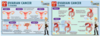
Describe how ovarian cancer is staged from Stage I to Stage IV [4]
A
13
Q
What are the 3 types of uterine pathology? [3]
A
- endometrial cancer
- adenomyosis
- fibroids
14
Q
Fibroids
- Who gets it? [1]
- Presentation [3]
- Features on ultrasound [1]
- Features on CT [1]
A
- premenopausal women (usually found incidentally)
- presentation:
- pain
- infertility
- menorrhagia
- hypoechoic (dark) mass on ultrasound
- bulky/lobulated uterus on CT scan
15
Q
Adenomyosis
- Definition/Cause? [1]
- Presentation [5]
- Features on imaging [1]
A
- endometrial tissue migrating into the myometrium
- presentation:
- asymptomatic or…
- dysmenorrhoea
- menorrhagia
- dyspareunia
- chronic pelvic pain
- thickening of junctional zone seen on imaging
16
Q
Endometrial Cancer
- Who gets it? [1]
- Presentation [1]
- Features on transvaginal ultrasound [1]
- Features on MRI [1]
- Features on CT [1]
A
- post-menopausal women
- vaginal bleeding
- endometrial thickening (>5mm) on transvaginal ultrasound
- local invasion on MRI
- distant metastases on CT
17
Q
Cervical Cancer
- Who gets it? [1]
- Presentation? [3]
- What is the parametrium? [1]
- What defines stage 2b and above in cervical cancer and how does this affect treatment given? [3]
A
- women under 35yrs, mostly associated with HPV virus
- presentation:
- vaginal bleeding
- vaginal discharge or
- picked up with abnormal cervical screening test
- parametrium = fibrous band that separates the cervix from the bladder
- stage 2b and above in cervical cancer = parametrial invasion
- if not invaded → surgery
- if invaded → chemotherapy/radiotherapy
18
Q
Vaginal Cancer
- Presentation? [3]
- What conditions is it often associated with? [2]
A
- presentation:
- bleeding
- lump
- itch
- all of which will not go away
- often associated with:
- cervical cancer metastasis
- HPV virus
