Quiz 2 Web-Only Edit Flashcards
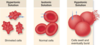
Simple stain (3A)
Consists of one dye that stains a component of the microbial cell
Staining in general is used to enhance contrast in the normally colorless tissue sections, tissue sections are commonly stained. For light microscopic examinations, colored agents (chromophores) are used.
What is the most commonly used basic dye? (3A)
Methylene blue
(shown: Saccharomyces cerevisiae wet mount stained with methylene blue, prepared in lab 3A)

Why is methylene blue a commonly used dye? (3A)
Because it is a basic dye (a cation when in its dissociated, blue coloured form), it dyes the more acidic (negativly charged) components of the cell like DNA and metachromatic granules.

What are two common things from the lab manual that are smeared for examination with methylene blue? (3A)
- Raw milk before microscopic examination
- Throat smears to diagnose diphtheria
What yeast was used in exercise 3A?
Saccharomyces cerevisiae
Briefly describe how to prepare a simple stain (3A)
- Place a dot of water on the slide
- Smear in a bit of the bacteria/yeast/etc
- Let it air dry, then fix it by passing through flame
- Apply several drops of methylene blue; let sit for about a minute
- Rinse with water, dry, and examine
Was a coverslip used to examine the simple stains prepared with methylene blue in experiment 3A?
No, stained organisms were examined without the use of a coverslip
How is methylene blue used to differentiate certain microbes? What microbes does it test for and give two examples from the lab manual (3A):
Eosin Methylene Blue (EMB, also known as “Levine’s formulation”) is a selective stain for Gram-negative bacteria. EMB contains dyes that are toxic for Gram positive bacteria and bile salt which is toxic for Gram negative bacteria other than coliforms. EMB is the selective and differential medium for coliforms. It is a lactose agar base containing a blend of two stains, eosin and methylene blue in the ratio of 6:1. A common application of this stain is in the preparation of EMB agar, a differential microbiological medium, which slightly inhibits the growth of Gram-positive bacteria and provides a color indicator distinguishing between organisms that ferment lactose (e.g., Escherichia coli, Enterobacter aerogenes) and those that do not. Organisms that ferment lactose display “nucleated colonies” – colonies with dark centers.

About how large is a yeast cell? (3A)
7 x 15 microns
Differential stain (3B)
Uses two or more dyes that can be used to categorize cells into groups
(shown: Gram’s stain)

What are the four chemicals used in a Gram stain? Which are stains? Which are mordants? (3B)
i) Crystal violet (stain)
ii) Gram’s iodine (mordant)
iii) Alcohol wash (generally EtOH, decolorizor)
iv) Safranin (counter-stain)

What color do Gram-positive cells stain? (3B)
Purple – hold onto the crystal violet b/c of the thicker peptidoglycan layer, the decolorizer does not penetrate quickly enough to wash it out

What color do Gram-negative cells strain? (3B)
Pink – Gram-negative organisms appear pink because they are counterstained. Because of presence of higher lipid content, after alcohol-treatment, the porosity of the cell wall increases, hence the CVI complex (crystal violet – iodine) can pass through. Thus, the primary stain is not retained. Also, in contrast to most Gram-positive bacteria, Gram-negative bacteria have only a few layers of peptidoglycan and a secondary cell membrane made primarily of lipopolysaccharide.

What type of cells does safaranin stain?
Both Gram+ and Gram- cells are stained pink by safaranin. The Gram+ cells also retain crystal violet due to their thicker layer of peptidoglycan, the lighter safaranin is masked.

mordant
A substance that binds to the dye and makes it less soluble. Most mordants are polyvalent metal ions. In Gram staining, the iodine acts as a mordant or trapping agent.
Why might a cell be Gram-variable? (3B)
- Depending upon how long the culture has been growing, it might appear Gram-positive or Gram-negative.
- For example, many Gram-positive bacteria will appear Gram-negative in later stages of growth.
- Young growths shouldn’t be Gram-variable (less than 12-18 hrs).
What three bacteria were used in the gram-staining procedure? (3B)
- Staphylococcus epidermis - Gram-positive
- Bacillus subtilis - Gram-variable (technically Gram-positive)
- Escherichia coli - Gram-negative
Briefly describe the Gram staining method (3B)
- Apply water to slide, apply bacterial strains to the water (wet mount), let air-dry, and heat-fix
- Stain with crystal violet for ~1 min, rinse w/ water
- Stain with iodine for ~1 min, rinse w/ water
- Decolorize for 2-5 sec
- Stain w/ Safranin for ~30 sec, rinse, dry; examine
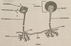
Cell capsule (3C)
- Usually composed of polysaccharides, size varies with environmental conditions
- Protects the cell, but is not essential for cell function, however there may be a correlation in some pathogenic bacteria between virulence and capsule production
- Often destroyed in some methods of staining
- India ink is often used to do a negative stain; stain the background, leaving the cells clear so we can see the capsule
Name a genus that contains a capsule (3C)
- Klebsiella*
(shown: negative stain of Klebsiella from demo 3C)

Name two genus that make spores (3D)
Bacillus; Clostridium
What type of stain would you use to view a capsule?
A negative stain:
Negative staining is an established method, often used in diagnostic microscopy, for contrasting a thin specimen with an optically opaque fluid. In this technique, the background is stained, leaving the actual specimen untouched, and thus visible. This contrasts with ‘positive staining’, in which the actual specimen is stained.
For bright field microscopy, negative staining is typically performed using a black ink fluid such as nigrosin or India ink. The specimen, such as a wet bacterial culture spread on a glass slide, is mixed with the negative stain and allowed to dry. When viewed with the microscope the bacterial cells, and perhaps their spores, appear light against the dark surrounding background.

Negative stain
Negative staining is an established method, often used in diagnostic microscopy, for contrasting a thin specimen with an optically opaque fluid. In this technique, the background is stained, leaving the actual specimen untouched, and thus visible. This contrasts with ‘positive staining’, in which the actual specimen is stained.
For bright field microscopy, negative staining is typically performed using a black ink fluid such as nigrosin or India ink. The specimen, such as a wet bacterial culture spread on a glass slide, is mixed with the negative stain and allowed to dry. When viewed with the microscope the bacterial cells, and perhaps their spores, appear light against the dark surrounding background.

Schaeffer–Fulton stain
The Schaeffer–Fulton stain is a differential staining technique designed to isolate endospores by staining any present endospores green, and any other bacterial bodies red. The primary stain is malachite green, and the counterstain is safranin, which dyes any other bacterial bodies red.