Psych EMQs Flashcards
A. Electroconvulsive therapy (ECT)
B. The “squeeze” technique
C. Sensate focus therapy
D. Cognitive behavioural therapy (CBT)
E. Hormone replacement therapy
F. Phosphodiesterase 5 inhibitors
G. Vaginal trainers
H. Vacuum pump
A painful, involuntary spasm of the vaginal muscles when penetration is attempted.
G. Vaginal trainers
The problem described is vaginismus.
A. Electroconvulsive therapy (ECT)
B. The “squeeze” technique
C. Sensate focus therapy
D. Cognitive behavioural therapy (CBT)
E. Hormone replacement therapy
F. Phosphodiesterase 5 inhibitors
G. Vaginal trainers
H. Vacuum pump
Low sex drive
C. Sensate focus therapy
This problem is also called low libido. Other causes of low libido (such as depression) should be excluded and treated (e.g. with CBT / antidepressants).
A. Electroconvulsive therapy (ECT)
B. The “squeeze” technique
C. Sensate focus therapy
D. Cognitive behavioural therapy (CBT)
E. Hormone replacement therapy
F. Phosphodiesterase 5 inhibitors
G. Vaginal trainers
H. Vacuum pump
Unintentional ejaculation, leaving one or both partners unsatisfied.
B. The “squeeze” technique
This is premature ejaculation. Squeezing the glans penis postpones orgasm.
A. Electroconvulsive therapy (ECT)
B. The “squeeze” technique
C. Sensate focus therapy
D. Cognitive behavioural therapy (CBT)
E. Hormone replacement therapy
F. Phosphodiesterase 5 inhibitors
G. Vaginal trainers
H. Vacuum pump
“Sex addiction”
D. Cognitive behavioural therapy (CBT)
This is hypersexuality or high libido. It can be difficult to treat, but CBT may help.
A. Electroconvulsive therapy (ECT)
B. The “squeeze” technique
C. Sensate focus therapy
D. Cognitive behavioural therapy (CBT)
E. Hormone replacement therapy
F. Phosphodiesterase 5 inhibitors
G. Vaginal trainers
H. Vacuum pump
Vaginal dryness and shrinkage of oestrogen-dependent tissues
E. Hormone replacement therapy
The problem is atrophic vaginitis and is a problem in post-menopausal women.
A. Parkinson’s disease
B. Delirium
C. Frontotemporal lobar degeneration
D. Huntington’s disease
E. Creutzfeldt-Jakob disease
F. HIV encephalopathy
G. Wilson’s disease
H. Tertiary neurosyphilis
I. Dementia with Lewy bodies
J. Alzheimer’s Disease
Instructions: For each of the pathological features below, choose the single most likely diagnosis from the above list of options.
Accumulations of insoluble prion protein

E. Creutzfeldt-Jakob disease
This is a florid plaque from the brain of a patient with variant Creutzfeldt-Jakob disease. It is not a common disease in old age, but should be considered in patients with early onset dementias.
A. Parkinson’s disease
B. Delirium
C. Frontotemporal lobar degeneration
D. Huntington’s disease
E. Creutzfeldt-Jakob disease
F. HIV encephalopathy
G. Wilson’s disease
H. Tertiary neurosyphilis
I. Dementia with Lewy bodies
J. Alzheimer’s Disease
Instructions: For each of the pathological features below, choose the single most likely diagnosis from the above list of options.
B-amyloid protein aggregates into insoluble clumps surrounded by dystrophic neurites

J. Alzheimer’s Disease
This is a plaque from the brain of a patient with Alzheimer’s disease.
A. Parkinson’s disease
B. Delirium
C. Frontotemporal lobar degeneration
D. Huntington’s disease
E. Creutzfeldt-Jakob disease
F. HIV encephalopathy
G. Wilson’s disease
H. Tertiary neurosyphilis
I. Dementia with Lewy bodies
J. Alzheimer’s Disease
Instructions: For each of the pathological features below, choose the single most likely diagnosis from the above list of options.
Eosinophilic, intracytoplasmic neuronal structures form in the cingulate gyrus and neocortex.

I. Dementia with Lewy bodies
This is a cortical Lewy body from a patient with Dementia with Lewy Bodies.
A. Parkinson’s disease
B. Delirium
C. Frontotemporal lobar degeneration
D. Huntington’s disease
E. Creutzfeldt-Jakob disease
F. HIV encephalopathy
G. Wilson’s disease
H. Tertiary neurosyphilis
I. Dementia with Lewy bodies
J. Alzheimer’s Disease
Instructions: For each of the pathological features below, choose the single most likely diagnosis from the above list of options.
Deposits of abnormal protein cause atrophy of the basal ganglia and thalamus, cortical neurone loss, especially affecting frontal regions. Caudate nucleus atrophy may be visible on MRI / CT scan.
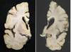
D. Huntington’s disease
The picture on the left is normal; that on the right is from a patient with Huntington’s disease, showing the flattened caudate nucleus.
A. Parkinson’s disease
B. Delirium
C. Frontotemporal lobar degeneration
D. Huntington’s disease
E. Creutzfeldt-Jakob disease
F. HIV encephalopathy
G. Wilson’s disease
H. Tertiary neurosyphilis
I. Dementia with Lewy bodies
J. Alzheimer’s Disease
Instructions: For each of the pathological features below, choose the single most likely diagnosis from the above list of options.
Neurones contain ‘Pick bodies’ and neurofibrillary tangles

C. Frontotemporal lobar degeneration
Frontotemporal lobar degeneration is an umbrella term for a number of disorders united by asymmetrical frontal and / or anterior temporal lobe atrophy. The picture shows Pick bodies, seen in the Pick’s disease subtype. These dementias tend to affect younger patients (under 60).
A. Dementia with Lewy Bodies
B. Metastatic breast carcinoma
C. Subdural haematoma
D. Vascular dementia
E. Normal pressure hydrocephalus
F. Extradural haematoma
G. Subarachnoid haemorrhage
H. Alzheimer’s disease
I. Normal CT head
J. Huntington’s disease

D. Vascular dementia
Non-contrast CT head in a patient multi-infarct dementia. Note the areas of lower density adjacent to the anterior and posterior horns of the lateral ventricles, due to chronic ischaemia.
A. Dementia with Lewy Bodies
B. Metastatic breast carcinoma
C. Subdural haematoma
D. Vascular dementia
E. Normal pressure hydrocephalus
F. Extradural haematoma
G. Subarachnoid haemorrhage
H. Alzheimer’s disease
I. Normal CT head
J. Huntington’s disease

C. Subdural haematoma
Non-contrast CT head in a patient with a subdural haematoma. Note the collection of blood adjacent to the left frontal lobe.
A. Dementia with Lewy Bodies
B. Metastatic breast carcinoma
C. Subdural haematoma
D. Vascular dementia
E. Normal pressure hydrocephalus
F. Extradural haematoma
G. Subarachnoid haemorrhage
H. Alzheimer’s disease
I. Normal CT head
J. Huntington’s disease

E. Normal pressure hydrocephalus
Head CT in a patient with normal pressure hydrocephalus. Note the dilated ventricles, out of proportion to the degree of involutional changes or cortical atrophy.
A. Dementia with Lewy Bodies
B. Metastatic breast carcinoma
C. Subdural haematoma
D. Vascular dementia
E. Normal pressure hydrocephalus
F. Extradural haematoma
G. Subarachnoid haemorrhage
H. Alzheimer’s disease
I. Normal CT head
J. Huntington’s disease

B. Metastatic breast carcinoma
Head CT with contrast of a patient with metastatic breast carcinoma: note the ring-enhancing lesion and surrounding oedema causing a mass effect and some midline shift.
A. Dementia with Lewy Bodies
B. Metastatic breast carcinoma
C. Subdural haematoma
D. Vascular dementia
E. Normal pressure hydrocephalus
F. Extradural haematoma
G. Subarachnoid haemorrhage
H. Alzheimer’s disease
I. Normal CT head
J. Huntington’s disease

I. Normal CT head
Non-contrast CT Head showing a normal brain. Note the difference between the grey and white matter appearances on CT, along with the symmetry of the ventricles.
A. Depressive disorder
B. Postnatal depression
C. Baby blues
D. Schizophrenia
E. Puerperal psychosis
F. Bipolar affective disorder
G. Schizoid personality disorder
H. Adjustment reaction
I. Schizotypal disorder
J. Cyclothymia
Symptoms usually respond to treatment within a month but may persist.
B. Postnatal depression
The treatment of postanatal depression is the same as for regular depression - but extra care in the choice of antidepressants should be taken if the mother is breast-feeding.
A. Depressive disorder
B. Postnatal depression
C. Baby blues
D. Schizophrenia
E. Puerperal psychosis
F. Bipolar affective disorder
G. Schizoid personality disorder
H. Adjustment reaction
I. Schizotypal disorder
J. Cyclothymia
Most patients recover within 6-12 weeks
E. Puerperal psychosis
Puerperal psychosis is a frightening and devastating illness, and treatment should match the presentation (delirium / affective / schizophreniform).
A. Depressive disorder
B. Postnatal depression
C. Baby blues
D. Schizophrenia
E. Puerperal psychosis
F. Bipolar affective disorder
G. Schizoid personality disorder
H. Adjustment reaction
I. Schizotypal disorder
J. Cyclothymia
Mary is a 36 year old woman who had her third child 3 days ago, her husband is concerned as she seems irritable and cries often. Her aunt suffered from depression.
C. Baby blues
Given the time scale the “blues” is the most likely diagnosis.
A. Depressive disorder
B. Postnatal depression
C. Baby blues
D. Schizophrenia
E. Puerperal psychosis
F. Bipolar affective disorder
G. Schizoid personality disorder
H. Adjustment reaction
I. Schizotypal disorder
J. Cyclothymia
The risk of this condition is increased by younger maternal age, poor social support, recent adverse life events or a personal history of depression.
B. Postnatal depression
These are all risk factors for post natal depression.
A. Depressive disorder
B. Postnatal depression
C. Baby blues
D. Schizophrenia
E. Puerperal psychosis
F. Bipolar affective disorder
G. Schizoid personality disorder
H. Adjustment reaction
I. Schizotypal disorder
J. Cyclothymia
1/3 of patients will relapse in subsequent pregnancies.
E. Puerperal psychosis
A third of mothers who have suffered a puerperal psychosis will relapse in future pregnancies - so being aware of this history is important for the team caring for her and her baby in the puerperium.
A. Epilepsy
B. Obsessive compulsive disorder
C. Foetal alcohol syndrome
D. Fragile X
E. Neglect
F. Asperger’s syndrome
G. Autism
H. Down syndrome
I. Schizophrenia
J. Mild depression
Elongated face, high arched palate, “autistic type” behaviour, large testes.
D. Fragile X
These are all symptoms associated with Fragile X. Others include prominent ears and hyperextensible joints.
A. Epilepsy
B. Obsessive compulsive disorder
C. Foetal alcohol syndrome
D. Fragile X
E. Neglect
F. Asperger’s syndrome
G. Autism
H. Down syndrome
I. Schizophrenia
J. Mild depression
Single palmar crease, early-onset Alzheimer’s
H. Down syndrome
These are associated with Down syndrome. Other features include upward-slanting palpebral fissures, epicanthic folds, protruding tongue and hypotonia.
A. Epilepsy
B. Obsessive compulsive disorder
C. Foetal alcohol syndrome
D. Fragile X
E. Neglect
F. Asperger’s syndrome
G. Autism
H. Down syndrome
I. Schizophrenia
J. Mild depression
Poor language and social skills, usually reversible
E. Neglect
Neglect in early childhood will stunt the social and linguistic development of a child. Except in the most severe and prolonged cases, this is usually recoverable if the child is placed with a loving and supportive family.
A. Epilepsy
B. Obsessive compulsive disorder
C. Foetal alcohol syndrome
D. Fragile X
E. Neglect
F. Asperger’s syndrome
G. Autism
H. Down syndrome
I. Schizophrenia
J. Mild depression
Reduced social functioning, speech disorders, auditory hallucinations.
I. Schizophrenia
These would fit with a diagnosis of schizophrenia. Auditory hallucinations differentiate this from autism, where hallucinations would be unexpected.
