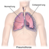
Pneumothorax Flashcards
(6 cards)
What is a pneumothorax?
Collection of air in the pleural space that causes an uncoupling of the lung from the chest wall

Describe the clinical presentation of a pneumothorax
- May be aymptomtic in young patients with a small pneumothorax
- May present with a sudden onset of unilateral pleuritic pain, with progressive breathlessness
- There may the physical signs:
- Reduced expansion
- Increased resonance to percussion
- Decreased breath sounds
- Reduced vocal resonance
What are the causes of a pneumothorax divided into?
What are the causes?
- Spontaneous pneumothorax
- Primary: lung parenchyma otherwise normal, caused by rupture of the apical bleb, often in tall, thin young men
- Secondary: underlying lung disease/abnormalitiy, e.g. COPD, pneumonia, cystic fibrosis, asthmatic, malignancy
- Traumatic pneumothorax
- penetrating trauma e.g. rib fractures
- Iatrogenic pneumothorax
- lung biopsy
- endoscopy
- subclavian cannulation
- postive pressure ventilation
What is the difference between a simple and tension pneumothorax?
Tension pneumothorax
- Air in the pleural space leading to cardiac compromise
- Caused by a valvular mechanism allowing air entry to the pleural space during inspiration, but no exit during expiration
- The intrapleural pressure is very high, deflating lung and decreasing venous return to the heart
- Suggested by:
- tracheal deviation away from the affected side
- respiratory distress
- pallor
- haemodynamic compromise
- distended neck veins
What investigations would you do for a patient with a suspected pneumothorax?
- Expiratory CXR
- trace outline for areas devoid of lung markings and always look for tracheal deviation
- ABG:
- Signs of respiratory distress or chronic lung disease
What is the emergency treatment for a pneumothorax?
Aspiration:
Insert large bore needle attached to syringe partially filled with saline into the 2nd intercostal space in the mid-clavicular line on the side of the suspected lesion. Pull back on the syringe to allow air the bubble out until a chest drain can be inserted. Alternatively, insert a cannula at the same location, and allow air to flow out.
Chest drain:
Pneumothoraces may be aspirated using a chest drain. Chemical pleurodesis with talc is used for patients with contraindication to surgery. Video-assisted thoracoscopic approach may be used to resect a bleb and achieve pleurodesis.


