Pictures Flashcards
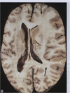
What is shown here?

Ischemic Infarct (appears this way several days after stroke)
probably from embolus lodging in middle cerebral artery
What is shown here?

this is the bifurcation of the common carotid artery into the external & internal carotid arteries
shown here is an atherosclerotic plaque that is obstructing the lumen
part of it is calcified, part of it has hemorrhaged
parts of it are easily broken off–could cause emboli & ischemic stroke
if it becomes completely occluded–could cause a thrombotic stroke
What causes this pathology?

MCA occlusion
ischemic infarct
note the right hemisphere infarct, with swelling & focal dusky discoloration
What is shown here?

this histo slides shows the aftermath of an acute stroke.
only a few hours afterwards
note the edema from cell injury
red neurons (hypereosinophilic & pyknotic)
perivascular neutrophils
dead neurons & glial cells
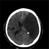
What does this imaging show?

large stroke (dark area)
lots of cerebral edema causing increased ICP & deviation of brain
possible herniation
What does this histo pic show?

acute cerebral infarction
see neutrophils around vascular supply coming to war!
What is shown in this histo pic?

10 days after CVA
macrophages (microglia) in the brain
some reactive gliosis
What is shown in this histo pic?

old cerebral infarction
see gliosis (scarring of the brain)
also see areas of tissue loss
What did this autopsy reveal?

area of cystic degeneration, consistent with old cerebral infarct
What is shown here?

vegetations
not calcifications, but can break off & cause stroke.
caused by infective endocarditis
tannish-reddish
What is shown here?

many small infarcts showing cystic degeneration. perhaps even some bacteria in these cysts.
consistent with vegetations in heart secondary to infective endocarditis

