Pictures Flashcards

1
Q
A

eiosinophils from nasal smear of allergic rhinitis

2
Q
A

nasal polyps

3
Q
A

nasal polyp histology

4
Q
A
  • allergic mucin -sloughed epitheial cells, esionophils, charcot layden crystals, fungal hyphae
    • allergic fungal sinusitis
5
Q
A

frontal sinus mucocele (empyema) secondary to sinusitis

6
Q
A

mucomycosis

7
Q
A

mucorycosis

8
Q
A

asteroid body & shaumann bodies

sarcoidosis

9
Q
A

non necrotizing epitheloid granulomas

sarcoidosis

10
Q
A

accumulation of macrophages in alveolar spaces

desquamative interstitial pneumonia

11
Q
A

stellate shaped nodule formed by collections of langerhans cells near bronchioles

langerhans cell histiocytosis
12
Q
A
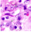
  • langerhans cell histiocytes
  • arrows pointing to nuclear gooves
13
Q
A

birbeck granules in langerhans cells (seen on EM)

langerhans cell histiocytosis

14
Q
A
15
Q
A

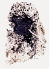
complicated CWP

16
Q
A

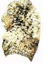
simple CWP

17
Q
A

silicosis

18
Q
A
19
Q
A

asbestos bodies

20
Q
A

pleural plaques

asbestos

21
Q
A

nasopharyngeal angiofibroma (staghorn antlers)

22
Q
A

schneiderian papilloma

23
Q
A

schneiderian papilloma, endophytic type

24
Q
A

olfactory neuoroblastoma

(blue cells)

25
Q
A

squamous cell - naropharyngeal carcinoma

26
Q
A

epstein barr virus (EBV) - nasopharyngeal carcinoma

27
Q
A

gohn complex (red complex) and caseating necrosis (blue arrow)

primary tuberulosis

28
Q
A

cavitation of upper lobes

secondary tuberulosis

29
Q
A

Ziehl-Neelsen stain

m. tuberulosis stains acid fast

30
Q
A

saddle embolus

PE in the righ the pulmonary trunk bifurcation (into r and left main pulmonary arteries)

31
Q
A

bone marrow embolus

32
Q
A

amniotic fluid pulmonary embolism

33
Q
A

fat pulmonary embolism

34
Q
A

marked medial hypertrophy - seen in Pulmonary Artery Hypertorphy (PAH)

35
Q
A

microscopy of diffuse pulmonary hemorrhage syndrome (goodpastures, idiopathic pulm. hemisiderosis, wegners)

  • intra-alveolar RBCs
  • intra-alveolar hemisiderin laden macrophages (prussian blue iron stain)
36
Q
A

goodpastures syndrome

  • intravleolar RBCs + hemosidirin macrophages
  • type 2 pneumocyte hypertrophy
  • thickened alveolar walls
  • second pic: capillaritis (PMNs in capillaries)
37
Q
A

goodpastures syndrome - linear IgG deposits

38
Q
A

wegners (polyangitis with granulomatosis)

necrotizing granulomas in “geographic” pattern

39
Q
A

mucous gland hyperplasia (increased # of goblet cells)

chronic bronchitis

40
Q
A

emphysema

expanded alveoli and blind-end appearing (thinned) septae

41
Q
A

asthma

mucus gland hypertrophy + eisonophils

42
Q
A

charcot-layden crystals

product of excess esionophiol degranulation - dx asthma

43
Q
A

curschmann spirals - from extruded mucous plugs found obtained from BAL specimens

asthma/chronic bronchitis

44
Q
A

squamous papilloma (laryngeal, on true vocal cords)

45
Q
A

laryngeal carcinoma

large, ulcerated fungating lesion

46
Q
A

thyroglossal duct cyst (anterior neck)

cyst surrounded by CT containing lymphoid aggregates & or remnant of thyroid tissue

47
Q
A

warthrin tumor (bilateral parotid gland neoplasm)

mucous filled cysts

48
Q
A

warthrin tumor

cystic spaces lined by a double layer of eosionophillic epithelial cells embedded in lymphoid stroma

49
Q
A

zellballens: nests of oval chief cells (neurectodermal in origin, stain w/ chromagranin) surrourded by delicate vascular septae

paraganglioma (carotid tumor)

50
Q
A

salivary mucocele

51
Q
A

polymorphic adenocarcinoma - minor salivary glands

resemble normal ductal tissue

52
Q
A

adenoid cystic carcinoma - minor aslivary glands

“cribiform” cystic glands - share epithelial wall (mouse in cheese)

53
Q
A

mucoeperimoid tumor (m/c malignant salivary gland tumor)

mucous cells w/ eccentric nuclei + squamoid cells + transition cells

54
Q
A

squamous papilloma of larynx (HPV-6, HPV-II)

stratified squamous epithelium

55
Q
A

brachial cyst (lateral neck, on SCM)

fibrous wall surrounded by lymphoid tissue containing germinal centers

56
Q
A

acinic carcinoma (parotid gland tumor)

granular cytoplasm looks like that of normal acini

57
Q
A

serptentin (crysotile) asbestos - flexible, curved

less pathogenic asbestos

58
Q
A

amphibole asbestos (amosite and crocidolite)

more pathogenic asbestos

59
Q
A

abestos bodies

60
Q
A

dense CT comprising pleural plaques seen in asbestos

61
Q
A

asbestos bodies (blue stain)

asbestos

62
Q
A

asbestos bodies

(asbestos)

63
Q
A

granuloma with central caseation

64
Q
A

high power zoom of a casesating granuloma in mycobacterium tuberlosis

giant cells & histiocytes can be seen

65
Q
A

histoplasmosis

monomorphic, narrow based buds

only effects immunocompromised (tree bark granulomas(

66
Q
A

histoplasmosis

monomorphic, narrow based buds

only effects immunocompromised (tree bark granulomas)

67
Q
A

blastomycosis

  • broad-based, pleormorphic
  • effects immunocompromised AND immunocompetent: suppurative granulomas filled w/ neutrophils
68
Q
A
  • broad-based, pleormorphic
  • effects immunocompromised AND immunocompetent: suppurative granulomas filled w/ neutrophils
69
Q
A

coccoides immitus

HIGHLY infectious

southwest US / mexico / central & south america

organisms are:

  • INTRACELLLUAR (within giant cells)
  • large, round thick walled spherules (these ones contain endospores)
70
Q
A

coccoides immitus

HIGHLY infectious

southwest US / mexico / central & south america

organisms are:

  • INTRACELLLUAR (within giant cells)
  • large, round thick walled non-budding spherules (these ones do not contain endospores)
71
Q
A

CMV pneumonia

owl eyes

72
Q
A

cryptococcus

arrow pointing to capsule

73
Q
A

cryptococcus

will stain pink with mucicarine stain due to its CAPSULE (vs histoplasmosis, blastomycosis, coccidoies - have no capsule)

74
Q
A

oral hairy leukplakia

“balloon cells”

75
Q
A

verrucous carcinoma

(extremely thick tumor)

76
Q
A

ameloblastoma (benign oral tumor)

palisading columnar cells with polarization (piano keys)

77
Q
A

amleblastoma (benign oral mouth tumor)

expansive multiloculated soap bubble appearance

78
Q
A

silicosis

upper lobe concentrated near hilum

79
Q
A

usual interstitial pneumonia

80
Q
A

anthracosis (CWP)

scattered carbon laden macrophages amongst intact parenchyma