Physiology/Pathophysiology Flashcards

1
Q

Draw the simple cascade model of clotting.

A
2
Q

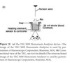
Explain the importance of membrane glutamic acid residues in coagulation.

A
  • Gla residues allow for binding of the protein to a membrane surface via interaction between calcium and membrane phospholipids
  • The Gla residues must be fully carboxylated via the vitamin-K cycle in the liver or they cannot bind the calcium, which prevents binding to the activated cell membrane surface
  • Therefore, vitamin K antagonists prevent carboxylation of the Gla residues and interfere with coagulation
3
Q

What is the normal distribution of PC, PS, PE on the resting cell membrane? What occurs to these phospholipids with cellular injury?

A
  • In a resting cell, PC is expressed on the external leaflet (being neutral) and PS and PE are on the inner leaflet
  • To maintain this resting state:
    • Flippase actively transports PS (and sometimes PE) from the external to the internal leaflet
    • Floppase moves PC from the internal to the external leaflet
  • With injury/activation of the cell:
    • Scramblase shuffles the phospholipids between the two membranes
    • Occurs in response to increased cytosolic calcium
    • Results in appearance of PS/PE on the EXTERNAL membrane
4
Q

What role does the shuffling of the PS/PE and PC play in promotion of coagulation?

A
  • The expression of PS on the outer membrane of a cell surface essentially causes the membrane to become pro-coagulable
    • Cells that do not have PS on their surface are essentially incapable of supporting the coagulation cascade,, generation of enzymes is very slow and cannot lead to thrombin or fibrin generation
  • With PS expressed (and PE, which makes coagulation occur even faster) following injury, the cell surface is a procoagulant surface
5
Q

In addition to the membrane composition/distribution of PC/PS, resting endothelial cells also have a number of other anticoagulant properties. List 3 of these properties.

A
  • Endothelial cells produce HSPGs
    • Binding site for anti-thrombin, which can inactivate any thrombin produced in the area
  • Endothelial cells express thrombomodulin
    • After thrombin binds to TM, it becomes an anti-coagulant molecule through interaction of the thrombin-TM complex with protein C
    • Protein C (+protein S) irreversibly cleaves factors Va and VIIIa, which prevents their participation in generation of any additional thrombin
  • Endothelial cells express TFPI
    • TFPI acts as an upstream inhibitor of FXa and FVIIa
6
Q

What 2 cell types must be present for cell-based coagulation to occur?

A
  • A cell bearing TF
  • Platelets
7
Q

What is the sole relevant initiator of coagulation?

A

Tissue factor

**Cells expressing tissue factor are generally localized outside the vasculature; with an injury to the endothelium, flowing blood is exposed to these tissue factor bearing cells**

8
Q

What are the 3 phases of cell based coagulation?

A

Initiation

Amplification

Propagation

9
Q

Describe what occurs during the initiation phase of cell based coagulation.

A
  • TF on the TF bearing cell interacts with FVIIa (which is the ONLY protein floating already active in the circulation)
    • The TF-FVIIa complex can interact/activate with Factor X:
      • Factor Xa can either interact with it’s cofactor, FV, and thereby generate small amounts of prothrombin and thrombin OR
      • Any factor Xa that dissociates free is inactived by TFPI or AT in the region
    • The TF-FVIIa complex can interact/activate with factor IX
      • Factor IXa can freely dissociate and interact with the surface of platelets and other cells

**Coagulation ONLY progresses beyond the small amount of thrombin generated during initation IF the injury allows platelets and other proteins to leave the vascular space and adhere to the TF bearing cells!!**

10
Q

Describe what occurs during the amplification phase of cell based coagulation.

A
  • After the thrombin has diffused away from the TF-bearing cell during initiation, the thrombin can now be used to activate platelets that have leaked from the vasculature to the site of injury
  • Thrombin:
    • Binds to factor V on the platelet surface, activating it to factor Va
    • Binds to factor XI on the platelet surface, activating it to factor XIa
    • Cleaves the factor XIII+vWF factor complex, resulting in factor XIIIa and free vWF which promotes platelet adhesion
    • Activates the platelet by triggering shuffling of the membrane phospholipids, creating a pro-coagulant surface and release granule contents
11
Q

Describe what happens during the propagation phase of cell based coagulation.

A
  • After a few platelets are activated in the amplification phase, release of granule contents results in recruitment of additional platelets to the site of injury–this is where propagation occurs.
  • Factor IXa
    • Is either present due to the release of IXa during initiation OR
    • Can be created by interaction of XIa (from amplification) with FIX on the platelet surface
  • Factor IXa and factor VIIIa (from amplification) bind together to form the “tenase” complex
    • The tenase complex activates factor X to Xa
    • The majority of Xa must be generated directly on the platelet surface (as the Xa that was generated during initiation is inhibited if it diffuses away from the TF bearing cell)
  • Factor Xa along with FVa cleaves prothrombin to thrombin
  • Prothrombinase (FXa+FVa) leads to a burst of thrombin generation, cleaving fibrinogen into fibrin
  • When there is a critical mass of fibrin, the soluble molecules will polymerize into fibrin strands and create a fibrin matrix
12
Q

Draw the complete pathway of cell based coagulation.

A
13
Q

Describe the additional roles of thrombin in clot formation/structure (aside from formation of fibrin from fibrinogen).

A
  • Thrombin activates factor XIII to XIIIa, which promotes cross-linking between fibrin strands
  • Some thrombin will bind to thrombomodulin on the endothelial cell surface
    • TM bound thrombin activates thrombin activatable fibrinolysis inhibitor (TAFI)
      • TAFI modifies fibrin molecules by removal of terminal lysine residues
      • This makes fibrin markedly more resistant to fibrinolysis
    • TM bound thrombin will also activate protein C
      • aPC complexes with protein S
      • aPC+proS cleave factors Va and VIIIa, which prevents further cofactor activity of either protein
      • Consequently shuts down generation of any new thrombin!!!
14
Q

How long is the average canine platelet lifespan

A

6 days

15
Q

What substances are contained in platelet alpha granules?

Dense granules?

A
  • Alpha granules
    • Platelet derived growth factor
    • Fibronectin
    • TGFBeta
    • Fibrinogen
    • Factors V and VIII
    • VWF
  • Dense granules
    • ADP, ATP
    • Histamine
    • Epinephrine
    • Serotonin
    • Calcium
16
Q

Briefly describe platelet adhesion and aggregation under high shear conditions.

A
  • Under high shear conditions, adhesion to exposed subendothelium is primarily mediated by collagen and vWF
    • vWF is produced by megakaryocytes and endothelial cells
    • Stored in Weibel-Palade bodies in endothelial cells and in alpha granules
  • After endothelial damage, vWF attaches to the exposed collagen, releasing factor XIII
  • Platelets can then roll along the endothelium, mediated by the platelet GP1b-alpha receptor attaching to the vWF on the subendothelium
  • After platelets attach to the endothelium via vWF and collagen, undergo a conformational change, exposing the integrin alphaIIbBeta3 receptor
    • Causes platelets to expose and assemble membrane glycoproteins which can bind fibrinogen and vWF, promiting platelet aggregation

At high shear rates, vWF mediates platelet aggregation!!!

17
Q

Briefly describe platelet adhesion and aggregation under low shear conditions.

A
  • Adherence occurs via collagen, fibronectin and laminin
  • Fibrinogen is the primary ligand of thrombus growth!!
18
Q
A
19
Q

What are the 2 major roles of vWF?

A
  • Carrier protein for factor VIII, protecting it from proteolysis by protein C
  • Mediates adhesion of platelets to damaged endothelium
20
Q

What are the 3 forms of vWD?

A
  • Type I vWD
    • All multimers of vWF are present, but in decreased numbers
  • Type II vWD
    • The large multimers are absent
  • Type III vWD
    • All multimeres are absent
21
Q

List clinical signs seen with primary hemostatic disorders.

List clinical signs seen with secondary hemostatic disorders.

A
22
Q

Describe clot retraction as a test for platelet function.

A
  • Influenced mainly by the number and function of platelets and the fibrinogen concentration in plasma
    • Other influences may change it–i.e. reduced in anemia, prolonged in polycythemia
  • Place 5ml whole blood into a nonadditive tube, insert a wooden applicator and incubated
  • The assessment of clot formation and clot retraction is noted over 8-24 hours
  • Within 2-4 hours, a normal clot should retract markedly
  • To measure % clot retraction, 1ml of whole blood is placed into tubes and incubated
    • At 1 hour, the accumulated serum from around the clot can be removed and measured
    • Volume multipled by 50 to obtain the percent clot retraction (normal is 25-60%)
23
Q

Describe the BMBT as a test for platelet function.

A
  • Measure the time for a stable platelet plug to form following a standardized incision on the upper lip
  • Normal BMBT is less than 3 minutes in dogs
  • A prolonged result is suggestive of thrombocytopenia, thrombocytopathia or vWD
    • Best as a screening tool for further more detailed assays
24
Q

Describe the utility of the PFA100 as a test for platelet function.

A
  • Simulates primary hemostasis by aspirating citrate-anticoagulated whole blood under a high shear rate through a small apearture in a collagen membrane coated with platelet agonists (ADP or epinephrine)
  • Provides a “closure time”–CT, which is the time it takes for a platelet plug to form and occlude flow
    • CT is highly sensitive to qualitative and quantitative defects in platelet receptors that mediate adhesion (GPIb-V-IX) and aggregation (GPIIbIIIa)
  • May be inaccurate in anemic patients, patients with high hematocrits, or platelet counts
  • Doesn’t tell specifically WHY/what specific platelet function defect is present, just identifies that there is one
25
Q

Discuss platelet aggregometry as a test for platelet function.

A
  • Gold standard for diagnosis of primary hemostatic defects
  • Requires platelet rich plasma or washed platelet preparations
    • Add agonist (ADP, alpha/gamma thrombin, collagen)
    • Agonists cause activation/exposure of GPIIbIIIa; fibrinogen binds to adjacent platelets to facilitate aggregation
    • As aggregation continues, the PRP suspension becomes more clear and thus increases light transmission
  • It does not mimic in-vivo responses, requires large volumes of blood and expertise
26
Q

Discuss the “Impact-R” device as a means for testing platelet function.

A
  • Machine simulates blood flow under high shear conditions over and extracellular matrix
  • Sample stained and optically analyzed; can measure average size of platelet aggregates and total surface coverage of the aggregates
27
Q

What types of viscoelastic testing are available? What is the purpose of viscoelastic testing?

A
  • Viscoelastic testing measure changes in viscosity or elasticity of a blood sample during clot formation
  • Good for BROAD understanding of patient’s global coagulation status, but less useful as a test for specific platelet function
  • Sonoclot
  • TEG
  • ROTEM
28
Q

Describe how the sonoclot works.

What does the typical curve look like and what 3 values are generated?

A
  • Sonoclot senses changes in blood viscosity/clot elasticity via a highly sensitive probe that vibrates within the blood sample placed in a cuvette and generates a curve.
    • As blood clots and fibrin strands form between the probe and cuvette, the drag increases, generating a curve
  • Generates activated clotting time, clot rate and PF
    • The initial portion of the curve, typically flat, starts as soon as the probe is placed into the blood and remains straight until fibrin begins to form and blood is no longer liquid
    • The time elapsed before an increase in impedance is seen is the ACT
    • The rate of fibrin formation is defined as the clot rate and is the primary slope
    • The next plateau occurs as platelets initiate contraction of the fibrin strand
    • The secondary slope reflects continued fibrinogenesis, fibrin polymerization, platelet-fibrin interaction
    • The peak clot signal represents initial clot retraction
    • The time to peak and peak clot strength are combined into a unitless factor, PF, with the amplitude of the peak being an index of fibrinogen concentration
29
Q

Describe the principle behind TEG and ROTEM. What is the difference between the 2 tests?

A
  • Based on the measurement of the physical viscoelastic characteristics of clots
  • As fibrin forms, fibrils link the pin to the cup and this is measured
  • ROTEM holds cup stationary with the pin rotating
  • TEG holds the pin stationary and the cup rotates
  • ROTEM traces are produced from a deflection in the angle of light directed at the pin/wire system
  • TEG senses the rotational movement of the pin via the mechanical electrical transducer and converts it into an electrical signal for display
30
Q

What are the 5 variables measured by TEG?

What do these correspond to in ROTEM?

A
  • TEG
    • R (initial fibrin formation)
    • K (Speed of clot formation)
    • Alpha angle (Speed of clot formation)
    • Maximum amplitude (Maximal clot strength)
    • Lysis (LY30, LY60) (Fibrinolysis)
  • ROTEM
    • Clot time (CT) (initial fibrin formation)
    • Clot formation time (CFT) (speed of clot formation)
    • Alpha angle (speed of clot formation)
    • Maximum clot firmness (MCF) (maximal clot strength)
    • Clot lysis (CL30, CL60) (fibrinolysis)
31
Q

What does the R value (CT) signify?

A

Reaction time; latency from the time the blood is placed in the TEG/ROTEM until initial fibrin formation

Typically reflects coagulation factor levels, but doesn’t always correlate with PT/PTT

32
Q

What does the K (CFT) value signify?

A

Measurement to a predetermined level of clot strength (minutes)

33
Q

What does the alpha angle signify?

A

Measure of the speed of fibrin and cross-linkage, thus the speed of clot strengthening

Both the K and alpha angle are affected by:

  1. Availability of fibrinogen
  2. Factor XIII, which facilitates fibrin formation into a stable clot
  3. Platelets (to a much lesser extent than fibrinogen/factor XIII)
34
Q

What does the MA (MCF) signify?

A

Represents the ultimate strength of the clot; direct function of the maximum dynamic properties of fibrin and platelet bonding.

Assessment of the combination of the platelet count and function , as well as fibrinogen activity

35
Q

What is the G value and what does it signify?

A

The G value can be calculated from the MA and is another measure of clot firmness.

Calculate by: G=5000 x MA/ 100-MA)

36
Q

What are the LY30/LY60 (CL30/CL60) variables and what do they signify?

A

Measure of clot stability, measuring lysis of the clot at 30 or 60 minutes after the MA has been identified.
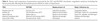
37
Q

Fill in the table.

A
38
Q

Discuss the best practices for sample collection, handling, and storage of blood for a TEG sample.

A
  • Collect from a large vein (jugular preferred)
  • Sodium citrate tubes
  • Complete after a 30 minute stabilization period of the blood at room temperature
  • Avoid vibration, shock, or rapid shifts in temperature; do NOT place on ice as these will all activate and alter PF
39
Q

What are activators that can be utilized with TEG and what is the purpose?

A
  • Tissue factor, kaolin
  • Activators serve for recalcification and activation of the blood sample
  • The activators CANNOT be used interchangeably, as there is a large variation between them
40
Q

Label the tracing.

A
41
Q

Label the TEG tracings with the appropriate abnormality.

A
42
Q

What pathways do the following test:

ACT

PT

PTT

A

ACT: extrinsic and common

PT: extrinsic and common

PTT: intrinsic and common

43
Q
A
44
Q

What are fibrin split products? What are d-dimers?

What do elevations of each signify?

A
  • FSPs or FDPs
    • Formed by dissolution of fibrin/fibrinogen by plasmin, implying increased thrombus formation
    • Elevated levels may be also present due to increased fibrin production (inflammation/neoplasia/trauma) or decreased hepatic clearance
  • D-dimers
    • Unique FSPs that are ONLY present after cross-linking and subsequent lysis of a fibrin clot
    • Elevations in D-dimers indicate ACTIVE thrombosis, since D-dimers originate from the cleavage of the cross-linked fibrin in the final clot
    • Autoagglutination, hypoalbuminemia, increases in corticosteroids can all result in elevated d-dimer levels
45
Q

What are the four stages of DIC as assessed by TEG?

A
  • Stage 1:
    • Hypercoagulable phase
  • Stage 2:
    • Decreased MA value (due to platelet consumption) although coagulation factors are still normal
  • Stage 3:
    • Prolongation of R and K due to consumption of coagulation factors
  • Stage 4:
    • Fibrinolytic/spindle (hypocoagulable) tracing due to excessive fibrinolysis
46
Q

Discuss the role of the endothelial glycocalyx in promotion of a hypercoagulable state. What role does ADAMST13 play?

A
  • With injury, synthesis of the GAG layer is decreased, which decreases the function of the key anti-coagulants that rely on the endothelium (TM, protein C, TFPI)
  • Affter the endothelial cells are activated, they release ultralarge multimers of vWF (UL-vWF) from the Weibel-Palade bodies.
  • The UL-vWF can bind platelet GPIbalpha receptors, inducing platelet tethering and activation.
  • UL-vWF are rapidly cleaved in health to smaller multimers by ADAMST13; the smaller multimers circulate freely and have much less platelet aggregation activity than the UL-vWF molecules.
    • The UL molecules stay tethered at the site of injury
  • Decrease/absence of ADAMST13 can lead to high concentrations of ULvWF, resulting in systemic platelet aggregation and thrombosis.
47
Q

What role do microparticles play in systemic coagulation?

A
  • MPs are circulating small vesicles released from activated/apoptotic cells (platelets, endothelial cells, leukocytes, RBCs, noeplastic cells)
  • Can provide a phospholipid membrane for thrombin generation
    • Can express TF on their surface and those that express PS and TF are characterized as procoagulant MPs
    • May also display vWF-binding sites and UL-vWF multimers
48
Q

What are the 3 primary anticoagulant proteins?

A
  • Antithrombin
  • Protein C
  • TFPI
49
Q

Discuss the role of antithrombin.

A
  • Acts primarily to inhibit TF and factor Xa
  • Most effective when it is bound to heparin-like GAGs of the glycocalyx
    • Typically decreased in systemic inflammation by:
      • Consumption (d/t increased thrombin generation)
      • Decreased production (negative APP)
      • Degradateion by neutrophil elastase
    • Can also be lost via the urine in animals with glomerulonephritis…
50
Q

Discuss the role of the protein C system.

A
  • Activated when trace amounts of thrombin bind TM located on the endothelium
  • When cofactor protein S is present, APC inhibits factors Va and VIIIa
  • By binding thrombin, TM helps generate APC and prevents thrombin from acting on fibrinogen and platelets
  • Reaction also generates TAFI, which inhibits fibrinolysis
  • Protein C system less functional during systemic inflammation resulting from decreased hepatic synthesis of proteins C and S
51
Q

Discuss the role of TFPI.

A
  • TFPI rleased primarily from endothelial cells
  • Acts to inhibit FVIIa and TF complexes as well as Xa
  • Protein S serves as a cofactor for inhibition of FXa by TFPI
52
Q

Discuss fibrinolysis and alterations in the process that occur in the face of systemic inflammation.

A
  • Circulating plasminogen is incorporated into forming fibrin clots and is converted to plasmin by TPA and urokinase
    • TPA and urokinase derived largely from the endothelium; released upon activation/injury
  • Plasmin breaks down the fibrin meshwork of the formed clot and allows for recannulation of blood vessels
  • The effects of plasminogen are decreased by endogenous plasminogen activator inhibitor (PAI-1)
  • In the presence of inflammation (TNFalpha, IL1B), there is a delayed but more sustained increaed in PAI-1, decreasing fibrinolysis and resulting in persistence of thrombi
53
Q

What is Virchow’s triad?

A
  • Endothelial injury
  • Blood stasis
  • Hypercoagulability
54
Q

What 3 cancers are associated with the highest rates of DIC?

A

Hemangiosarcoma

Mammary carcinoma

Adenocarcinoma

55
Q

What are the vitamin K dependent clotting factors?

What is the most appropriate coagulation test for evaluating a vitamin K associated coagulopathY?

A

2,7,9,10 protein C, protein S

PT–because factor VII has the shortest half life

56
Q

Does hypofibrinogenemia result in prolongation of standard coagulation tests?

A

No; PT, PTT typically remain normal until hypofibrinogenemia is SEVERE (less than 50-100mg/dL)

57
Q

What effect does polycythemia have on TEG tracings?

Anemia?

A

Polycythemia produces hypocoagulable tracings

Anemia produces hypercoagulable tracings

**TEG affected by blood viscosity!!**

58
Q

A factor VIII deficiency is what?

Factor IX?

Factor XII?

A

Factor VIII: hemophilia A

Factor IX: hemophilia B

Factor XII: Hagemann factor deficiency (non-pathologic)

**Factor XII is involved in contact factor activation, but is NOT essential for in-vivo hemostasis. Deficiency results in significant prolongation of aPTT without a hemorrhagic tendency**

59
Q

Define massive transfusion.

A

Transfusion of the patient’s full blood volume in 24 hours or more than half its blood volume in 3 hours.

Administration of 1.5ml/kg/min of blood products over 20 minutes

60
Q

What criteria must be met for a diagnosis of DIC?

A

A underlying condition that could lead to DIC

3 or more:

Thrombocytopenia

Prolongation of PT, PTT or TT

Elevated D-dimers

Hypofibrinogenemia

Reduced AT activity

Schistocytes

61
Q

What is the mechanism of action of vincristine in treatment of thrombocytopenia? What evidence supports its use in management of ITP?

A
  • Causes release of platelets from megakaryocytes
  • Impairs phagocytosis of opsonised platelets by impairing microtubule assembly in macrophages
  • Given with standard doses of glucocorticoids, vincristine has been shown to decrease hospitalization time without changing prognosis in dogs with ITP.
62
Q

Compare extrinsic versus intrinsic platelet disorders.

A

Extrinsic disorders are characterized by lack of a functional protein needed for platelet adhesion and aggregation.

Intrinsic disorders are those inherent to platelets.

63
Q

What is methemoglobin and when does clinical methemoglobinemia occur?

A
  • Hemoglobin is made up of four globins, each attached to a heme molecule which contains a central iron molecule
    • The iron must be maintained in its ferrous state for hemoglobin to bind oxygen
  • MetHb is an inactive form of HgB, created when the iron is oxidzed to the ferric state secondary to oxidative damage within the RBC
  • MetHb is incapable of carrying oxygen
    • Increases the affinity of oxygen in the remaining ferrous moeities of the hemoglobin molecule which decreases release of O2 to the tissues and shifts the O2 HgB curve to the left
  • Dusky cyanotic or chocolate brown colored mucous membranes
64
Q

What are Heinz-Bodies?

A
  • Aggregates of denatured precipitated Hgb within RBCs that form as Hgb with oxidative damage is metabolized
  • Oxidation of the SH gropus of Hgb causes a conformational change in the globin chain that leads to precipitation of the denatured globin and aggregates clump into the HZBs, coalescing until pale structures can be seen within the RBC
  • Formation of MetHb is thought to be necessary for development of HZBs
  • Cats are more sensitive as they have 8 SH groups on the globin as opposed to just 4 as dogs do
  • Numerous HZBs can disrupt the RBC membrane sufficiently to result in ghost cells–empty RBCs with just membrane and HZBs remaining, and are associated with oxidation induced intravascular hemolysis
65
Q

List some causes of metHb?

HZBs that lead to anemia due to hemolysis?

A
  • MetHb
    • acetaminophen, benzocaine products, skunk musk, nitrates/nitrites, dapson, metoclopramide, sulfonamides
  • HZBs (can cause some degree of MetHb, but signs typically attributed to hemolytic anemia associated with the HZBs)
    • Onions, garlic
    • Propylene glycol
    • Zinc
    • Methylene blue
    • propofol repeatedly in cats
    • Phenothiazines
66
Q

What is the toxic dose of acetaminophen in cats?

Dogs?

A

Cats: 10mg/kg

Dogs: 150-200mg/kg

67
Q

What are the typical pathways of acetaminophen metabolism and how does toxicity ensue?

A
  • Metabolized in the liver
    • Conjugated to sulfate compound by phenol sulfotransferase
    • Congugated to glucuronide compound by a uridine transferase
    • Can be transformed and oxidized by CP450 system that converts it to NAPQ1
      • GSH reacts with NAPq1 to form mercapturic acid which is excreted in urine
    • PAP (para-aminophenol) also produced by deacylation of acetaminophen
  • Low doses are metabolized readily to nontoxic products, but higher doses can overwhelm the sulfate/glucuronide conjugate systems and deplete GSH stores
  • NAPQ1 and PAP build up and unmetabolized acetaminophen accumulates
68
Q

Why are cats less able to metabolize acetaminophen?

A
  • Cats lack glucoronyl transferase which is necessary for conjugation of acetaminophen
  • Also have somewhat limited sulfate binding capacity
  • Have 1/10 capacity to eliminate acetaminophen as compared to dogs
69
Q

What are the consequences of NAPQ1 and PAP accumulation?

A
  • NAPQ1 leads to hepatocellular damage
  • Both NAPQ1 and PAPcan cause intracellular oxidative damage converting Hgb to metHgb–PAP may be more to blame
  • After acute episode of MetHb production, HzBs begin to form and cause enough changes to trigger hemolysis
  • **Dogs significant hepatic insult, cats HzB anemia—can do both however!!**
70
Q

What are expected clinical signs in a patient with MetHb?

A
  • Signs begin with MetHb level of 20%
    • Tachycardia, tachypnea, dyspnea, lethargy, ataxia, stupor…
  • Cyanosis appears at metHb levels of 12-14% or more
  • Coma and death with metHb >80%
  • Head, neck, limb edema, facial edema (more common in cats)
71
Q

What is the “saturation gap”?

A
  • Comparing pulse oximeter O2 saturation to an arterial blood gas saturation
  • Pulse oximeter determines the ratio of oxyhemoglobin to deoxyhemoglobin; the presence of MetHb distorts this ratio
  • If MetHb levels >30%, the pulse oximeter reading plateaus at 85% regardless of true O2 content
  • Methemoglobinemia should be suspected if the stauration gap is >5%
72
Q

How does NAC aid in treatment of acetaminophen toxicity?

A
  • NAC augments endogenous glutathione and directly interacts with NAPQ1 to form a nontoxic conjugate
  • Increases the fraction of acetaminophen excreted as the sulfate conjugate
    • Half life in cats is halved in cats treated with NAC
  • Initial dose 140mg/kg IV followed by 7 x 70mg/kg q6h
73
Q

What changes are ONLY seen with intravascular hemolysis?

A

Hemoglobinemia and hemoglobinuria

74
Q

What is neonatal isoerythrolysis?

A

Type A or AB kittens born to type B queen; develop HA after absorption of alloantibodies via colostrum

75
Q

What antibodies are most commonly identified in canine IMHA?

A

IgG, alone or in combination with IgM

IgG antibodies are typically poor activators of complement and are eliminated through extravascular hemolysis

IgM antibodies are typically eliminated through a combination of intravascular hemolysis (through activation of classical complement pathway) and extravascular phagocytosis

76
Q

Briefly describe the pathogenesis of IMHA.

A
  • Phagocytosis of opsonized RBC occurs following binding of the Fc fragment of the Ig molecule to Fc receptors on cells of the mono-nuclear phagocytic system
    • The primary site of extravascular erythrophagocytosis is the spleen
  • Interaction of RBC-bound autoantibody with macrophages results in either complete phagocytosis or partial phagocytosis of a portion of the RBC membrane, resulting in formation of a spherocyte
  • Complement fixation can also be initiated, leading to formation of a transmembrane pore and intravascular osmotic hemolysis
77
Q

Why does ingestion of an anticoagulant rodenticide lead to coagulopathy?

A
  • Activation of the vitamin K dependent clotting factors depends on reduced vitamin K (hydroquinone)
  • After the factors are activated, the reduced vitamin K becomes an inactive epoxide
  • Vitamin K epoxide reductase converts the inactive epoxide back to active hydroquinone
  • Anticoagulant rodenticides antagonize the action of vit K epoxide reductase so that levels of hydroquinone decreae and activation of the vitK dependent clotting factors cannot occur
  • Lag period of 3-5 days from exposure to development of signs as factors become depleted
78
Q

Where do the antiplatelet drugs work:

Clopidogrel

Aspirin

Abciximab

A
79
Q

What effect does Clopidogrel have on platelets?

A
  • Decreases ADP and collagen induced platelet aggregation and platelet serotonin release
  • Decreases the release of the procoagulant molecules from platelet granules and may slow in vivo thrombus growth by delaying additional platelet activation
80
Q

What effect does Aspirin have on platelets?

A
  • TXA2 is a potent vasoconstrictor and platelet agonist, produced by activated platelets via COX in the cytosol
    • Uses AA from the platlet membrane
  • Irreversible blockade of platelet COX-1 (thereby blocking TXA2 production), resulting in long lasting platelet inhibition
81
Q

How do the fibrinogen receptor antagonists work?

A
  • Abiciximab (eptifibatide as well)
  • Block the final common pathway of platelet activation–GpIIb/IIIa fibrinogen receptor
  • Interferes with/displaces platelet/fibrinogen binding
82
Q

What is the difference between a “red clot” and a “white clot”?

A

Red clots are clots formed within the venous system under low-shear conditions, consist mostly of fibrin and RBCs

White clots are clots formed within the arterial system under high-shear conditions and consist of platlets held together by fibrin strands

83
Q

What group of medications is appropriate for prevention of arterial thrombi? Venous thrombi? Why?

A
  • The antiplatelet medications are best for arterial thrombi because of their composition (fibrin strands+platelets)
  • The anti-coagulants are best for venous thrombi
  • To target both arterial and venous thrombi, may need a combination of both
84
Q

Why does warfarin potentially have both procoagulant and anticoagulant properties?

A
  • Inhibition of II, VII, IX, X makes it anticoagulant (through inhibition of vitamin K epoxide reductase)
  • Also inhibits proteins C and S which are actually anticoagulant molecules, therefore giving it a brief procoagulant property (period of procoagulant action seen in people immediately after starting warfarin, heparin is given when first starting to counteract this)
85
Q

How is an INR calculated and what is done with that information?

A

INR= [PT patient/PT reference)

Goal is to anticoagulate with warfarin therapy until the INR is 2-4.0 in people

Has not been validated in veterinary patients

86
Q

Compare UFH to LMWH.

A
  • UFH
    • 30000Da
    • Binds to AT, inhibits IIa, IXa, Xa, XIa, XIIa with highest affinity for II and X
    • Considered indirect anticoagulant bc it works by enhancing pre-existing properties of AT
    • Direct anticoagulant effect by causing release of TFPI from the endothelial cell surface
    • Highly protein bound, binds to endothelial cells, macrophages, etc; unpredictable bioavailability
  • LMWH
    • 5000 Da
    • Can bind to AT and IIa
    • Exert primary effect on FXa; associated with fewer major bleeding events than UFH
    • NOT highly protein bound and can’t bind to endothelial cells–more predictable bioavailability
    • Longer elimination half-life, require dosing only every 12-24 hours
87
Q

How is heparin therapy usually monitored?

A

aPTT; goal of obtaining a value 1.5 to 2.0 times normal (newer study with conservative target of 1.2 to 1.5 times baseline aPTT)

Anti-Xa activity can also be measured, however, assays not readily available in vet med

88
Q

How do the direct thrombin inhibitors work?

A
  • Dabigatran
  • Bind to thrombin; both fibrin bound and circulating, do not require AT as a cofactor
  • $$$
  • No studies on dabigatran in vet med
89
Q

How do the FXa inhibitors work?

A
  • Direct inhibition of FXa without the need for AT
  • Apixaban, rivaroxaban
  • ROCKET AF study in humans comparing rivaroxaban to warfarin and found it to be non-inferior for prevention of stroke and systemic TE, with less risk of intracranial hemorrhage and fatal bleeding episodes
90
Q

How does streptokinase work?

A
  • Combines with plasminogen to form a complex that converts plasminogen to plasmin
    • Converts circulating and fibrin-bound plasminogen, therefore considered a nonspecific activator of plasmin
  • Results in systemic proteolytic state that may predispose to bleeding from loss of coagulation factors and fibrinogen and an increase in fibrin degradation products
  • Half life is short, but fibrinogenemia can persist up to 24 hours
  • No longer commercially available
91
Q

How does urokinase work?

A
  • Similar activity to streptokinase (increasing plasmin) but is more fibrin specific because it binds preferentially to the lysine-plasminogen form that differentially accumulates within thrombi
  • Not currently available in the US
92
Q

How does TPA work?

A
  • Primary activator of plasmin in vivo, but does not readily bind circulating plasminogen and does not induce a systemic proteolytic state at physiologic levels
  • High affinity for fibrin and form an intimate relationship within thromi, resulting in a relatively fibrin-specific conversion of plasminogen to plasmin
  • Half life is short (2-3 minutes)–but sustained fibrinolytic state may persist and at high doses, systemic proteolytic state and bleeding can be seen
93
Q

What are the anti-fibrinolytic agents and how do they generally function?

A
  • Aminocaproic acid, tranexamic acid–synthetic lysine analogues
  • Both reversibly inhibit the lysine binding site on plasminogen, which is essential for binding to fibrinogen.
    • Consequently blocks the activation of plasminogen on the surface of fibrinogen and thereby prevents the breakdown of fibrin, although plasmin generation does occur
94
Q

Compare EACA and TXA.

A
  • EACA
    • Competitive inhibition of plasminogen activation; higher doses may also directly inhibit plasmin
    • 2 studies of postop administration to greyhounds significantly reduced prevalence of postop bleeding following gonadectomy and limb amputation
  • TXA
    • Also competitive inhibitor of plasminogen activation; also will inhibit trypsin and thrombin
    • 6-10 times more potent than EACA in-vitro with higher and more sustained anti-fibrinolytic activity
    • CRASH-2 trial; if given with 3 hours of trauma in patients with/at risk for significant bleeding, significantly reduced mortality. Given after 3 hours, seemed to increase risk of death due to bleeding
95
Q

What is the function of DDAVP as a hemostatic agent?

A
  • Stimulation of endothelial release of factor VIII and vWF via stimulation of V2 receptors
  • Plasma concentrations of VIII and vWF double/quadraple within an hour…
  • Patients with hemophilia A or type I vWD benefit from administration of DDAVP along with blood products
  • Does NOT shorten bleeding times in patients witih type II or type III vWD
  • Also enhances platelet function in uremic throbocytopathia/congenital defects of platelet function
  • Tachyphylaxis reported after repeat administration within 48 hours probably because all available factor VIII and vWR has been mobilized from the endothelium
96
Q

How does recombinant factor VIIa work?

A
  • Formation of TF-fVIIa complex at the site of endothelial damage which initiates coagulation, production of thrombin and clot formation
  • TF-independent mechanism whereby rFVIIa at supraphysiologic doses binds directly to the phospholipid membrane of activated platelest activating factor X and leading to a massive rise in thrombin generation at the platelet surface
  • High doses of rFVIIa can compensate for lack of factor VIII or IX in hemophilia A and B patients; termed the “bypass” effect
  • Not used in vet med…
97
Q

What is the difference between FFP and FP?

A
  • FFP contains all the clotting factors
  • FP does NOT contain factors V, VIII and vWF
98
Q

What are the blood types in dogs?

A
  • More than a dozen blood groups–DEAs
  • Most important blood type is DEA 1
    • Strong alloantibody resonse after sensitization of a DEA 1 negative dog by a DEA 1 positive transfusion
      • Can lead to acute hemolytic transfusion reaction in a 1.1 neg dog previously transfused with 1.1 positive blood
  • DEA 1.1 negative considered universal donors for dogs who have never been transfused
  • Also is the Dal antigen
99
Q

What are the blood types in cats?

A
  • A, AB, B
  • AB cats are “universal recipients”
  • All cats with type B blood have very strong naturally occurring anti-A alloantibodies
    • Neonatal isoerythrolysis in A or AB kittents born to B queens mated with A toms
  • Mik RBC antigen; cats negative for Mik have the potential to develop transfusion reaction even if given type specific AB blood
    • No way to test for Mik, therefore, crossmatching even type-matched cats before a transfusion is prudent
100
Q

What is the purpose of the crossmatch? What does the major crossmatch test? The minor?

A
  • Detects serologic compatibility between the recipient and the donor
  • Major crossmatch tests for alloantibodies in recipient’s plasma against donor RBC
  • Minor crossmatch tests for alloantibodies in recipient’s RBC against donor plasma
101
Q

What additive is used in preparation of blood products?

A

CPDA

Citrate

Phosphate

Dextrose

Adenine

102
Q

What is the formula for provision of whole blood?

Packed RBCs?

A

Whole blood (ml) = 2 x desired rise in PCV (%) x kg

pRBCs (ml) = desired rise in PCV (%) x kg

ALSO:

80 x kg x [(Desired-Recipient PCV)/PCV of product] for dogs (whole blood)

60 x “ “ for cats (whole blood)

103
Q

What is the normal RBC lifespan in dogs? Cats?

A

110 days dogs, 70 days cats

104
Q

What are the types of transfusion reactions?

A
  • Immunologic
    • Febrile non-hemolytic (type I):
      • Develops due to binding of antigen and Ig-E complexes with mast cells leading to inflammatory mediator release. Signs include fever, facial swelling and gastrointestinal upset.
    • Acute hemolytic (type II):
      • develops due to interaction between specific IgG/IgM antibodies and red blood cell surface antigen. Signs include those associated with intravascular hemolysis (fever, vomiting, dyspnea, hypotension, hemoglobinemia/hemoglobinuria) and extravascular hemolysis (fever, icterus and declining PCV)
    • Delayed immunologic reaction (48 hours-3 weeks)
      • Cytotoxic reactions (type II) can develop secondary to IgG antibody and red blood cell surface antigen interaction, leading to signs associated with extravascular hemolysis (fever, icterus, declining PCV). Immune complex deposition (type III) can also occur, with deposition of antigen/antibody complexes in the tissues with complement activation and inflammation, associated with signs such as fever, vasculitis, arthralgia, myalgia, glomerulonephritis and lymphadenitis.
  • Non-immunologic
    • Infectious disease transfusion, hypocalcemia, hyperammonemia, volume overload
105
Q

What is a red blood cell storage lesion?

A
  • Multiple deleterious changes in RBC during storage
  • Microparticle accumulation, cytokine accumulation, free iron, increased ammonia levels
106
Q

What is the benefit of leukoreduction?

A
  • In dogs, shown to eliminate the inflammatory response to transfusion, decrease microparticle formation compared with non-LR units, and decrease cytokine production compared with non-LR units.
    *
107
Q

What electrolyte disturbances may develop following massive transfusion?

A
  • Hypocalcemia and hypomagnesemia
    • Arise secondary to chelation with citrate in the blood products
  • Hyperkalemia
    • In people, potassium in stored blood rises, however, not the case in dogs
    • Hyperkalemia thought to arise secondary to potassium leakage into the bloodstream from damaged tissues, extracellular potassium shifting secondary to acidosis, and reduced potassium excretion associated with oliguria
108
Q

What hemostatic defects may arise secondary to massive transfusion?

A
  • Most commonly thrombocytopenia, hypofibrinogenemia, and dilutional coagulopathy
  • Thrombocytopenia
    • Primarily from blood loss and dilution of platelet poor products, fluids
  • Clotting defects
    • Clotting factor consumption secondary to tissue injury
109
Q

Why does a severe metabolic acidosis often arise following massive transfusion?

A

When blood is stored, glucose metabolism leads to an increase in lactic and pyruvic acids; the pH of stored blood may be as low as 6.4 to 6.6, which can lead to systemic consequences.

110
Q

List complications that may arise in association with massive transfusion.

A
111
Q

What are the two major abnormalities associated with ATC?

A

Hypocoagulation

Hyperfibrinolysis

112
Q

List the 3 main hypotheses for the development of ATC.

A
  1. DIC with a fibrinolytic phenotype
  2. Enhanced thrombomodulin-thrombin-protein C pathway
  3. Marked sympathoadrenal response leading to catecholamine induced endothelial damage
113
Q

Discuss the DIC with a fibrinolytic phenotype hypothesis of ATC.

A
  • DIC classically is initially more prothormbotic and hypercoagulable, which then progresses into a consumptive, hypocoagulable, hemorrhagic disorder
    • In patients with ATC, it is proposed that during the first 34-48 hours post injury, patients suffer from DIC with a fibrinolytic phenotype which later progresses to the thrombotic phenotype
  • Immediately after trauma/shock, severe endothelial damage, hypoxia and ischemialeads to marked thrombin generation with subsequent systemic fibrin formation
    • Concurrently there is massive release of TPA into circulation, resulting in large amounts of plasminogen being converted to plasmin
    • Simultaneous events lead to the hyperfibrinolytic and hypocoagulable expression
  • As the patient is resuscitated, then the later prothrombotic state develops subsequent to greater expression of PAI-1 as compared to TPA.
    *
114
Q

Discuss the hypotheisis that ATC develops secondary to increased TM-thrombin-protein C pathway.

A
  • The primary initiators of ATC in this hypothesis are considered to be severe tissue injury and hypoperfusion.
  • BInding of thrombin to thrombomodulin switches thrombin to an anticoagulant agent via activation of protein C.
  • Activated protein C inhibits factors Va and VIIIa, exerting anticoagulant properties, and exerts fibrinolytic protperties via suppresion of PAI-1 and TAFI formation.
115
Q

Discuss the neurohormonal hypothesis of ATC development.

A
  • Trauma with tissue injury induces a dose-dependent sympathoadrenal response and subsequent release of catecholamines into circulation
  • Circulating catecholamines directly damage the endothelial glycocalyx in a dose-dependent fashion, changing the endothelium from a more-antithrombotic to pro-thrombotic state.
    • Change in endothelial function allows for local hemostasis to take place at the site of injury
  • In order to prevent systemic coagulation and maintain local perfusion, the body then attempts to counterbalance the effects of the prothrombotic endothelium with an anticoagulable and fibrinolytic response in the whold blood
  • As the degree of tissue trauma and endothelial damage increases, the counterregulatory response rages out of control, resulting in systemic hypocoagulation and hyperfibrinolysis
116
Q

List 6 key factors shown to influence ATC.

Which 2 have been globally accepted as the 2 main initiators?

A

Tissue injury

Hypoperfusion

Systemic inflammation

Metabolic acidosis

Hypothermia

Hemodilution

**Shock due to ongoing tissue hypoperfusion and the severity of tissue injury**

117
Q

The exacerbation of ATC from which three factors has been termed trauma-induced coagulopathy? (Think lethal triad…)

What does the presence of these abnormalities cause in order to confound ATC?

A

Acidosis

Hypothermia

Hemodilution

Leads to coagulation factor and platelet dysfunction as well as increased fibrinogen consumption and decreased platelet count.

118
Q

Patients with severe tissue injury, but no other major physiological derangements do not commonly present with ATC and have lower mortality. Therefore, tissue injury alone may not be enough to initiate ATC. What sites/types of injury may be more commonly associated with the development of ATC?

A

Traumatic injury to brain and long bones

Presence of a penetrating injury

119
Q

Viscoelastic testing has multiple benefits, however, what is the one central advantage they offer over traditional coagulation assays when it comes to diagnosis of ATC?

A

Allow for identification of the hyperfibrinolysis that is central to ATC, which would allow for institution of appropriate antifibrinolytics.

120
Q

Due to its dynamic nature, ATC is difficult to diagnose. The presence of which things would be highly suggestive of ATC?

A
  • History of sustaining severe trauma with marked tissue injury and presence of makred hypoperfusion (i.e. low systolic BP, BD <-6mmolL, lactate >5mmol/L)
  • Prsence of hemorrhagic shock, uncontrollable intracavitary hemorrhage, or spontaneous bleedign from wounds, catheters
  • VIscoelastic tracing displaying a persistently decreased clot strength of <40% of reference value or prolonged aPTT/PT of >1.5 x normal.
121
Q

What is a contraindication to a delayed/hypotensive resuscitation approach?

A

The presence of TBI or spinal cord injury.

CPP is dependent upon MAP (CPP=MAP-ICP)

Therefore, underresuscitation with a delayed or hypotensive approach may result in delayed CPP.

122
Q

Discuss damage control resuscitation.

A
  • Resuscitative approach designed to systematically and rapidly (within 24-48h) reverse hypothermia, acidosis and coagulopathy and therefore “optimize” the patient before pursuing definitive resuscitation or surgical repair.
  • Involves:
    • Direct treatment of coag abnormalities with appropriate ratios of blood component products
    • Limited fluid resuscitation to maintain an SBP of 80-90mmHg until major hemorrhage has been halted
    • Early prevention, correction of hypothermia
    • Utilization of whole blood/blood products as the primary source of volume resuscitation (hemostatic resuscitation)
    • Abolishment of excessive use of isotonic crystalloids
123
Q

What is the 3-tiered approach to damage control surgery?

A
  1. Initial abbreviated surgery to control hemorrhage, address contamination (<60-90miknutes)
  2. Intensive care to correct physiological derangements
  3. Definitive surgical repair of injuries if stable in 24-72 hours