Physiology Flashcards

1
Q

Boyle’s Law

A

At any constant temperature the pressure exerted by a gas varies inversely with the volume of the gas

(As the volume of a gas INCREASES the pressure of the gas DECREASES)

2
Q

Forces holding the Thoracic Wall and Lung

A

INTRAPLEURAL FLUID COHESIVENESS - the water molecules in the intrapleural fluid are attracted to each other. This means that the pleural membranes stick to each other

NEGATIVE INTRAPLEURAL PRESSURE- the subatmospheric intrapleural pressure creates a transmural pressure gradient across the lung and chest wall.

3
Q

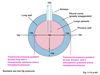
Pressures important in Ventilation

A
  • Atmospheric Pressure
  • Intra-alveolar Pressure
  • Intrapleural Pressure
4
Q

External Respiration

A
  1. Exchange between the Atmosphere and Alveoli
  2. Exchange of O2 and CO2 between the air in alveoli and the blood
  3. Transport of O2 and CO2 from the lungs to the tissues
  4. Exchange of O2 and CO2 between the blood and tissues
5
Q

Inspiration

A
  • Active
  • The thorax is increased vertically by the contraction of the diaphragm which flattens
  • The external intercostal muscle contraction lifts the ribs and moves out the sternum which increases the thorax anteriorly
  • Increase in lung size = intra alveolar pressure falls so air moves into the lungs down the pressure gradient
6
Q

Expiration

A
  • The inspiratory muscle relaxes
  • The chest wall and lungs recoil as elastic properties: this increases intra-alveolar pressure
  • Air leaves the lungs down a different pressure gradient
7
Q

Transpulmonary Pressure

A
  • Transmural pressure gradient in the lungs
  • The difference between alveolar and intrapleural pressure in the lungs
  • Pneumothroax (air in pleural space) removes transmural gradient
8
Q

Alveolar Surface Tension

A
  • Allows the lugns to recoil
  • Attraction between the water molecules which act on the surface of lung tissue
  • Prevents stretching of the lungs, if too strong then alveoli will collapse
9
Q

Surfactant

A
  • Reduces the alveolar surface tension
  • A mixture of lipids and proteins: secreted by Type II alveoli
10
Q

LaPlace’s Law

A
  • Smaller alveoli have a higher tendency to collapse as the have a smaller radius
  • The surfactant lowers the surface tension of smaller alveoli more
11
Q

Respiratory Distress Syndrome

A
  • Developin foetal lungs = unable to produce surfactant until late in development
  • Premature babies may not have enough surfactant
  • Baby makes very strenuous inspiratory efforts to overcome high surface tension
12
Q

Alveolar Interdependence

A
  • Keeps the alveoli open
  • If an alvelolus starts to collapse then the surrounding alveoli are stretched and recoil
  • This exerts expanding force
13
Q

Forces keeeping the Alveoli OPEN

A
  1. Transmural Pressure Gradient
  2. Pulmonary Surfactant
  3. Alveolar Interdependence
14
Q

Forces keeping the Alveoli CLOSED

A
  1. Elasticity of stretched connective tissue
  2. Alveolar Surface Tension
15
Q

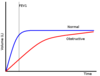
Obstructive Pulmonary Disease

A

Results from an obstruction or blockage in the airways

16
Q

Restrictive Pulmonary Disease

A

Results from the lungs being affected

17
Q

Tidal Volume (TV)

A

Volume of air entering/leaving the lungs during a single breath

Average: 500ml

18
Q

Inspiratory Reserve Volume (IRV)

A

Extra volume of air that can be maximally inspired over and above the typical resting tidal volume

Average: 3000ml

19
Q

Inspiratory Capacity (IC)

A

Maximum volume of air that can be inspired at the end of a normal quiet expiration

IC = Tidal Volume + Inspiratory Reserve Volume

Average: 3500ml

20
Q

Expiratory Reserve Volume (ERV)

A

Extra volume of air that can be actively expired by maximal contraction beyond the normal volume of air after resting tidal volume

Average: 1000ml

21
Q

Residual Volume (RV)

A

Minimum volume of air remaining in the lungs even after a maximal expiration

Average: 1200ml

22
Q

Functional Residual Capacity (FRC)

A

Volume of air in the lungs at the end of a normal passive expiration

FRC = Expiratory Reserve Volume + Residual Volume

Average: 2200ml

23
Q

Vital Capacity (VC)

A

Maximum volume of air that can be moved out during a single breath following a maximal inspiration

VC = Inspiratory Reserve Volume + Tidal Volume + Expiratory Reserve Volume

Average: 4500ml

24
Q

Total Lung Capacity

A

The maximum volume of air that the lungs can hold

Vital Capacity + Residual Volume

Average: ~5700ml

25
Q

Changes in Residual Volume (RV)

A

The RV increases as the elastic recoil of the lungs is lost

e.g. Emphysema

26
Q

Dynamic Lung Volumes

A
  • The use of a volume/time graph to determine;
  • FVC (Forced Vital Capacity)
  • FEV1 (Forced Expiratory Volume in 1 Second )
27
Q

Forced Vital Capacity

A

Maximum volume that can be forcibly expelled from the lungs following a maximum inspiration

28
Q

Forced Expiratory Volume in 1 Second (FEV1)

A

Volume of air that can be expired during the first second of expiration (when determining the FVC)

29
Q

FEV1/FVC Ratio

A

The proportion of the Forced Vital Capacity that can be expired during the first second of expiration in FVC determination

Usually more than 70%, useful diagnostic tool

30
Q

Obstructive Spirometry Curve

A

The FEV1/FVC ratio is lower than 70% because the FEV1 is lower than in normal lungs, but the FVC is the same

31
Q

Restrictive Spirometry Curve

A

The FEV1/FVC ratio is the same as in a normal person, but only because both the FEV1 and FVC are lowered

32
Q

Airway Resistance

A
  • Resistance in the airway is normally very low so air moves with a small pressure gradient
  • Primary Factor = radius of the conducting airway

BRONCHOCONSTRICTION (parasympathetic) increases air flow resistance

BRONCHODILATION (sympathetic) decreases air flow resistance

33
Q

Dynamic Airway Compression

A
  • Dynamic airway compression makes active expiration more difficult in patients with an obstruction
  • The increasing pleural pressure during expiration compresses the alveoli, which allow air to move out of the lungs
34
Q

Dynamic Airway Compression- NORMAL

A
  • The increased airway resistance causes an increase in airway pressure upstream
  • Air travels down the concentration gradient and out of the lungs
35
Q

Dynamic Airway Compression- OBSTRUCTION

A
  • If there is an obstruction the driving force between the alveolus and airway is lost
  • This causes a fall in airway pressure along the airway downstream
  • This is as a result of airway compression from the rising pleural pressure during active expiration
36
Q

Pulmonary Compliance

A
  • During inspiration the lungs are stretched
  • Compliance is a measure of the effort needed to distretch or distend the lungs
  • The less compliant the lungs are, the more work is required to produce a given degree of inflation
37
Q

Decreased Pulmonary Compliance

A

Diseases: Pulmonary Fibrosis, Oedema, Lung Collapse, Pneumonia

  • A greater change in pressure is needed to produce a given change in volume
  • Causes shortness of breath and a restrictive pattern of lung volumes
38
Q

Increased Pulmonary Compliance

A

Diseases: Emphysema

  • May become abnormally increased if the elastic recoil of the lungs is lost
  • Patients have to work harder to expel air
  • Compliance increases with age
39
Q

Work of Breathing

A

Increased in the following ways;

  1. Pulmonary Compliance is DECREASED
  2. Airway Resistance is INCREASED
  3. Elastic Recoil is DECREASED
  4. When a need for INCREASED ventilation arises
40
Q

Peak Flow Meter

A

Assess pulmonary function, useful in obstructive lung disease (best of three attempts is used)

41
Q

Structure of the Alveoli

A

The walls consist of a single layer of flattenes Type I alveolar cells

42
Q

Pulmonary Ventilation

A

Tidal Volume x Respiratory Volume

The volume of air breathed in AND out per minute

Resting Conditions: 6L/min

  • To increase the pulmonary ventilation both the depth and rate of breathing increase
43
Q

Alveolar Ventilation

A
  • Always less that pulmonary ventilation as some inspired air remains in the airway (anatomical dead space)

(Tidal Volume- Dead Space Volume) x Respiratory Rate

Resting Conditions: 4.2L/min

  • The volume of air exchanged between the atmosphere and alveoli per minute: respresents ‘ new air’ available for gas exchange with the blood
44
Q

Breathing Changes

A
45
Q

Ventilation Perfusion

A
  • The transfer of gases between the body and atmosphere depends on:
  • VENTILATION- the rate at which gas is passing through the lungs
  • PERFUSION- the rate at which blood is passing through the lungs
46
Q

Ventilation: Blood Flow Ratio

A
  • The blood flow and ventilation vary from bottom to the top of the lung
  • The V:BF ratio is low at the bottom of the lungs and high at the top of the lungs
47
Q

Alveolar Dead Space

A
  • The match between the air in alveoli and the blood in the pulmonary capillaries isn’t always a perfect match
  • This results in some ventilated alveoli which are not adequately perfused with blood
  • This is termed as alveolar dead space
48
Q

Ventilation Perfusion Match

Perfusion is GREATER than Ventilation

A
  • Increased CO2 results in relaxation of smooth muscle in airways which leads to decreased airway resistance
  • This increases the airflow
  • Decreased O2 causes increased contraction of local pulmonary arteriolar smooth muscle and increases vascular resistance
  • This decreases blood flow
49
Q

Ventilation Perfusion Match

Ventilation is GREATER than Perfusion

A
  • Decreased CO2 leads to increased contraction of local airway smooth muscle and increased airway resistance
  • Decreased airflow
  • Increased O2 causes relaxation of local pulmonary arteriolar smooth muscle and decreased vascular resistance
  • Increased blood flow
50
Q

Gas Exchange across Alveolar Membranes

A

Four factors which influence the rate of gas exchange;

  1. Partial Pressure of O2 and CO2
  2. Diffusion Coefficient for O2 and CO2
  3. Surface Area of Alveolar Membrane
  4. Thickness of Alveolar Membrane
51
Q

Dalton’s Law

A

The total pressure exerted by a gaseous mixture is the sum of the total partial pressures of each individual component in the gas mixture

52
Q

Partial Pressures

A
  • The pressure that one gas in a mixture of gases would exert if it were the only gas present in the whole volume
  • The total atmospheric pressure is 760 mm Hg, most of which comes from Nitrogen
53
Q

Measuring Pressure

A
  • In the UK, the unit kPa (kilopascals) is used
  • It can be found from mm Hg units by dividing by 7.5
54
Q

Alveolar Gas Equation

A

PA O2 = partial pressure of oxygen in alveolar air

Pi O2 = partial pressure of oxygen in inspired air

Pa CO2 = partial pressure of CO2 in arterial blood

0.8 is the respiratory exchange ratio (RER)

55
Q

Water Vapour Pressure

A
  • The air in the respiratory tract is saturated with water
  • This water contributes about 47 mm Hg to the total pressure of the lungs
56
Q

Partial Pressures across Pulmonary Capillaries

A
  • The O2 partial pressure gradient from the alveolus to the capillaries is 60mm Hg
  • The CO2 partial pressure gradient from the capillaries to the alvelous is 6mm Hg
57
Q

Partial Pressures across Systemic Capillaries

A
  • The O2 partial pressure gradient from the capillaries to the tissues is >60mm Hg
  • The CO2 partial pressure gradient from the tissues to the capillaries is >6mm Hg
58
Q

Pressure Gradients

A
  • The partial pressure gradient for CO2 is much smaller than that for oxygen
  • This is offest by the fact that CO2 is much more soluble in membranes that oxygen
  • It is this solubility that is known as the diffusion coefficient: the diffusion coefficient of CO2 is x20 that of O2
59
Q

Alveolar PO2 and Arterial PO2

A
  • The gradient between PAO2 and PO2 should be small
  • A large difference would indicate problems with gas exchange in the lungs or a right to left shunt in the heart
60
Q

Surface Area and Membrane Thickness of Alveoli

A
  • The lungs provide a very large surface area with thin membranes
  • They also have the alveoli and a very extensive pulmonary capillary network
61
Q

Fick’s Law of Diffusion

A

The amount of gas that moves across a sheet of tissue in unit time is proportional to the area of the sheet BUT is inversely proportional to it’s thickness

62
Q

Henry’s Law

(In the context of O2)

A

O2 dissolved in blood ∝ partial pressure of O2

63
Q

Dissolved Oxygen

A
  • There partial pressure of oxygen and diffusion coefficient restricts much O2 from being transported free
  • Only ~1.5% is transported this way
64
Q

Haemoglobin

A
  • Most O2 is bound to Hb, which has 4 haem groups: each of these binds reversibly to one O2 molecule
  • A Hb is considered fully saturated when it is carrying it’s maximum O2 load
  • The PO2 is the primary factor which determines the % saturation of Hb with oxygen
65
Q

Oxygen- Haemoglobin Curve

A
  • 5.3 kPa is the average resting PO2 of systemic capillaries
  • Even if the ‘number’ of Hb molecules DECREASES the oxygen saturation will remain close to 100%
  • This is because all Hb present is carrying it’s maximum oxygen load- there are now just fewer oxygen molecules before
66
Q

Oxygen Delivery Index

A

DO2 I = CaO2 x CI

CaO2 = oxygen content of arterial blood (ml/L) which has the following formula;

CaO2= 1.34 x [Hb] x SaO2 (% saturated with O2)

CI = cardiac index (L/min/metres2)

67
Q

Cardiac Index

A

The cardiac output: body surface area ratio

Units: L/min/metres2

Normal: 2.4 - 4.2

68
Q

Oxygen Delivery

A
  • Oxygen delivery to the tissues can be impaired by;
  • Decreased partial pressure of inspired oxygen (affected by altitude)
  • Respiratory Disease can decrease arterial PO2 and hence decrease Hb saturation with O2
  • Anaemia decreases the Hb concentration and so the oxygen content of the blood
69
Q

Haemoglobin Binding

A
  • Binding of one O2 to Hb increases the affinity of Hb to O2
  • This is called co-operativity and creates a sigmoid curve
  • The curve flattens when all sites are becoming occupied

FLAT = moderate fall in PO2 will not have much affect

STEEP = tissues get a lot of oxygen for a small drop in PO2

70
Q

Bohr Effect

A
  • The Oxygen Dissocation Curve shifts to right in response to raised PO2, H+
  • Also in response to increased 2,3- Biphosphoglycerate
71
Q

Foetal Haemoglobin

A
  • Different structure, with two alpha and two gamma subunits
  • Interacts less with 2,3 BPG in red blood cells
  • Foetal haemoglobin has a higher affinity for oxygen, so transfer can occur even if the the PO2 is low
72
Q

Myoglobin

A
  • Present in cardiac and skeletal muscles
  • 1 Hb group per myoglobin and no cooperative binding of O2
  • It release all O2 at a very low PO2, providing short term storage of O2 for anaerobic conditions
  • Not usually found in blood: if detected then there is muscle damage
73
Q

CO2 transport

A
  • By blood (10%)

- Bicarbonate (60%)

  • Carbamino compounds (30%)
74
Q

Bicarbonate

A

HCO3- is removed from red blood cells by swapping with Cl- ions, this is the chloride shift

The H+ is buffered by Hb, to form HbH

  • Both of these steps allows the favouring of the equilibrium reaction in this direction
75
Q

Carbamino Compounds

A
  • Formed by combination of CO2 with terminal amine groups in blood proteins
  • Rapid reactions which occur even in the absence of an enzyme
  • Reduced Hb (once O2 has been delivered) can bind more CO2 than Hb
76
Q

Haldane Effect

A
  • The Haldane Effect is removing O2 from Hb to increase the ability of Hb to pick up CO2 and CO2 generated H+
  • The Bohr and Haldane Effects work together in synchrony to liberate O2 and take up CO2 etc. at tissues
  • Oxygen shifts the CO2 dissociation curve to the right, as at a higher partial pressure Hb has a weakened ability to bind CO2 and H+
77
Q
A