Physiological Consequences of Airway Obstruction Flashcards
What factors determine the work of breathing?
Load & Drive

What factors affect the load of breathing?
- Stiff lungs
- Narrow airways
- Chest wall
- Diaphragm
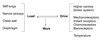
What factors influence the drive to breathe?
- higher centres (limbic system)
- mechanoreceptors
- irritant receptors
- chemoreceptors
- baroreceptors
- temperature

In airflow obstruction, the increased sensation of breathing is due to
- Increased load due to increased friction in the tubes - an increase in the resistive work of breathing
- expiration becomes active

What are the consequences of increased WOB?
- recruitment of accessory muscles (scalene, sternomastoids)
- increased O2 consumption by respiratory muscles (40-50%, compared to 2% in normal)
- risk of respiratory muscle fatigue (severe obstruction)
- can lead to type II respiratory (ventilatory) failure
In normal people, the work done to ventilate is a combination of
- small amount of friction
- small amount of expanding the lungs via elastic tissue
What is type I respiratory failure?
- decreased PaO2 (< 60mmHg)
- decreased PaCO2
i.e. hyperventilation is clearing CO2
What is type II respiratory/ventilatory failure?
caused by inadequate ventilation, it is not necessarily a gas exchange problem
- decreased PaO2
- increased PaCO2
i.e. hypoventilation; occurs when respiratory muscles fatigue
What is the management for type II respiratory/ventilatory failure?
- O2 administration if breathing (will not impact CO2)
- bronchodilators to manage obstruction
- ventilatory support if above are not working or immediate urgent tx required (will lower CO2)
- will automatically +O2 if no gas exchange problem occuring simultaneously
The elastic work of breathing of someone with asthma or COPD is
relatively low
Active exhalation occurs by
contraction of the abdominal and internal ICMs
normal during exercise and in abnormal situations such as significant airway obstruction
During inspiration,
intra-alveolar P ___ Patm
less than
during expiration,
intra-alveolar pressure ___ Patm
greater than
at the end of inspiration and expiration,
intra-alveolar pressure ____ P atm
equals
intra-pleural pressure is always ___ intra-alveolar pressure because
intrapleural pressure is always less than intra-alveolar pressure
due to elastic recoil of the lungs and the chest wall
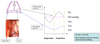
Why is systolic BP normally lower on inspiration than expiration at rest?
- inspiration generates negative intrapleural pressure
- becomes negative transpleural pressure
- lowers BP by reducing pulmonary return to the left side of the heart
What is pulsus paradoxus?
- in severe airflow obstruction
- contraction of inspiratory muscles to generate more negative intrapleural (tf transpleural) pressure to suck air in
- much greater drop in systolic BP on inspiration relative to expiration
- disappears on respiratory muscle fatigue (type II resp/vent failure)
Spirometry measures
- mechanical lung function
- FEV1 (~80% of FVC comes out in the first second, total within 1-3s)
- produces volume vs. time curve (rate of flow)

What is a normal FEV1 and FEV1 ratio?
- > 70% FVC (younger >80%)
- FEV1 decreases with increasing severity of obstruction
- FEV1/FVC > 70% (>80%)
- if
- if

What does a flow-volume loop measure?
- flow rate vs. volume during a forced expiration (upper loop) followed by a forced inspiration (lower loop)
- can distinguish lower bronchial obstruction (asthma, COPD) from higher tracheal obstruction (tumour, stenosis)

What is the general altered breathing pattern of airflow obstruction?
deep, slow breaths (lower frequency)
to minimize resistive work of breathing

What is the general altered pattern of breathing when the lungs are stiff (i.e. increased elastic WOB)?
e.g. pulmonary fiborosis, oedema
small, rapid breaths to minimize elastic WOB

What is maximum minute ventilation, and what are the consequences in chronic obstructive disease?
- same term as maximum ventilation (MV), ~100L/min (minute ventilation)
- 35x FEV1
- FEV1 ~ 4-5L tf MV 100-200L/min
- in severe chronic obstruction, FEV1 < 1 tf MV ~ 20-30L/min
- rest: requires 8-10L/min
- exercise: requires 15-20L/min
- tf limiting factor in severe airflow obstruction; cannot increase ventilation to supply O2 or clear CO2
- in COPD, emphysema, MV is significantly reduced (50, 30, 20 etc.)
In normal individuals, exercise is limited by
- HR (220 - age)
- CO
- O2 metabolism by peripheral muscles
- at maximal exercise, 30% maximum ventilation (MV) is unused


