Physical Assessment of the Newborn Flashcards
When should the initial assessment be done?
in the delivery room to detect significant anomalies, birth injuries, and cardiorespiratory disorders that may compromise a successful adaptation to extrauterine life.
When should the next physical assessment be done?
A more detailed exam should take place in the nursery before the infant’s status is discussed with the parents, within 12-18 hours after birth.
When should a final physical assessment be performed
within 24 hours of discharge.
Skin Assessment
The skin should be soft, smooth and opaque. Vernix caseosa may be present. In a post-term infant, the skin may be dry and peeling. The skin should be warm to touch. Petechiae may be seen over the head/neck or above nipple line secondary to pressure from a nuchal cord.
Vernix Caseosa
whitish greasy material that covers the fetus’s body after 35 weeks gestation, then it decreases in quantity as it is shed into the amniotic fluid with increasing gestational age.

What does discolored vernix caseosa mean
it occurs with hemolytic disease and meconium stating as a result of in utero fetal distress.
skin color plethora
deep rosy red more common in infants with polycythemia, but also in an infant who is over oxygenated or overheated. Obtain a venous HCT.

jaundice
yellow color - bilirubin levels in the blood are usually > 5 mg/dl. This abnormal in infants <24 hr old and may signify a blood incompatibility or infection.

pallor
washed-out or pale skin color; may be secondary to anemia, birth asphyxia, or shock.
Cyanosis
blue or dusky appearance of the skin; reduce O2 sats < 85%; most difficult task with cyanotic neonates is recognizing the cyanosis; best observed when they are quiet, sleeping in a thermoneutral environment under a white light; examine the tongue, oral and buccal mucosa, and peripheral skin.

Peripheral cyanosis
cyanosis of the extremities; “vasomotor instability”; acrocyanosis; may be due to a cold environment, high HCT, local obstruction, or may persist for days, even weeks.

Central cyanosis
cyanosis of the mucous membranes and periphery due to the presence of 3g/dL or more of reduced Hg in arterial blood. May indicate a pathologic condition; persistent central cyanosis always requires immediate eval. Causes of cyanosis can range from trivial to life-threatening.
Extensive bruising at birth
is associated with traumatic birth and may result in early jaundice.

Milia
multiple yellow or pearly white 1 mm papules, scattered on the chin, nose, forehead, and cheeks. Benign, representing tiny epidermal cysts in connection with the sebaceous follicle. Disappear within a few weeks.

sebaceous gland hyperplasia
tine white or yellow lesions visible at the opening of each pilosebaceous follicle. More prominent on the nose, upper lip, and malar regions. Represent hyperplastic sebaceous glands. Spontaneously diminish in size after birth and no longer visible.

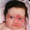
erythema toxicum
Numerous small areas of red skin with a yellowish white papule in the center. Noticeable at 48 hours but can appear as late as 7 days. Resolves spontaneously within 4-5 days after the appearance. (Can be confused with candida dermatitis, miliaria rubra and pustular melanosis)

Mongolian spot
dark blue or purple macular spots resembling bruises, usually located over the lumbosacral area. 90% of African Americans, Native Americans, and Asians 85% of Hispanics 5-10% of Caucasians Fade during late infancy, or at least by 4 yrs. Somer persists to adulthood.

Macular hemangioma
telangiectatic nevus nevus flammeus salmon patch stork bite A true vascular nevus is normally seen on the occipital area, eyelids, and glabella (forehead area over the eyebrows).

When do macular hemangioma’s disappear?
spontaneously within the first year of life EXCEPT those on the nape of the neck, many of which persist The most common type of birthmark, macular hemangioma occurs in 60-70% of all infants.
Harlequin phenomenon
A clear line of demarcation with an area of redness and an area of paleness. Cause is usually unknown. Most likely related to vasomotor instability. It is benign and transient, could indicate shunting of blood is occurring (as in sepsis or persistent pulmonary HTN)

Cutis marmorata
Mottling, lacy red pattern that may be seen in a normal infant or on with cold stress, hypovolemia, or sepsis. Persistent mottling is seen with a variety of chromosomal abnormalities.

Miliaria
Transient lesions that commonly occur in a warm environment as the result of obstruction of the sweat gland ducts.
Miliaria crystallinia
clear superficial tiny vesicles without inflammation

Miliaria rubra
small erythematous, grouped papules on a red base

Petechiae
small pinpoint hemorrhagic skin lesions frequently seen on the presenting part and on the face, especially if there was a nuchal cord. Petechiae on the torso is more likely to be associated with thrombocytopenia or congenital infection.

Pustular melanosis
transient condition of unknown cause, pustules, vesicles, hyperpigmented macules, presenting individually or in combination, single or in clusters, at birth. New lesions do not occur. They may form a brownish crust or rupture, producing a fine white scale around the lesion. Resolve in several days. No associated erythema.

Birthmarks
are very common and occur in 99% of neonates
Salmon patches and Mongolian spots are 100 times more common than all other birthmarks
Port wine stain
nevus flammeus
Deep red or purple in color, usually present at birth, blanch minimally and do not disappear with time.
Can be treated with laser therapy
Sturge-Wener syndrome
Port wine stain that appears over the forehead and upper eyelid, distributed over the first branch of the trigeminal nerve. It is associated with glaucoma, seizures, and mental retardation.

Hemangioma
may not be visible at birth, but 90% are visible by 1 month of age. Proliferation occurs for 6-8 months, but involution may take years
50% at 5 years
90% at 9 years
Periocular lesions require ophthalmologic consultation and aggressive treatment.
Cavernous hemangioma
usually appears as a large red firm ill-defined mass resembling a cyst
can be located anywhere on the body

Strawberry hemangioma
appears within the first few days after birth as a raised pink or red macule that is sharply demarcated
most commonly found on the face
enlarge the first 5-6 months of life
blanch incompletely
usually regress spontaneously

CEPHALOHEMATOMA
This skull deformity is the result of a hemorrhage, usually from a traumatic or forceps delivery. It does NOT cross suture lines.

HYPOSPADIUS
This congenital defect occurs in one in 3000 males where the placement of the urethral meatus is anywhere between the tip of the glans and the perineum.

Epicanthal Folds
This fold over the medial aspect of the eye may be familial but occurs in less than 1% of the population. It is also seen as a common feature of Down syndrome and other syndromes.

Syndactyl
This is the name for the abnormal fusion of the digits (fingers or toes)

Epstein Pearls
These small white inclusion cysts generally cluster around the juncture of the hard and soft palates. This is a normal finding that generally resolves with sucking.

Tonic Neck Reflex or Fencing Position
The reflex demonstrated below occurs by placing the infant in the supine position and turning the head to one side. The upper extremity on the side that the head is turned toward should extend, and the upper extremity on the opposite side should flex.

Brushfield Spots
These “salt and pepper” spots of the iris are often seen with Down syndrome.

Pectus Excavatum
This congenital structural depression of the sternum is usually of no clinical concern.

Hypertelorism
This is the term for widely spaced eyes as depicted below. The diagnosis should be made on the measurement of the inner canthal distance.

Natal Teeth
Present in approximately one in 3,500 live births

Caput Succedanum
A diffuse edematous swelling of the soft tissues of the scalp, which may extend over suture lines

Cleft Palate
Results from incomplete fusion of the palate

Super Numary Nipple
Raised, pigmented areas found vertical and medial to the true nipple

Erb-Duchenne Palsy
The arm is in the position of tight adduction and internal rotation of the shoulder with arm extension and pronation at the elbow.

Tongue Tie (Ankyloglossia)
The frenulum on the underside of the tongue prevents complete tongue protrusion.

Club Feet (Talipes Equinovarus)
The feet are turned downward and inward, and the sole is directed medially

Diastasis Recti
Nonunion of the two rectus muscles from the umbilicus to the xiphoid causing a mild herniation in the midline

Metatarsus Adductis
Adduction of the forefoot, correctable with active ROM. Most common congenital foot deformity.

Simian Crease
A single transverse palmar crease. Highly associated with congenital abnormalities.

Rocker Bottom Feet (Congenital Vertical Talus)
Associated with genetic syndromes, particularly trisomies

UMBILICAL HERNIA

Sacral Dimple

Resting Posture
- Lies with hips abducted and partially flexed, knees flexed
- Arms adducted and flexed
- Fists loosely clenched, thumb rests in the palm or lying adjacent to the fingers
- Abnormal posture: neck extension, flaccid, obligate flexion of thumb, frog-leg positioning

Spontaneous Movement
IS IT:
- Smooth
- Symmetric
- Limbs move in alternating fashion
- Gross movement with discomfort/stimuli
- Abnormal: persistent tremors, seizure activity

Muscle Tone
- Phasic tone—resistance of the upper and lower extremities to movement, tendon reflexes, clonus
- Postural tone—resistance to gravity, traction response, head lag assessment
- Abnormalities: hypotonia, hypertonia

Hypotonia
HYPOTONIA

