Phys exam 2 Flashcards

1
Q

what action is responsible for the heart sounds

A

the valves closing makes the sound of the heart

2
Q

which valves are wider and why?

  • AV valves
  • semilunar valves
A
  • AV valves are wider.
  • semilunar valves are narrow because they need to shoot out with more pressure.
3
Q

The heart does not contract simultaneously.

  • What direction does it contract?
  • What timing stays the same?
A
  • it contracts from top to bottom
    • then bottom to top
  • Left & right stay the same, though
4
Q

Define cardiac output

  • what’s remains the same
  • what may be different

How much does each portion get (%):

  • renal
  • GI
  • muscles
  • cerebral
  • coronary + skin
A
  • cardiac output = volume/minute
    • pressure may change
    • volume stays the same
  • each portion
    • renal, GI, muscles = 25%
    • cerebral = 15%
    • coronary + skin = 5%
5
Q

who discovered that veins have one-way valves

A

william harvey

6
Q

amongst the vessels (artery, arteriole, capillary, vein), which has the highest:

  • velocity
  • surface area
  • volume
  • control over BP
A
  • velocity = aorta
  • surface area = capillaries
  • volume = veins
  • control over BP = arterioles

Arterioles = greatest site of BP drop off

7
Q

what determines the resistance of an arteriole?

  • what medications constrict it?
  • what medications dilate it?
A

smooth muscle

  • constriction = alpha1 agonists
  • dilation = beta2 agonists
  • NOTE: arterioles are the only** part of the vasculature that **moves
8
Q

what is the only thing that determines total peripheral resitance?

A

arterioles

9
Q

Capillaries

  • are they thin/thick
  • are they fast/slow
  • how many layers of endothelial cells
  • are they muscular/not
A

Capillaries are

  • thin
  • slow
  • 1 layer of endothelial cells
  • not muscular
10
Q

what function do the capillaries serve?

  • what happens to lipid soluble material
  • what happens to water soluble material
A
  • capillaries are used for diffusion and picking up substances
  • lipid soluble stuff goes THROUGH cells (O2, CO2)
  • water soluble goes AROUND cells
11
Q

what controls flow in an arteriole?

A

sphincters control arteriole size

  • ex) pre-capillary sphincters
12
Q

how many layers do arteries and veins have?

A

3

  • endothelium
  • external layer
  • smooth muscle layer
13
Q

what moves blood forward in veins?

name 2 differences between veins and arteries

A

surrounding muscles move blood forward in veins

3 differences

  • they have more capacity than arteries
  • they have one-way valves
14
Q

name 5 arteriole dilators (NNHPP)

name 3 arteriole constrictors (AVN)

A

dilating substances

  • NO
  • nitroglycerin
  • histamine
  • prostaglandins
  • prostacyclins

constricting substances

  • angiotensin 2
  • vasopressin
  • nor-epi
15
Q

what’s the formula for velocity of blood flow?

A

Q = VA

  • Q=flow
  • V=velocity
  • A=area (diameter)

if you take the same volume through a larger area, flow will be slower (ex: capillaries)

16
Q

what happens to flow if….

  • resistance goes up
  • resistance goes down
  • pressure at beginning goes up

what’s the major way to change flow

A
  • high resistance = low flow
  • low resistance = high flow
  • high pressure = high flow

changing resistance = how to change flow

17
Q

what’s Ohm’s law

A

Q=ΔP/R

  • Q = flow
  • P = pressure
  • R = resistance
18
Q

how does dobutamine work for patients with pulmonary embolism?

A

dobutamine increases pressure, therefore decreasing flow

  • Q=ΔP/R

(…is this right?)

19
Q

What is Poiseuille’s equation?

  • what happens to resistance with
    • higher viscosity
    • longer tube
    • narrower tube
A

Resistance = (Viscosity x Length) / r^4

  • greater viscosity = more resistance
  • longer tube = more resistance
  • narrower tube = more resistancee
20
Q

Using Pouisseulle’s equation for resistance, explain

  • dobutamine vs. congestive failure
  • dilating mitral stenosis
  • angioplasty
A
  • dobutamine increases pressure (force of contraction)
    • this decreases TPR
    • this increases the flow
  • dilating mitral stenosis
    • increased diameter (radius)
      • -> decreased resistance
      • -> increased flow
  • angioplasty
    • use thrombolytics to increase flow
21
Q

with flow, what happens if you…

  • double length
  • double viscosity
  • double pressure
  • double radius
A

flow when…

  • double length = 1/2 flow
  • double viscosity = 1/2 flow
  • double pressure = 2x flow
  • double radius = 16x flow

Only need 19% increase in radius to double the flow

22
Q

series and parallel resistance

  • what happens to total resistance with
    • resistors in series
    • resistors in parallel

what are all blood vessels in? (series/parallel)

A
  • resistance in series ADDs
  • resistance in parallel DIVIDEs

All blood vessels are in SERIES

  • aorta to arteries to capillaries to veins
  • cardiac output goes through them sequentially
  • greater resistance in ONE means added resistance to the entire system
23
Q

What blood vessel has the greatest variation in diameter (and thus, resistance)?

Thus, what do most BP medications target?

A

arterioles

  • this is why most medications target arteriole diameter
    • calcium blockers
    • ACE inhibitors
    • angiotensin receptor blockers
  • beta blockers are hard to understand
    • beta2 stimulation dilates
    • but then why do beta blockers decrease BP?
      • beta blockers decrease HR and lower renin
24
Q

what will more parallel resistors do to the total resistance of a system?

A

more parallel resistors = lower total resistance

  • adding a new ORGAN will decrease total resistance
  • ex) the liver is a resistor in parallel

it’s like many toll booths

  • does not mean slower flow
  • does not always mean narrowing
25
Q

what is “laminar flow”?

  • is it faster or slower flow
  • where within a vessel would you see the fastest flow of blood
    • by the walls
    • center of the vessel
A
  • laminar flow = faster flow
    • smooth layers flow quickly
    • quickest flow is in the center, without the turbulence from the walls
26
Q

R______’s number measures turbulence

what happens to turbulence as you increase…

  • density
  • diameter
  • velocity
  • viscosity
A

Reynold’s number = turbulence

HIGH turbulence

  • more dense
  • more diameter
  • more velocity

LOW turbulence

  • more viscosity

think NASCAR (wider/faster road, dense traffic = racing)

quicksand = not racing

27
Q

what are “shearing forces”

A
  • Means “breaking flow into layers”
  • More shear = greater difference in velocity
    • If everyone’s the same speed, shear is low
  • Shear is huge along the walls of a vessel
  • Flow is slowest along the walls of a vessel
28
Q

what is compliance?

A

compliance = volume / pressure

  • “how easy going you are without feeling pressure”
  • high compliance = a gallon of liquid goes in and pressure doesn’t change
  • low compliance = a gallon of liquid goes in and pressure shoots up
29
Q

which are high compliance (fill quickly with no pressure change)?

which are low compliance (fill slowly with pressure change)?

  • lungs
  • veins
  • skull
  • old arteries
  • lymphatics
  • stomach
A

HIGH compliance

  • lungs
  • veins
  • lymphatics
  • stomach

LOW compliance

  • old arteries
  • skull
30
Q

What are the dangers of LOW compliance?

  • blood pressure
  • vessels
  • which body part is this dangerous for
A

LOW compliance

  • causes hypertension
  • causes rupturing of blood vessels
  • SKULL = dangerous
    • emptying a little bit causes a BIG pressure change
    • decompressing skull is dangerous
31
Q

What is good regarding compliance of:

  • skull
  • arteries
  • lung
A
  • compliant skull = BAD
  • compliant arteries = GOOD
    • don’t want small volume changes to make HTN
  • lungs
    • too compliant = bad (emphysema)
    • too non-compliant = bad (fibrosis)
32
Q

Compliance of blood vessels

  • what’s more compliant?
    • arteries
    • veins
  • why?

what happens to compliance artery>capilllaries>veins

A
  • veins are more compliant
  • arteries = non-compliant because they need to be able to push
  • compliance goes UP, pressure goes DOWN as you go from artery > capillary > vein
33
Q

what happens if compliance decreases in blood vessels?

A
  • high blood pressure
  • strokes
  • myocardial infarction
34
Q

Interpret the attachment

A

N/A

35
Q

What kind of vessel is the site of pressure and compliance change?

  • aka what kind of vessel controls TPR
A
  • Site of pressure change
  • Site of compliance change
  • Controls vascular resistance aka TPR

BP = 90-100 mmHg going INTO arterioles

BP = 30 mmHg LEAVING arterioles (60-70% drop)

36
Q

Why is there a big pressure change with blood entering/leaving capillaries?

A

Pressure

  • 30 mmhg entering
  • 5 mmhg leaving

BIG percentage change

Because capillaries need to push nutrients out

37
Q

More elasticity = ___________ compliance

A
  • More elasticity
  • LESS compliance
    • ex) arteries

LESS springy would mean better compliance

38
Q

With blood pressure

  • greater pressure = _______ stroke volume

Which phase lasts longer (systolic/diastolic)?

  • therefore, the mean BP is closer to which?

What is “pulse pressure”?

  • With high compliance, would you see a large or small pulse pressure?
  • What’s the formula for mean arterial pressure?
A
  • greater pressure = greater stroke volume
  • diastolic phase lasts longer
    • you spend 2x as much time in diastole!
    • mean BP is closer to diastolic value
  • pulse pressure = difference between sys/dias
    • high compliance = small pulse pressure
    • MAP = diastole + 1/3 pulse pressure
39
Q

what causes the “dicrotic notch” in a heartbeat?

A
  • Dip down in BP after systole
  • The springy aortic valve “rebounds” after closing and pushes up, pushing pressure up.
    • Like jumping down and up on a trampoline

Image:

  • Closure of aortic valve (A2) makes pressure go down
  • Rebounding back UP of aortic valve pushes pressure UP
40
Q

What is the MOST “physiologically accurate” measure of organ perfusion

A

Mean arterial pressure (MAP)

41
Q

What happens to compliance with atherosclerosis?

  • what happens to BP/MAP
  • Does stroke volume change?
A

The harder the arteries, the less compliant

Greater pressure from the same original stroke volume

Raises Systolic BP, MAP

42
Q

What’s aortic stenosis?

What chamber experiences higher pressure as a result?

A
  • Crunchy, clogged aorta
  • Blocks exit of blood from Heart
  • Normal = NO gradient (no difference) between LV and aorta pressure. Both 120
  • Stenosis = LV pressure > aorta pressure
43
Q

Which valve(s) lesions give you

  • SOB
  • CHF
A

All valves can

44
Q

What happens to the pulse as a result of aortic stenosis?

  • What happens to timing of aortic valve closure
    • What is the timing in relation to the pulmonary valve closure
  • How will this sound on exam?
A
  • Delays the pulse
    • Decreases perfusion of the brain
    • Can result in syncope
  • Results in delayed closure of the aortic valve
    • Was supposed to be before the pulmonary valve
  • Systolic crescendo-decrescendo = murmur (delayed)
45
Q

A long term smoker comes with increasing swelling of his legs. There is ascites and enlargement of the liver and spleen. Which is most likely to be present?

A.Right ventricular hypertrophy

B.Patent foramen ovale

C.Left ventricular atrophy

D.Pulmonary hypotension

E.Increased cardiac output

A

A.Right ventricular hypertrophy (fyi, the disease is COPD)

46
Q

What happens to the blood in the heart with aortic regurgitation?

  • What happens to pulse pressure
  • Which chamber is affected?
A
  • Regurgitation = insufficency
  • Blood spills backward
    • Heart creates an extra high EF to compensate for spillage
    • Greater pulse pressure
    • LV gets enlarged (bad)
47
Q

What is compliance of the lungs (high/low)

How about the pulmonary artery?

A
  • Pulmonary Artery = very compliant
    • to allow for it to be filled with air volume
  • Low pressure
  • Lungs are “Soft and Squishy”
48
Q

1Which part of vascular system has the greatest CHANGE in pressure?

a. Aorta
b. Arteries
c. Arterioles
d. Capillaries
e. Veins

A

c.Arterioles

49
Q

What is the main reason Mean arterial pressure is closer to diastolic pressure?

a. Arteries are less compliant than veins
b. Elasticity of Arterioles
c. Loss of fluids by hydrostatic forces in capillaries
d. Two-thirds of cardiac cycle is in diastole

A

d.Two-thirds of cardiac cycle is in diastole

50
Q
  1. Which is more elastic?
    a. Arteries of older persons
    b. Capillaries of young people
    c. Veins of older persons
    d. Lymph channels at any age
A

a.Arteries of older persons

  • Elasticity opposes compliance
  • Thick elastic walls = arteries
51
Q
  1. What is the site of action of treatment of Aortic regurgitation?
    a. Aorta
    b. Pulmonary artery
    c. Arterioles
    d. Capillaries
    e. Veins
A

c.Arterioles

  • All of our drugs target arterioles
    • Aorta = on LEFT
    • Pulmonary artery = on RIGHT
52
Q

What is the most accurate test of aortic stenosis?

a. Electrocardiogram (EKG)
b. Chest Xray
c. Left heart catheterization
d. Right heart catheterization
e. Echocardiogram

A

c.Left heart catheterization

It’s the only way to get the most specific pressure measurements

53
Q

Why do aortic stenosis and regurgitation cause dyspnea?

a. Increased hydrostatic pressure in peripheral capillaries
b. Loss of oncotic pressure
c. Pulmonary hypertension
d. Increased pulmonary capillary hydrostatic pressure
e. Increased venous return to right heart

A

d.Increased pulmonary capillary hydrostatic pressure

  • Peripheral capillaries are your hands, and you don’t breathe from there
  • Oncotic pressure = plasma proteins and that doesn’t deal with this
  • D is correct because blood & fluid gets backed up into the lungs, then pushing fluid back into the alveoli, making you SOB
54
Q

A man with shortness of breath that has been happening for months with a systolic murmur going to his neck. What test will you do first to show the diagnosis?

a. Xray
b. Cardiac Catheterization
c. EKG
d. Echocardiogram

A

d.Echocardiogram

55
Q

Injection drug user with fever and a murmur. Blood cultures grow Staphylococcus. He develops sudden shortness of breath, lung congestion and rales. Murmur worsens. What structure broke?

a. Chordae/Papillary muscle
b. Atrial septum
c. Aorta
d. Pulmonary artery

A

a. Chordae/Papillary muscle

56
Q

A woman is in intensive care with overwhelming infection and septic shock. Her blood pressure is 70/40 and pulse 120/minute

Which of these will help her?

a. Nitric Oxide
b. Nitroglycerin
c. Histamine
d. Prostaglandins
e. Norepinephrine

A

e.Norepinephrine

57
Q

54 female, in emergency department with severe hypertensive crisis. Headache. Dyspnea. Confused. BP 210/140mm hg. Drugs affecting which of these vascular structures will lower blood pressure best?

a. Aorta
b. Arteriole
c. Capillaries
d. Vein

A

b.Arteriole

58
Q

How the heart is special (not a question)

A
  • Nodal tissue that initiates its OWN beating
  • Unique conduction system (Bundles and Purkinje fibers and second modulating AV node
59
Q

Which node is the fastest in the heart (SA or AV)?

A

SA is the fastest

60
Q

What is the sequence of the electrical system in the heart?

  • SIABBPM
A
  • SA node
  • internodal tracts
  • AV node
  • bundle of his
  • bundle branches
  • purkinje fibers
  • myocardium (muscle cells)
61
Q

Does nodal tissue need an outside stimulus to depolarize?

What happnens with the presence of an outside stimulus?

What’s the first conduction structure that’s in the ventricles?

A
  • No outside stimulus needed to depolarize the heart
    • however, an outside stimulus can speed/slow the heart
  • The first fiber in the ventricles is the bundle of His
62
Q

SA node

  • nickname?
  • what gives it automaticity?
  • what’s it’s rate per minute
  • what structures does it split into
A
  • pacemaker
    • is the FASTEST one
  • “pacemaker cells” give it automaticity
  • rate = 60-100/min
  • splits into internodal pathways
63
Q

How many internodal pathways are there and what do they lead to?

  • what’s their function?
A
  • 3 internodal pathways
  • leading to the AV node
  • there are 3 because it offers multiple detours to AV node in case one is compromised
64
Q

Atrial fibrillation

  • what is “fibrillation”
  • why is it atrial fibrillation?
    • is ventricle filling active or passive?
    • how much of CO is done by atrial systole?
  • what’s the greatest risk factor
    • why
  • what’s the greatest risk of complication
A
  • fibrillation = seizure of the heart
    • happens when SA and internodal pathways aren’t working
  • atrial because it stops before it hits the venricles
    • ventricle filling is passive
    • approx 10% of CO is from atrial systole
  • greatest risk factor = HTN
    • because it leads to cardiomyopathy
    • stretching the heart leads to short circuiting
  • greatest complication = stasis > clots/emboli > stroke
65
Q

AV node

  • is conduction relatively fast or slow?
  • what is it responsible for
  • what’s its rate per minute
A
  • AV conduction is relatively slow
  • responsible for regulating rate
  • rate is 50-60 per minute
66
Q

SA or AV node?

  • which is the site of more treatments?
  • which is the site of more dys-rhythmias?
  • what’s less severe fibrilliaton called?
  • name a few drugs (x3)
A
  • AV node is the site of treatments
  • AV node is the site of dysrhythmias
  • atrial flutter is treated
    • calcium blockers
    • beta blockers
    • digoxins
67
Q

as you go down the heart, does conduction get faster, slower, or stay the same?

  • is a left or right bundle branch block more dangerous?
A
  • signal gets slower as you go down
  • LEFT bundle branch block is way more dangerous
    • because we LIVE off of the LV
68
Q

what element is cardiac resting membrane potential tied to?

  • why this specific element?
  • what equation ties to this

what’s the key concept difference between cardiac action potential and nerve/muscle action potential?

A
  • resting membrane potential is tied to potassium
    • because it’s ungated
  • nernst equation explains this
    • balance of…
    • concentration forces vs. electrical forces

Difference between cardiac vs. nerve/muscle

  • it’s SLOWER. there’s a prolonged plateau that allows time to move the blood in the heart
  • you don’t need time for this in nerve/muscle
69
Q

How long does depolarization take in the SA node versus…

  • the purkinje fibers?
  • skeletal muscle?

What’s the point of longer refractory periods?

  • what’s junctional rhythm?

What happens during the plateau?

A
  • Depolarization times
    • 150 ms = SA node
    • 300 ms = purkinje fibers
    • 1 ms = skeletal muscle
  • longer refractory period = allows heartbeat to be sequential
    • otherwise, heart pumps in both directions at the same time (junctional rhythm)
  • the plateau = prolonged deporalization
    • isovolumetric contraction
    • allows blood to move around
    • it’s the unique feature of cardiac action potential
70
Q

When you cardiovert someone, do you do it

  • before the refractory period
  • after the refractory period
  • during the refractory period

What’s the wrong time and what would happen if you did it at the wrong time?

A
  • you cardiovert any time when you’re NOT in the refractory period
  • if you did it during the refractory period, you would cause v-fib or asystole
71
Q

What phase (1,2,3,4) is the plateau?

What occurs during this time?

What’s the status of the membrane potential?

A

Plateau = phase 2

  • Time for
    • fluids to move out of heart
    • Wave of electricity to spread
  • Caused by Ca++ going INTO cells
  • Positive IN keeps membrane potential UP!
72
Q

In the heartbeat, what happens to ion channels (Na, K, Ca) during…

  • Phase 1
  • Phase 2
  • Phase 3
  • Phase 4
  • Phase 5
A
  1. Na channels open
  2. Na channels close, some K channels are open
  3. Ca2+ channel open. Positive charges going in maintain plateau
  4. Voltage gated K channels (Big ones!) open
  5. Back to resting membrane potential
73
Q

Phase 0

  • what’s this called? the ________
  • what ion gates open?
  • what happens to the polarity?
A

Phase 0 = the upstroke

  • aka atrial, ventricular, purkinje

Na gates open, shoots up to +20mV

  • Na inactivation** gates close before then, because they’re **pre-timed
  • EQ potential would be +65mV, but we never reach that

when membrane is most negative, that’s when you have the greatest voltage change per time

74
Q

What happens at Phase 1 of the heartbeat?

  • what channels close
  • what channels open
  • what signals the end of phase 1

What happens at Phase 2 of the heartbeat?

  • what channels open
  • what stops repolariztion
  • what ions are L-type channels and what do they do
  • name some calcium channel blockers (x3)
    • does this slow a sinus heart rate? why?
A

Phase 1

  • Sodium channels have closed
  • Un-gated potassium channels open
  • (K+) OUT, brings the action potential DOWN towards repolarization, until it hits plateau

Phase 2

  • Ca channels open
  • Positive going IN stops repolarization (the plateau)
  • •The “L-type” (or LONG) Ca channels PREVENT repolarization
    • They are blocked by calcium channel blocking medications (which allow repolarization)
      • (nifedipine, amlodpine, diltiazem)
    • They do not SLOW a sinus heart rate because if you’re blocking the Ca channel, it repolarizes sooner
75
Q

What happens during phase 3?

  • which ion gates open/close
  • what ion action allows RE-polarization

What happens during phase 4?

  • what is the final resting membrane potential?
A

Phase 3 repolarization

  • Voltage gated K+ channels open
  • Ca channels CLOSE
  • Voltage gated channels are way bigger, more volume
  • Positive K+ OUT allows repolarization

Phase 4

  • Back at resting membrane potential (-85 mV)
  • Based on Un-gated K+ channels
76
Q

Final summary of heartbeat (phase 0-4)

  • Does this happen in nodal or non-nodal tissue only?
  • What heart structures does the depolarization travel through?
A

Final summary

  • This is unique to the part of the conduction system in the heart that is NOT NODAL TISSUE (NOT THE SA NODE)
  • This is what goes through the atrium, ventricles, and purkinje fibers.
  • Plateau is unique. And it is not automaticity.
77
Q

Learn the attached image

A
78
Q

Learn the attached image

A
79
Q

How is the SA node different than the other neural tissue in the heart?

A
  • Un-stable phase 4
  • Phase 4 is constantly depolarizing Na+
  • Upstroke (Phase 0) is based on CALCIUM (not sodium)
  • No phase 1 or 2
  • Automaticity unstable Phase 4
  • -65 mV to -45mV
  • “Funny sodium channels”
    • Funny = Constantly Depolarizing
    • Funny channels shut off as it rises to -45mV to allow calcium to go in
80
Q

Learn the following image regarding heart depolarization

A
81
Q

Regarding heart depolarization, what speeds/slows phase 4?

  • sympathetic ____ phase 4
  • parasympathetic _____ phase 4
A
  • ex) NE/epi = sympathetic
  • ex) acetylcholine = parasympathetic
  • sympathetic SPEEDS phase 4
  • parasympathetic slows phase 4
    • “vagal stimulation”
82
Q

Effects of parasympathetic (vagal) and sympathetic nerve activation on AV nodal action potentials

  • which speeds and which slows?
A
83
Q

What are latent pacemakers, in general?

  • what are the three latent pacemakers
  • what are their rates/minute
A

Latent pacemakers = backup emergency pacemakers

  • AV node: 40-60/minute
  • Bundle of His: 40/minute
  • Purkinje fibers: 20-30/minute
84
Q

Solve the attached question

A

Where in the heart is the defect?

a. Internodal Pathways

85
Q
A
86
Q
A
87
Q
A
88
Q

What’s the definition of conduction velocity?

  • what tissue in the heart is the fastest
    • which is the slowest
      • why??
    • what are the speeds
  • how does resistance relate to conduction

What are some types of abnormally FAST conduction (x3)?

  • what syndrome is associated with this
  • how is it cured
A

conduction velocity = speed at with AP is propagated (m/sec)

  • purkinje fibers = fastest (2-4 m/s)
  • AV node = slowest (0.01 m/s)
  • lower resistance = faster conduction

AV node must be slow to allow ventricles to fill

  • prevents contraction of ventricles before the heart gets a chance to fill completely

types of abnormally FAST conduction

  • A-fib
  • supraventricular tachycardia
  • WPW syndrome = pre-excitation syndrome
    • sends you into either sVT or VT
  • cured by ablation
89
Q

Propagation of action potential in the heart

  • What’s the relationship between upstroke and current
  • What does this mean for voltage per until of time
  • What’s the resistance of
    • the membrane (high/low)
    • internally (high/low)
A

Velocity is the size of the inward current

  • More upstroke = More current=dVoltage/dTime
  • High membrane resistance + low internal resistance = SUPER FAST! (keeps it all inside)
  • Tunnel wall = high membrane resistance (thick and insulated to take the electricity forward)
  • No traffic/all green lights = low internal resistance
  • Gap junctions also speed things up
90
Q

What is excitability in regards to AP in the heart?

  • definition
  • what ion channels does this relate to
A

Excitability

  • Capacity to generate an action potential
  • Amount of current needed to get to threshold
  • How easy it is to open sodium activation channels
91
Q

In the heart…

  • What’s a refractory period?
  • What’s an absolute refractory period?
  • What’s a relative refractory period?
A

Refractory period: Depolarization will…

  1. Open Na activation gates
  2. Close inactivation gate (slower)
  • Closed inactivation gates make the cell “refractory”
  • Allows one-way flow. Time delay allows time to push the blood out

Absolute refractory period

  • You can’t depolarize while you’re already depolarized
    • Sodium channels closed
    • No stimulus can create an action potential
  • Includes
    • Upstroke (phase 0, 1)
    • Plateau (phase 2)
    • Most of repolarization (phase 3)
  • ARP ends when it comes below -50 mV

Relative refractory period

  • Can be depolarized by an extra big stimulus
  • Some Na channels have recovered
  • In “effective” refractory period, impulse cannot propagate
92
Q

Learn the attached image

  • Autonomic cardiac effects
A
93
Q

Learn the attached image

  • Dromotopic effects
    • What is “dromo” latin for?
    • Which is fast? (sym/parasym)
    • Which is slow? (sym/parasym)
    • What drug class creates a positive dromotopic effect?
A

DROMO = RACE

Sympathetic

  • Positive dromotopic effects
  • AV fast!
  • Beta-1
  • Alpha does not touch heart!

Parasympathetic

  • Negative dromotopic effects
  • AV slow!
94
Q

Acetylcholine effect on the heart

  • What effect does this have on HR?
  • What effect does it have on the polarization of the SA node?
  • What effect does this have on the special Gk protein?
A
95
Q

Electrocardiogram: define…

  • P wave
  • PR interval
  • QRS
  • T wave
  • ST wave
A
  • P Wave = Atrial depolarization
  • PR interval = Time for atrial depolarization to hit ventricles (from SA -> AV node)
  • QRS = Ventricular depolarization
  • T Wave = Ventricular repolarization
  • ST wave = Important for ischemia
    • depression implies myocardial ischemia (decreased blood)
    • elevation is even worse, implies MI
96
Q

Why are we unable to see the atrial repolarization in the QRS?

What’s the QTc?

A

Atrial repolarization is buried in the QRS because the atrium is much smaller in terms of muscle, and we don’t actually see it in the repolarization

QTc = a QT corrected for the heart rate, tells you who will have a fatal arrhythmia

97
Q

Heart Rate Calculation

  • What does each box represent?
  • How many boxes makes a heart rate of 60 (aka 1bps)?
A
  • Each box is 0.2 seconds
  • 5 boxes makes a HR of 60
98
Q

A 37 year old man with a history of testicular cancer comes to the office for a follow up.

EKG shows this. What is it?

  • a. Tachycardia
  • b. Normal
  • c. Bradycardia
A

c. Bradycardia

  • This is a harmless sinus bradycardia because you see a P wave for each QRS.
  • No treatment required.
  • 7 big boxes apart (5+) is very slow
99
Q

A patient is lightheaded with low blood pressure

What is the treatment?

  • a. Atropine
  • b. Acetylcholine
  • c. Muscarine
  • d. Tricyclic antidepressant
A

a. Atropine (speeds HR. anti-cholinergic)
* Tricyclic antidepressants have mild anti-cholinergic effects, but we can’t use these therapeutically

100
Q

64 year old woman with light headedness.

What is the MECHANISM of the treatment?

a. Faster opening of Voltage gated K+ channel
b. Slowing Na+ channel
c. Speeding/shortening Phase 4
d. Opening Ca++ channels in SA node

A

Speeding/shortening Phase 4

  • atropine is anti-cholinergic
  • so, it speeds up phase 4
  • you hit threshhold faster by inhibiting Ach on SA and AV nodes
101
Q

A patient has myasthenia gravis. What is the mechanism of adverse effects of treatment on heart?

  • a. Closing Na+ channels
  • b. Prolonging Phase 2 (plateau) in purkinje fibers
  • c. Gk channels hyperpolarize SA node (by increasing extrusion of K)
  • d. Beta 1 stimulation
  • e. Loneliness
A

c. Gk channels hyperpolarize SA node

  • (by increasing extrusion of K)
    • Use pyridostigmine and neostigmine.
    • They inhibit Ach-esterase.
102
Q

Where is the location of the defect?

a. Internodal pathways
b. AV node
c. His Bundle
d. Purkinje

A

a. Internodal pathways
* The R to R is irregularly irregular

103
Q

What is the FASTEST way to fix (convert) this patient to a normal sinus rhythm?

  • a.Beta blockers
  • b.Calcium Channel Blockers
  • c.Digoxin
  • d.Warfarin
  • e.Electrical
A

e.Electrical (aka cardioversion)

104
Q

What is the mechanism of the medication for slowing this patient?

  • a. Beta ONE blockade
  • b. Beta TWO blockade
  • c. Potassium channel
  • d. Muscarinic receptors
  • e. Nicotinic receptors
A

Beta ONE blockade

  • This is another a-fib
  • The drugs all slow down the rate, they don’t convert the rhythm
  • Atrial Flutter
  • R to R intervals are much more regular
105
Q

Where is the this beat originating?

  • a. SA node
  • b. Internodal pathways
  • c. AV node
  • d. Bundle of His
  • e. Purkinje Fibers
A

e. Purkinje Fibers

Wide = slow (myocyte to myocyte)

This is ventricular tachycardia

106
Q

Why is this complex wide?

  • a. Decrease Phase 4
  • b. Increase Potassium release (hyperpolarization)
  • c. Slow conduction through myocardial tissue
A

c. Slow conduction through myocardial tissue

  • (how an artificial pacemaker looks)
  • Wide = slow
107
Q

Fill in the blanks–what happens at each stage?

A
108
Q

Fill in the values

  • Stroke volume
    • definition, number
  • LVEDV
    • definition, number
  • Ejection fraction
    • definition, number
  • Cardiac output
    • definition, number
    • How many mL/min, roughly?
A
  • Stroke volume
    • LV diastolic - LV systolic volume
    • Usually 70ml
  • LVEDV
    • LV end diastolic volume
    • usually 120 ml
  • Ejection fraction
    • SV/LVEDV
    • usually 55-70%
  • Cardiac output
    • SV x HR
    • usually 70 x 70 = 4900 ml/min
109
Q

If pressure and resistance stay the same…

  • does increasing LVEDV change CO?
  • what’s another word for LVEDV?
A
  • Yes, more LVEDV = more CO
  • another word for LVEDV = preload
    • more preload
      • = more CO
      • = more SV
      • = more work
  • any increase in SV or CO = more work needed!
110
Q

Define myocardial “work”

_____ x _____ = myocardial work

  • does more work mean more/less O2 consumed?
A

SV x aortic pressure = work

SV x afterload = work

  • high BP kills you
  • afterload reduction saves you
  • vasodilation good for CHF
  • lower work = less O2 consumed
111
Q

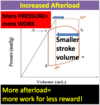
More afterload = (more/less) work?

More pressure = (more/less) work?

More afterload = (more/less) stroke volume?

A

More afterload = more work

  • More work = more oxygen consumed
  • more oxygen consumed = more ischemia
  • more ischemia = ARRYTHMIA & DEATH
112
Q
  • What’s harder on the heart?
    • Does it take more work to pump volume or pressure?
  • What’s the #1 symptom of aortic stenosis (greater afterload)?
A

1 symptom in afterload increase = chest pain (angina)

More pressure = more work required

  • greater volume isn’t as big of a deal
113
Q

More pressure = more work

More work = more O2 needed, heart dies

Congestive heart failure

  • ACE inhibitors ____ afterload
  • ACE inhibitors ____ mortality
  • Digoxin ____ afterload
  • Digoxin ____ mortality

WHY?

A

Congestive heart failure

  • ACE inhibitors decrease afterload
  • ACE inhibitors decrease mortality
  • Digoxin doesn’t change afterload
  • Digoxin doesn’t change mortality

WHY?

  • digoxin = making a heart work harder. afterload isn’t addressed
114
Q

Law of Laplace

  • wall tension
  • pressure
  • radius
  • What’s the relation?
  • How do you lower wall tension?
  • Why does a heart thicken with greater tension?
A

T = Pr

  • bigger radius = bigger tension (like a balloon)
  • lower wall tension by
    • decreasing radius
    • by lowering pressure
  • heart thickens to spread out the load
    • double thickness = half tension
115
Q

FIck principle

  • cardiac output
    • 1g/dl hgb = 1.33 mL oxygen
    • 15g/dl hgb = 20mL oxygen/dL
      • = 200 mL O2 /L blood
        • but only 50ml is released per L blood
  • artery sat = 100%
  • vein sat = 75%

If you consume 250mL O2 per minute, what’s the CO?

A

50ml O2 released per L of blood per minute

thus, 5L blood per minute = CO

116
Q

How much of CO does atrial systole contribute?

A

10-15%

117
Q

What’s first? atrial depolarization or ventricular depolarization?

A

atrial depolarization

118
Q

What’s S3?

  • rapid ____ filling
  • pathologic of _____ & ______
  • treatment is required? (y/n)

What’s S4?

  • caused by _____ systole into _______
  • pathologic of ______ & ______
  • treatment required? (y/n)
A

S3

  • rapid ventricular filling
  • pathologic of CHF & pulmonary edema
  • treatment is required

S4

  • caused by atrial systole into stiff non-compliant ventricle
  • pathologic of HTN & LV hypertrophy
  • treatment not required
119
Q

Other names for LVEDV

  • does LV depend on body size?
  • what’s CO adjusted for BMI?
  • what’s inotropy?
A
  • sarcomere length
  • ventricular filling
  • preload
  • RA pressure
  • filling pressure

LV is dependent on body size

  • cardiac index = adjusted CO
  • inotropy = contractility
120
Q

How does each respond to CHF?

  • kidney
  • carotid baroreceptors

Why is there even a reaction in the first place?

A

CHF = they sense less perfusion from less pumping, so they’re going to try to increase blood pressure to increase perfusion.

THESE ARE ALL TEMPORARY FIXES. YOU MUST REDUCE AFTERLOAD (otherwise heart will get tired and die if ony preload is increased)

Kidney

  • increase renin
  • increase angiotensin
  • increase aldosterone

Carotids

  • increase ADH
121
Q

What does more venous return do to…

  • RA pressure
  • LV filling
  • LVEDV
  • Stroke volume
  • CO
A

More venous return

  • more RA pressure
  • more LV filling
  • more LVEDV
  • more stroke volume
  • more cardiac output
122
Q

If you double the HR, do you double the CO?

A
  • not necessarily
  • because if you double the HR, you decrease the filling time
    • therefore although HR goes up
    • SV goes down
  • if you’re exercising, you’re also changing the pressure
    • that’s why when you exercise, HR and CO both go up–because you’ve changed the pressure
123
Q

What’s the maxiumum for the heart

  • RA pressure (what happens when exceeded?)
  • CO

What’s the minimum for the heart

  • vascular blood volume (‘unstressed’)
A

Maximum

  • RA pressure = 4mmhg
  • CO = 9L/min
  • after 4mmhg, RA blood will squirt out

Minimum

  • 4L of venous volume “unstressed”
    • any less than this and you wont have a blood pressure
    • “fill the tank before anything goes through the pipes”
124
Q

Arteries/veins

  • what is “stressed volume”
  • what is “unstressed volume”
A
  • filling the tank before anything goes through the faucet
  • 4L is unstressed venous volume
  • anything over 4L is arterial “stresssed” volume

Constriction of veins means decreasing vein compliance, which will shift volume from unstressed to stressed (raises BP)

125
Q

What’s the most important factor for cardiac output?

A
  • Venous return creates (RA) pressure
    • RA pressure creates LV filling
    • LV filling becomes LVEDV
    • LVEDV = Stroke Volume
    • Stroke volume x Heart rate = CO

Therefore! Venous Return = Cardiac Output

126
Q

What do you do for a patient in acute pulmonary edema?

  • What needs to be addressed immediately?
A

Patients with acute shortness of breath, rales, edema, S3 gallop need VOLUME removed from lungs FAST!

  1. Venous Dilation
  2. Increasing “unstressed” volume
  3. Diuretics
127
Q

What percent of the body’s bood is held in the venous system?

Name some venodilators

A

60%

  • nitrates/nitros/amyl nitrates
  • morphine (acute pulmonary edema)
  • furosemide
  • sildenafil (nitric oxide–Viagra)
  • prostacyclin
128
Q

What does venous dilation do to

  • unstressed volume
  • stressed volume
  • vein compliance
  • venous return
A

venous dilation

  • greater unstressed volume
  • less stressed volume
  • greater vein compliance
  • lower venous return
129
Q

TPR

  • what kind of vessel has greatest impact on TPR?
  • what happens to venous return with
    • high TPR
    • low TPR
A
  • arterioles control TPR
    • relaxing the arterioles allows blood to move forward
  • high TPR = low venous return
  • low TPR = high venous return
130
Q

Inotropic effects

  • What do positive inotropes do for…
    • Moving blood (backwards or forwards?)
    • contractility (up/down)
    • stroke volume (up/down)
    • RA pressure (up/down)
    • cardiac output (up/down)
A
131
Q

Decreased RA pressure’s effect on volume of venous return to the heart

  • increased/decreased?
A

Low RA pressure = more venous return

  • you’re making it easier for blood to re-enter the heart
132
Q

Increased blood volume leads to more/less…

  • stressed volume?
  • cardiac output
  • RA pressure

what change in venous compliance can achieve the same effect?

A

More blood volume equals

  • greater stressed volume
  • greater cardiac output
  • greater RA pressure

low venous compliance = same as more blood volume

133
Q

what’s the most effective way to increase stroke volume?

  • increase filling
  • increase contractility

what will increase stroke volume

  • sympathetic stimulation
  • parasympathetic stimulation
A

increase stroke volume by increasing contractility with a positive inotrope (which works via sympathetic stimulation)

  • sympathetic stimulation will always increase CO
134
Q
A
135
Q

What does increased TPR do to

RA pressure aka LVEDV

CO

A
136
Q
A

A

137
Q
A

D

138
Q
A

A

139
Q
A

D

140
Q
A
141
Q
A