peds Flashcards
Legg-Calvé-Perthes Disease (Coxa Plana)
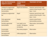
prognosis
goal is sphericity of femoral head
asperical - early DJD
poor prognosis w/ >6 years onset, female, lateral column C (regardless of age), adcreased abduction
Septic arthritis - aspiration results
>50K WBC, glucose 50 lower than serum level
Septic arthritis - who gets it from osteomyelitis?
neonates, in whom transphyseal vessels allow proximal spread into the joint in joints with an intraarticular metaphysis (hip, elbow, shoulder, ankle
Sprengel Deformity -associated diseases
Klippel-Feil syndrome (fused cervical vertebra w/ short neck; one third have Sprengel deformity) Kidney disease Scoliosis Diastematomyelia (split spinal cord)
Slipped Capital Femoral Epiphysis
technique
The pin should be started anteriorly on the femoral neck, ending in the central portion of the femoral head

Developmental Dysplasia of the Hip - associated problems and natural hx
other problems w/ positioning - torticollis (20%) and metatarsus adductus (10%) hip contracts and acetab becomes dysplastic and filled w/ pulvinar (fibrofatty tissue)
Developmental Dysplasia of the Hip - radiographic studies
dynamic u/s before ossification of femoral head at 4-6 months
Slipped Capital Femoral Epiphysis
xrays and grading
AP and frog-leg pelvic views
If the slippage is unstable, a cross-table lateral view is required
Grade I: 0% to 33% slippage
Grade II: 34% to 50% slippage
Grade III: more than 50% slippage
Brachial Plexus Palsy - what happens with significant IR contracture
progressive glenoid hypoplasia
Septic arthritis - treatment
aspiration, I&D
Legg-Calvé-Perthes Disease (Coxa Plana)
presentation
boys 4-8 years
pain (often knee), effusion, limp, decreased hip ROM (lack abd/IR)
Developmental Dysplasia of the Hip
-dynamic u/s angles
coronal view, the normal α angle is greater than 60 degrees, and the femoral head is bisected by the line drawn down the ilium.

Developmental Dysplasia of the Hip - risk factors in order
Breech>family hx>female >firstborn *left hip (67%) and girls (85%)
Osteomyelitis - why more common in kids?
rich metaphyseal blood supply and thick periosteum
Proximal Femoral Focal Deficiency- classification
A: femoral head present with normal acetabulum; B: femoral head present with dysplastic acetabulum; C: femoral head absent with markedly dysplastic acetabulum; D: both femoral head and acetabulum absent
Septic arthritis - joints with intraarticular metaphsysis prone to septic arthritis from osteomyelitis?

Proximal Femoral Focal Deficiency - associations
coxa vara, fibular hemimelia, ACL deficiency, knee contracture
Septic arthritis vs transient synovitis
Kocker criteria: 3/4 = >90% 1) WBC>12K 2) ESR>40 3) inability to bear weight 4) fever > 101.5/38.6
Rotational Problems of the Lower Extremities Femoral anteversion -features -treatment
3-6 years old, kids sit w/ legs in W position corrects by age 10 usually, no shoes/PT/braces are effective older children with less than 10 degrees of external rotation, femoral derotational osteotomy (intertrochanteric is best) may be considered for cosmesis, although this is not a functional problem
Osteomyelitis kids.- imaging findings
xray findings only after 5-7 days, MRI is key
Leg Length Discrepancy - when does bone growth stop?
age 16 in boys and age 14 in girls
open reduction for DDH
age?
approach and reason?
procedure steps?
6-18 months with failed closed reduction OR 18 months-3 years
Anterior approach (less risk to medial femoral circumflex) but may use medial if <12 months b/c less blood loss and direct access to obstacles for reduction, increased osteonecrosis
capsulorrhaphy, adductor tenotomy, femoral shortening + acetab procedure if dysplastic
Septic arthritis - when is an LP needed?
if H. influenzae because of association with meningitis
Brachial Plexus Palsy surgical treatment of plexus palsy
early - nerve late - deformity **ER = lat/teres maj tx **EF = pec tx, flexorplasty **IR contracture/glenoid hyperplasia = pec/sub scap release (if <5 yrs) OR prox hum rot osteotomy (>5 yrs)
Congenital Dislocation of the Knee - involved anatomy
quad contracture, anterior subluxation of hamstrings, tight collateral ligaments
Proximal Femoral Focal Deficiency - etilogy
sonic hedhehog gene
Congenital Coxa Vara
radiographic findings
Decreased neck-shaft angle
Triangular ossicifcation defect in inferomedial femoral neck in developmental coxa vara
Hilgenriner epiphyseal angle key to treatment (line b/w Hilgenreiner and prox femoral physis) guides treatment (nl <25)
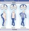
Brachial Plexus Palsy Total plexus palsy -roots -deficits
C5-T1 sensory and motor; flaccid arm
Slipped Capital Femoral Epiphysis
physical exam findings
obligate external rotation (decreased IR) with flexion of the hip
Developmental Dysplasia of the Hip - physical exam
ortolani is out (elevate/abduct to reduce) Barlow (adduct/depress to dislocate) Limited abduction as laxity resolves Galeazzi sign - short femur when feet together and knees flexed Asymmetric glut folds, trendelenburg in older kids, lumbar lordosis
Legg-Calvé-Perthes Disease (Coxa Plana)
classification
Herring for prognosis
based on lateral pillar invovlement during fragmentation stage
Legg-Calvé-Perthes Disease (Coxa Plana)
nonop Rx
traction, NSAIDs, PWB, PT for ROM
Osteomyelitis - most common bug?
Staphylococcus aureus (after H flu vaccine came out), Kingella more common in young kids and culture negative (need blood culture medium)
Slipped Capital Femoral Epiphysis
complications
1- osteonecrosis w/ unstable slip
2- chondrolysis - narrow joint space, pain, decreased ROM; pin penetration if anterior superior quadrant of head)
3 - DJD from pistol grip deformity of proximal femur
Congenital Coxa Vara
Treatment based on degree
Based on Hilgenreiner epiphyseal angle (nl < 25)
<45 - corrects spontaneously
45-60 close observation
>60 surgery
Proximal Femoral Focal Deficiency- treatment
A and B (femoral head present) - potential reconstructive procedure w/ lengthening/shortening; C and D (no head) - amputation, femoral-pelvic fusion, limb lengthening
Fibrotic Deltoid Problems - clinical feature - treatment
Short fibrous bands replace the deltoid muscle and cause abduction contractures Surgical resection
Septic arthritis - neisseria gonorrhoeae presentation
migratory polyarthraliga, small red papules, multiple joints
Congenital Coxa Vara
presentation
waddling gait (bilateral) vs painless limp (unilateral)
bilateral 35%
Brachial Plexus Palsy Klumpke palsy -roots -deficits
c8/T1 wrist flexors, intrincs horner syndrome
Poland Syndrome -clinical features -cause -treatment
-B syndractylyl of hands/feet; craniosynoostosis (early closure of cranial sutures) - flap skull w. broad forehead, radiolulnar synostosis -AD FGFr2 mutation -surgical release of border digits at 1 yr, turn middle 3 digits into 2 digits at 2 yerars old
Congenital Pseudarthrosis of the Clavicle -clinical features -causes -xray findings -treatment
-failure of union of medial/lateral ossicifcation centers of R clavicle (10% bilateral) -pulsations of subclavian artery -AP view shows rounded sclerotic bone -ORIF w/ bone grafting if impairment/cosemetics at age 3-6 yrs; predictable union (unlike cognential pseudoarthrosis of tibia)
closed reduction for DDH
age?
procedure steps?
post reduction step?
6-18 months OR if pavlik fails
general, arthrography (“thorn sign” indicates normal labral position), hip space w/ > 90 flexion and slight abduction
CT/MRI to confirm reduction
Brachial Plexus Palsy Erb Duchenne palsy -roots -deficits
c5/6 deltoid, cuff, EF, Wrist/Hand extensors – waiter’s tip deformity
Septic arthritis - causes
hematogenous spread, local osteomyelitis
Congenital Dislocation of the Knee - operative treatment
soft tissue release if failure to gain 30 flexion after 3 months of casting goal is 90 flexion via: quad lengthening, anterior capsular release, hamstring transposition, mobilize collateral ligaments
Congenital Dislocation of the Knee - treatment nonop
reduction with seriel casting if both hip and knee dislocated, knee first b/c pavlik requires reduced knee
Leg Length Discrepancy - measuring
blocks of set height under affected side; with scanography Lateral CT scanography—more accurate than conventional scanography if there are soft tissue contractures of hip, knee, or ankle
Osteomyelitis kids - diagnosis
aspiration OR clinical picture w/ convincing MRI
Brachial Plexus Palsy signs of poor prognosis in plexus palsy
total plexus injury lack of biceps fxn @ 6 months Horner syndrome
Osteomyelitis - Brodie abscess
Chronic bone abscesses may become surrounded by thick fibrous tissue and sclerotic bone
Slipped Capital Femoral Epiphysis
stabilty and risk of osteonecrosis
Stable: weight bearing with or without crutches possible (no risk)
Unstable: weight bearing not possible because of severe pain (~50%)
Leg Length Discrepancy - contribution by growth plate per year
proximal femur - 3 mm distal femur - 9 mm proximal tibia - 6 mm distal tibia - 5 mm
Proximal Femoral Focal Deficiency- what is it?
developmental defect of proximal femur
Septic arthritis - radiographs
widened joint space
Developmental Dysplasia of the Hip - teratologic
dislocated in utero and irreducible; associated with neuromuscular conditions and genetic abnormalities (athrogryposis and Larsens) most severe pseudoacetabulum at birth early surgery
Osteomyelitis kids - lab tests to order
blood cultures, CBC, ESR, CRP
Rotational Problems of the Lower Extremities -causes of In-toeing by age
metatarsus adductus (in infants) internal tibial torsion (in toddlers) femoral anteversion (in children < 10 years)
Brachial Plexus Palsy initial treatment of plexus palsy
maintain passive ROM, await return of motor fxn (up to 18 months) – 90% resolve
Proximal Femoral Focal Deficiency - appearance
short, bulky thigh; FABER position
Legg-Calvé-Perthes Disease (Coxa Plana)
containment operations
femoral osteotomy - PF varus O
pelvic - salter, triple, dega, pemberton
Septic arthritis - presentation
more acute than osteomyeltis; decreased ROM, severe pain w/ passive ROM +/- systemic symptoms
OR for DDH - obstacles to reduction?
transverse acetabular ligament, pulvinar, in-folded labrum, inferior capsular restriction, and psoas tendon
Legg-Calvé-Perthes Disease (Coxa Plana)
pathology
osteonecrosis of proximal femoral epiphysis from unk vascular insult
follwed by revascularization/resorption through creeping substitution that allows remodeling and fragmentation
Osteomyelitis kids - operative treatment when?
failure to respond to abx, frunk pus on MRI, sequested abscess
Septic arthritis - gonorrhoeae labs and treatment
usually <50K WBC, no surgical drainainge usually, large doses of penicillin
Rotational Problems of the Lower Extremities External tibial torsion -features -treatment
causes out-toeing that may worsen w/ age (tibia ER w/ growth); associated w/ increased femoral anteversion; knee pain rest, PT, supramalleolar osteotomy if >8-10 years and ER > 40 degrees
Transient synovitis - treatment
IF Kocker criteria not met then no aspiration; NSAIDs - improvement in 24-48 hrs
Slipped Capital Femoral Epiphysis
treatment? Who should get opposite side pinned?
pinning in situ (unstable severe SCFE may get surgical hip dislocation with modified Dunn technique - create priosteal flaps on femoral nek); endocrinopathy or in young children (<10 years) or with an open triradiate cartilage
Congenital Coxa Vara
surgical procedure
correctie valgus osteotomy of proximal femur plus transfer of GT for hip abductor mechanics if needed
DDH - treatment goal and by age
goal - concentric reduction to prevent DJD
birth - 6 months:
pavilk if ortolani positive (out), recheck at 3 wks w/ u/s .–> CR, athrography, spica vs rigid brace if still out
Barlow positive (in but comes out) - watch closely
6 - 18 months
hip arthrography, adductor tenotomy, CR, spica w/ postreduction CT/MRI; open if closed fails
18 m - 3 years
OR
3- 8 years
Osteotomy: Salter, Dega, Pemberton, Staheli
>8 years
Osteotomy if open plates –> steele, southerland, staheli; if closed –> ganz, chiari
Slipped Capital Femoral Epiphysis
treatment for SCFE that fails to remodel
intertroch (Kramer) or subtroch (Southwick) oseotomies; Femoral neck (cuneiform) high rates of AVN/OA
Pavlik harness
age?
position?
risks?
<6 months
100 degrees of flexion and mild abduction
AVN w/ excessive abduction via impingement of posterosuperior retinavular branch of medial femoral circumflex
Femoral nerve palsy from excessive flexion
Pavlik disease - eroision of pelvis (stop after 3 wks of failed reduction)
osteotomy for DDH - indications
toddlers and older for acetabular dysplasia IF congruent reduction; pelvic vs femur depends on side of dysplsia
Leg Length Discrepancy -treatment by projected discepancies
<2 cm observe 2-5 cm Epiphysiodesis of the long side Shortening of the long side (ostectomy) Lengthening of the short side >5 cm lengthening (1mm per day via ilizarov method which perserves medullary canal and blood supply)
Rotational Problems of the Lower Extremities -causes of out-toeing by age
external rotation hip contracture (in infants) external tibial torsion and external femoral torsion (in older children and adolescents)
Legg-Calvé-Perthes Disease (Coxa Plana)
surgery for who?
>8 years w/ lateral pillar B, B/C hips
Osteomyelitis kids - treatment
IV abx if no subperiosteal abscess, broad then narrow based on aspiration, monitor CRP (Should decline w/ing 48-72 hrs), treat 4-6 wks until ESR/CRP normal
DDH Osteotomy - diagram

Slipped Capital Femoral Epiphysis - pathology
weakness of perichondrial ring and slippage through hypertrophic zone of the growth plate
Congenital Dislocation of the Knee - associations
myelomeningocele, athrohryposis, Larsen syndrome
Sprengel Deformity -decription -functional deficit
Undescended scapula winging hypoplasia omovertebral connections (30% of cases) decreased abduction
risks factors for failed pavlik?
>7 wks at start
bilateral dislocation
absent ortolani sign
Rotational Problems of the Lower Extremities Metatarsus adductus -features -treatment
Forefoot adducted at tarsal-metatarsal joint (lateral border–> convex vice flat) ; 1st year of life; 85% resolve on own if correctable to neutral actively do nothing; passively then stretch; not correctable - serial casting. Surgery w/ lateral column shortening and medial column lengthening (rarely needed)
Brachial Plexus Palsy 3 types of brachial plexus palsies: roots, prognosis
Erb-Duchenne C5/6 - best prognosis , most common Total plexus C5-T1, worse prognosis Klumpke C8-T1, poor prognosis
DDH xray age, lines and normal values
age > 3 months
AI < 25 degrees
Ossific nucleus should be medial to Perkin’s and inferior to Hilgenreiner’s
Shenton’s line should be smooth
at 4-6 months may see delayed femoral head ossification on affected side

Septic arthritis - who gets it?
kids < 2 years
Slipped Capital Femoral Epiphysis
who needs additional w/u and what?
if <10 –> endocrine w/u for hypothyroidism (most common), growth hormone deficiency, or renal osteodystrophy
DDH OR Surgical risks:
osteonecrosis. Damage to medial femoral circumflex (w/ medial approach because close to psoas), fialed OR has high complication rate, dx after 8 years which gives acetab little chance to remodel
Legg-Calvé-Perthes Disease (Coxa Plana)
radiographic findings
cessation of growth of ossific nucleus
medial joint space widening
cresent sign – subchondrol fx
Septic arthritis - organism and abx by age

Osteomyelitis kids - MRSA association
DVT and septic emboli
Osteomyelitis in kids - how does it start?
hematogenous seeding of metaphysis w/ small arterioles, blood flow sluggish, bone abscess created
Transient synovitis - ESR levles
<20 usually
Herring classification chart

Slipped Capital Femoral Epiphysis
typical patient
obese adolescent African American boys during their rapid growth spurt (10-16 years of age)
Herring classification

Slipped Capital Femoral Epiphysis - direction of displacement
Epiphysis remains in the acetabulum, and the neck is displaced anteriorly and rotates externally.
Developmental Dysplasia of the Hip- Potential obstructions to obtaining a concentric reduction
Iliopsoas tendon pulvinar contracted inferomedial capsule transverse acetabulum ligament inverted labrum
Poland Syndrome -clinical features -cause -exam findings -treatment
-Unilateral chest wall hypoplasia (sternocostal head of pectoralis major absent); hypoplasia of hand/forearm; Symbrachydactyly and shortening of middle fingers -subclavian artery hypoplasia -chest wall deformity, hand deformity, no F/E tendons, radioulnar synostosis -syndactyl release; lack of soft tissue coverage requires FTSG
Legg-Calvé-Perthes Disease (Coxa Plana)
treatment goals
contain hip, restore ROM
Legg-Calvé-Perthes Disease (Coxa Plana)
salvage operations
valgus femoral osteotomy for hinge abduction
chiari and/or shelf osteotomies for hip not contained
Rotational Problems of the Lower Extremities Internal tibial torsion -features -treatment
most common cause of intoeing; 2nd year of life; BLE w/ excessive medial ligament tightness; transmalleolar axis is internal resolves w/ growth; surgery rarely required (supramalleolar osteotomy at 7-10 years)
normal leg alignment changes during childhood
genu varum as newborn to genu valgum at 2.5 yrs to straight at 4-6 years

physiologic genu varum
how old?
xray findings?
pathologic causes?
<2 years
flaring of tivia and femur in a symmetric pattern
pathological causes: OI, trauam, dysplasia, blount disease
genu valgum
- normal angles and limits
- differential for pathology
- treatment
up to 15 degrees normal in kids 2-6 yrs
differential: renal osteopdystropy (especially if BLE), tumors (osteochondroma), infection, trauma
surgery if > 15 degrees or 10 cm b/w medial mals after 10 yrs old; hemiepiphysiodesis of medial side
types of tibial bowing (based on apex of curve), cause, rx

Posteromedial tibial bowing
- cause
- associations?
- natural hx?
physiologic “PM - Probably Mild”
LLD, calcaneovalous feet, tight anterior structures
spontaneous correction but montior LLD (averages 3-4 cm, may need epiphysiodesis)
Anteromedial tibial bowing
- cause?
- association?
- genetics?
- treatment?
Fibular hemimelia (congenital longitudinal deficiency of fibula)
associated with ankle instability (due to ball and socket joint), equinovalgus foot, tarsal coalition, femoral shortening (PFFD, coxa vara), ACL insufficiency
Sonic hedgehog gene
shoe lift to bracing to syme amputation
Anterolateral tibial bowing
- cause
- association
- treatment
congenital pseudarthrosis “AL - Always Look”
50% have NF
w/u includes genetics for NF, total contact brace to protect from fx, intramedullary fixation, possible vascularize fibular graft
Osteochondritis Dissecans
- age, most common location
- MRI poor prognosis for non-op rx
- treatment
10-15 years, PL medial femoral condyle
Synovial fluid behind lesion; lateral femoral condyle and patella
nonop w/ bracing, NWB if open physes
op w/ adolescent w/ minimal growth left or loose lesion – drilling, fixation, bone grafting all used
Osgood-Schlatter Disease
- cause
- treatment
-tibial tubercle apophysisi
self limited, activity modification; ice and quads stretching; doesn’t resolve until growth has halted, late excision of separate ossicles rarely needed
Discoid Meniscus
- cause
- symptoms
- radiographic findings
- classification and treatment
abnormal development of lateral mensicus
mechanical block, pain, palpable click
wide cartilage space, squaring of condyles, mri w/ 3 sagittal images
complete, incomplete covering and wrisberg varient - lacks posterior meniscotibial attachment (unstable)
not torn, observe; torn and symptomatic, sauzerization; wirsberg, repair
Congenital Dislocation of the Knee
- associations
- structural compenents
- treatment and method
- myelodysplasia, arthrogryposis, Larsens + DDH, clubfoot, metatarsus adductus
- quads contracture, tight collateral ligaments, anterior subluxation of hamstring tendons
- nonop - reduction and serial casting (treat b/f Pavlik for DDH); OR if <30 degrees of flexion after 3 months, <90 degrees flexion
method: quads lengthening, hemstring transposition posteriorly, collateral ligament mobilization
Clubfoot (Congenital Talipes Equinovarus)
-deformity
Mnemonic: CAVE—cavus, adduction of forefoot, varus of hindfoot, equinus
Shortening or contraction of muscles (intrinsic muscles, Achilles tendon, tibialis posterior, flexor hallucis longus, flexor digitorum longus), joint capsules, ligaments, and fascia, which leads to the associated deformities

Clubfoot (Congenital Talipes Equinovarus)
-associations
arthrogryposis, myelomeningocele, hand anomalies (Streeter dysplasia), diastrophic dwarfism, prune-belly syndrome, tibial hemimelia, and other neuromuscular and syndromic conditions
Clubfoot (Congenital Talipes Equinovarus)
-radiographic findings
“Parallelism” of calcaneus and talus seen on radiographs
Clubfoot (Congenital Talipes Equinovarus)
-general treatment
penseti casting - first line
operative treatment - posteriomedial release if nonop fails, triple arthrodesis for children presenting late, talectomy for salvage
Clubfoot (Congenital Talipes Equinovarus) - ponseti casting
- method?
- sequence?
- post casting care
- complications
first line treatment, serieal weekly manipulation/casting w/ long leg cast
sequency: CAVE - cavus, adductus, varus, equinus
Cavus - supinate forefoot and dorsiflex 1st ray
Adduction/Varus - lateral pressure on distal talar head
equinas - perc achilees lengthening at end of casting
Last cast placed in 70 degrees of abduction
post cast bracing w/ foot-abduction brace fulltime x 3m, sleeping x 3y
recurreunce, rocker bottom deformity, flat op talus
Clubfoot (Congenital Talipes Equinovarus) - posteromedial release
-structures to be addressed

Clubfoot (Congenital Talipes Equinovarus): dorsal bunion afters surgery
- cause
- treatment
Strong tibialis anterior and flexor hallucis brevis/abductor hallucis contribute
May be iatrogenic if peroneus longus divided
Treatment is with capsulotomy, flexor hallucis longus lengthening, and transfer of the flexor hallucis brevis to become a metatarsophalangeal extensor.
Clubfoot (Congenital Talipes Equinovarus)
Dynamic supination
Common deformity after clubfoot treatment; occurs in up to 15% to 20% of patients
Proposed causes:
Overpull of the anterior tibialis, with a weak peroneus longus
Undercorrection of forefoot supination
Treated with transfer of the tibialis anterior laterally
common childhood foot disorders - diagram

Metatarsus Adductus - what is it?
Forefoot adduction with the hindfoot in normal alignment

Skewfoot - what is it?
adductus with hindfoot valgus, lateral subluxation of navicular on talus

Metatarsus Adductus and Skewfoot - treatment
stretching if peroneal muscle stim corrects adductus otherwise serial casting
Pes Cavus (Cavus Foot) - what is it?
cavus foot (elevated longitudinal arch) with varus hindfoot
Pes Cavus (Cavus Foot) - causes
2/3rds due to neurological disorder (CMY - defect in gene for peripheral myelin protein 22 (PMP22))
Pes Cavus (Cavus Foot) - muscle imbalance
strong peroneus longuys and PT overpull TA, PB to cause hindfoot varus and depressed 1st MT head. Plantar fascia shortens over time as a result of EHL use for dorsiflexion
Pes Cavus (Cavus Foot) - evaluation?
full neuro exam, MRI of neuraxis if unilateral, DNA testing, lateral block (coleman) test to assess highfoot flexiblity
Pes Cavus (Cavus Foot) - non optreatment
nonop doesn work once deformity present
Pes Cavus (Cavus Foot) - operative treatment

Congenital Vertical Talus - what is it?
irreducible dorsal dislocation of navicular on talus w/ fixed equinas hindfoot deformity
Congenital Vertical Talus - less severe form?
oblique talus, navicular reduced with plantar felxion
Congenital Vertical Talus - lateral xray
talus is vertical, calc in equinas; increased talocalc angle
Congenital Vertical Talus - treatment
serial manipulation/casting followed by perc achilles tenotomy and minimal TN capsulotomy and pin fixation
Tarsal Coalitions - what joints are usually involved and at what ages?
talocalcaneal (12-14 years) or calcaneonavicular (9-12 years)
Tarsal Coalitions - xray findings
lateral - elongated anterior process of the calcaneus “antearter” sign for calcnav
Tarsal Coalitions - treatment
immobilization initially; calcnavic - resection w/ EDB or fat interposition; talocalc <50% middle facet resect and interposition, >50% subtalar arthrodesis
Calcaneovalgus Foot - what is it?
neonatal condition due to positioning, dorsiflexed/abducted hindoot that is passively correctable, rx is stretching and observation
Juvenile Bunions - features
bilateral and familial, more severe than adult, adolesecent girls
Tarsal Coalitions - treatment
non op, avoid surgical if possible because of recurrence; if IMA> 10 and HVA > 20 then proximal MT osteotomy, distal capsular reefing, adductor tenotomy with bunionectomy (modified McBride)
Kohler disease - features and treatment
osteonecrosis of navicular bone at age 5; resolves spontaneously with decreased activity (with or without immobilization)
Flexible PES Planus - clinical features
foot is flat only when standing, arch fores when on toes (otherwise consider tarsal coalition)
Flexible PES Planus - rx
asymptomatic then observation; symptomatic - arch supports, if fails then calc lengthening osteotomy OR 3C (sliding calc osteotomy, opening-wedge cuboid osteotomy, plant flexion closing wedge osteotomy of medial cuneiform)
habitual toe walking - features and treatment
associated with neuro dx (autism, CMT, CP); contracture of achilles may be present; non- stretching and night splints, op - lengthening
accessory navicular - freatures, xray view, rx
PTT inserts, associated with flap feet, medial arch pain; external oblique view; usually resolves on own but may require casting or excision with repair of PTT
ball and socket ankle - associations
no treatment needed but associated with tarsal coalition, absence of lateral rays, fibular deficienecy, LLD
Adolescent Idiopathic Scoliosis -age?
after 10 years old
Adolescent Idiopathic Scoliosis - scoliometer referral threshold and coronal curve correlation?
7 degress –> 20 degree coronal curve
Adolescent Idiopathic Scoliosis - who should get an MRI?
Left thoracic curves Painful or rapidly progressing scoliosis Apical kyphosis of the thoracic curve Juvenile-onset scoliosis (onset before age 10 years) Neurologic signs or symptoms Congenital
Adolescent Idiopathic Scoliosis - scoli film measures - cobb, stable vert (moe), risser

Adolescent Idiopathic Scoliosis - risk factors for progression
curve size, remaining growth; >20 in young patients, >45 at maturity will continue to progress; Peak height velocity (PHV) best predictor, menarch, risser 0
Adolescent Idiopathic Scoliosis - treatment based on curve and risser stage

Adolescent Idiopathic Scoliosis - when to brace?
curve > 25, risser 0-2; goal is to halt/slow progression not reverse
Adolescent Idiopathic Scoliosis - boston brace: effective for apex at or below? Less effective in who? Effectiveness is?
apex at or below T8, boys and overweight, dose dependent >12 hrs
Adolescent Idiopathic Scoliosis - Crankshaft phenomenon
Continued anterior spinal growth after posterior fusion in skeletally immature patients avoided by anterior discectomy and fusion coupled with posterior spinal fusion
Early-Onset Scoliosis (EOS) - medical complications?
thoracic insufficiency syndrome; rx VEPTR (verticle expandable prosthetic titanium rib) but high complication rates
Early-Onset Scoliosis (EOS) - age?
<10 years old
Juvenile Idiopathic Scoliosis- age?
4-10 years old; idiopathic
Juvenile Idiopathic Scoliosis - side of curve?
right sided
Juvenile Idiopathic Scoliosis - differences from AIS?
higher risk of progression, less response to bracing; obtain MRI (25% abnormal); same treatment protocl
Juvenile Idiopathic Scoliosis - age for growing rods vs fusion?
10 years old - always fuse front and back
Infantile Idiopathic Scoliosis - age?
<4 years
Infantile Idiopathic Scoliosis - differences w/ AIS?
L thoracic curve most common as well as other defects
Infantile Idiopathic Scoliosis - natural hx?
90% resolve spontanesously
Infantile Idiopathic Scoliosis - risk for progression?
phase of ribs - phase I no overlap: RVA difference >20 high risk of progression; phase II - rib overlaps apical vertbra - high risk
Infantile Idiopathic Scoliosis - MRI?
if progressive IIS
Infantile Idiopathic Scoliosis - treatment?
RVAD < 20 observe otherwise try bracing/casting until old enough for OR
congenital spinal deformities - cause
developmental defect in spine during 5-8 week of gestation
congenital spinal deformities - associaitons?
intraspinal (get MRI), cardiac, GU
congenital spinal deformities - types of defect?
segementaion (block, bar), formation (hemivertbra), mixed

congenital spinal deformities - risks of progression
congenital spinal deformities - congenital kyphosis types
failure of formation (worst), segementation, mixed

congenital spinal deformities - treatment
posterior fusion alone: <5 years w/ curve <50 and nl neuro exam A/P fusion: older kids or more severe curves
neuromuscular scoliosis treatment - duchene MD
steroids reduce/delay scoliosis

neuromuscular scoliosis treatment - friedrich ataxia

neuromuscular scoliosis treatment - spinal muscular atrophy

neuromuscular scoliosis treatment - spina bifida

neuromuscular scoliosis treatment - CP

neuromuscular scoliosis treatment - NF

neuromuscular scoliosis treatment - arthrohryposis

neuromuscular scoliosis treatment - associated conditions
pulmonary and cardiac for duchene muscular dystrophy
neuromuscular scoliosis treatment - nutritional markers before surgery?
WBC<1500, albumin <3.5 then supplement or g tube feedings b/c longer hospital stay and more infections
neuromuscular scoliosis treatment - levels to fuse in nonambulatory patients?
usually T2-pelvis, goal is center trunk over pelvis with balanced spine
Neurofibromatosis - genetics, diagnosis, most common orthopedic site of disease?

Scheuermann disease - definition?
thoracic kyphosis >45 degrees w/ >5 degrees anterior wedging at 3 sequential levels
scheuermann disease - cause? who get its?
unknown
overweight boys, not postural as doesn’t correct with hyperextension
scheuermann disease - treatment
bracing: curves 50-75 if >1 yr growth left (risser 2 or below)
surgery: curve > 75 despite PT and skeletal maturity; all posterior w/ multiple ponte osteotomies; T2 to 1st lordotic disc and body touched by PSVL (up from posterior edge of s1)
postural hyphosis or round back - what is it? treatment?
no verterbal body changes, corrects with backward bend
Rx is hyperextension exercises
Klipper-Feil Syndrome - orthopedic manifestations? other associations? classic triad?
abnormal cervical spine formation, congenital scoliosis, sprengel deformity
renal disease/aplasia, congenital heart disease, spinal cord abnormalities (get MRI, renal and cadiac eval)
triad: low posterior hairline, short webbed neck, limited cervical ROM
Klippel-Feil syndrome - spine treatment?
conservative, avoid collision sports, surgery for myelopathy
atlantoaxial instability - associations and treatment?
Downs - avoid contact sports, if ADI>10 mm then posterior fusion
JRA, skeletal dysplasias
atlantoaxial rotatory displacement/subluxation - presentation? causes? dx? treatment?
presents w/ torticollis
causes: downs, retropharyngeal inflammation (Grisel disease), trauma
dx: CT C1/2 with head straight, left and then right
Rx: <1 wk: c-collar, nsaids, heat
1-4 weeks: halter/halo traction
>4 wks: surgical reduction and fusion
os odotoideum - when do you treat and how?
if ADI>10 mm or <13 mm available for cord; neuro symptoms
c1-2 fusion
spondylolysis - what is it? who gets it? treatment?
stress fx at parts interarticularis
athletes w/ hyperextension (gymnasts, football linemen, wrestlers)
rx - nothing if incidentla
symptomatic: rest, bracing, PT
if nonop fails: OR direct pars repair if hot bone scan and MRI w/out disc degeneration otherwise fuse
spondylolishtesis - what? location? types?
forward slippage of proximal vertbra
most common at L5-S1
isthmic (from spondylolysisi), dysplastic (congental abscence of facets - greater risk for profession)
spondylolisthesis - radiographic parameters?
grade 1-5 based on translation
Slip angle >50 greater risk of slip progression, instability, post op pseudarthrosis

pelvic incidence - how to measure? normal? affected by posture? predispses to what?
line from midpoint of sacral end plate to center of femoral heads and a line perpendicular to sacral end plate
nl = 50 degrees
not affected by posture
may predispase to spondylolisthesis

most common neurological complication from spondyloliesthesis reduction during surgery?
L5 root injury
pelvic measurement chart

Diastematomyelia - what is it? treatment?
fibrous/cartilage/bone bar that creates a longitudinal cleft in spinal cord –> may cause tethering and deficits
Rx - aysmptomatic then observe, resect before correction of spinal deformity
sacral agenesis - assocation?
materal diabetes
CP - onset? Progressive? Cause?
<2, nonprogressive, injury to immature brain (not identifiable but usually includes prematurity, perinatal infections), anoxic injuries, meningitis)
CP - UMN or LMN? Joint deformities?
UMN causes mix of muscle weakness and spasticity, initial dynamical defromity at joints but spasticity leads to contractures, subluxation/dislocation
CP - physiologic classification
Spastic - increased tone/reflexes, slow movements due to simulataneous contraction of agonists and antagonists; most common; good for OR intervention Athetosis – writhing movements, less common; not good for OR Ataxia - no coordination, wide based gait, not good for OR Mixed

CP - anatomic classification
hemiplegia - UE and LE on same side; handedness early; all walk diplegia - LE>UE; most walk; IQ may be normal quariplegia - total body, low IQ, high mortality, don’t walk
CP - GMFCS
gross motor and functional classification system

fractures pathognomonic for abuse?
corner (at jxn of metaphysis and physis) and posterior rib fxs
physeal fx zone
classically zone of provisional calficiation within zone of hypertrophy
Salter Harris Classification
graphs

what salter harris fxs usually require ORIF
III, IV b/c intrarticular
partial growth arrest - cause?
growth plate injury results in arrest of part of physis which causes angular growth and deformity
who should have physeal bridge resection? Method?
pt w/ >2 cm growth remaining and <50% plate involvement; resection bar and interpose fat graft
treatment of growth arrest if >50% of physis
ipsilateral completion of arrest and contralateral epiphysiodesis or ipslateral limb lengthening
proximal humerus fx deformity and causes
proximal fragment - abductions, ER 2/2 RTC; distal fragment - adduction and shortens 2/2 deltoid and pec major
proximal humerus contributes what percentage to humerus growth?
80% allows high fx tolerance
orders of ossification at elbow and ages
CRITOE

pediatric elbow - radiographic evaluation
Proximal radius should align with capitellum in all views. Long axis of ulna should align and be slightly medial to humerus on AP radiograph. Anterior humeral line should bisect capitellum on true lateral radiograph. Humeral-capitellar (Baumann) angle should be in valgus and fall between 9 and 26 degrees. Soft tissue shadows may demonstrate an anatomic anterior fat pad.

distal humerus physeal seperation xray, considerations, treatment?
xray intact relationship b/w radius and capitellum but distal humerus off, usually medial (unlike lateral elbow dislocation), eval for child abuse, CRPP
SCH nerve injury w/ extension type
AIN > radial, usually neuropraxia
SCH nerve injury w/ flexion type
ulnar
SCH xray findings
posterior fat pad always pathologic, anterior humeral line intersects middle third of capitellum, AP Baumann angle
SCH types and rx
graph

SCH pin placement
divergent and lateral
SCH Rx complications
ulnar nerve injury from medial pin OR cubitus valgus (tardy) compartment syndrome before cast pressure/swelling angular deformity: cubitus varus - no fxn effect, valgus - tardy ulnar nerve palsy, recurvatum - poorly tolerated
lateral condyle fx - best radiograph view?
internal oblique
lateral condyle fx - treatment
if displaced then CRPP, confirm with athrogram; if still displaced then ORIF (avoid posterior capsular distruption for AVN)
lateral condyle fx - complications
lateral overgrowth/osteonectroiss, delayed union/malunion, angular deformity
medial qpicondule fx association?
elbow dislocation 50% of the time
radial head/neck fx ORIF associations?
RH AVN, PIN injury, radioulnar synostosis
nurse maid’s elbow reduction
thumb over radial head with supination and flexion of elbow
Monteggia fx - definition
proximal ulna fx with RH dislocation
Monteggia fx reduction
restore ulna length to reduce radial head, long arm cast with 100 degrees of flexion and suppination
monteggia fx complications
PIN neuropraxia (10% of the time)
how should you immobilize apex dorsal forearm fxs?
suppination
how should you immobilize apex volar forearm fxs?
pronation
both bone shaft FFx - displacement for ORIF?
>20 degrees if less than 10; >10 degrees, bayonet apoosition, 30 malrotation if older than 10 years old
distal ulna/radius fxs acceptable displacement?
30 degrees sagital if >5 years growth (subtract 5 degrees per year less); cornonal 15 degrees if >5 years remaining
tile classification
A - stable B - rotationally unstable, vertically stable C - rotationally and vertically unstable
pelvic fxs - OR indications for peds
vertically unstable, intraarticularn
femoral shaft fx treatment by age
<6 - spica (short period of traction then spica if short >2 cm) 6-13 flex nails +/- spica, ex fix (higher refracture rate), plate (needs to be removed 2/2 scar) >13 IM nail (may cause AVN - avoid piriformis)
metaphyseal fracture fragment in SHII fxs named?
Thurston-Holland fragment
complication from tibial tubercle fxs?
compartment syndrome (anterior tibial recurrent artery) and recurvatum deformity
proximal tibial metaphyseal fxs -
heal rapidly but present with late genu valgum (Cozen phenomenon) wich resolves spontanesously over time
order of closure of distal tibial physis? Last to close?
central to medial; last is anterolateral (Tillaux fxs)
Tillaux fxs - SH type? Location? Eval?
SHIII of distal tibia, anterolateral tibia physis is last to closet picture, get a CT

triplane fxs - SH type? Orientation?
SHIV, epiphysis - sagital, physis - axial, metaphysis - coronal

