Patho Final Flashcards

1
Q

What is cholelithiasis?

A

Formation of the gallbladder stones dt cholesterol or concentrated pigments

2
Q

What is cholecystitis?

A

Inflammation of the gallbladder dt irritation of concentrated bile.

Can cause cholelithiasis

3
Q

What is choledocholithiasis?

A

Presence of gallstones in the common bile duct

4
Q

What is cholangitis? What conditions can cause cholangitis?

A

Inflammation of the common bile duct.

Maybe dt crohns or ulcerative colitis

5
Q

What is the normal volume held in the GB?

A

50-70mls

6
Q

What does the presence of >70mls of bile in GB mean?

A

Walls are failing to contract or gallstones are pressent

7
Q

What is the relationship of calcium with the presence of gallstones

A

When Ca salts decrease, bile becomes sludgy and the cholesterol can precipitate and form stones

8
Q

What is bile composed of?

A

Bile pigments (bilirubin), cholesterol, calcium salts and H2O

9
Q

What are yellow stones?

A

aka cholesterol stones. Dt poor lipid metabolism dt obesity or pregnancy. Most common sort. 75-80% of cases

Most often occurs in young females, multiparous, first nations

10
Q

What are black stones from? Who are most often affected?

A

Result from excess bilirubin that is stagnant (aka polybilirubin stones).

Most often occurs in hemolytic anemias - sickle cells in africans, enlarged spleen causing increased RBC breakdown

11
Q

What are brown stones?

A

combination of of black yellow. Has more cholesterol than black stones but less ca salts than yellow salts

Dt infections of bile duct. Happens in asian descent.

12
Q

What is the normal color of the gall bladder? What color is it with cholesterol deposits? What color is the GB in chronic cholecystitis with pigmentary stones?

A
  1. Normal: green
  2. Yellow
  3. Pink - no bile, red areas of hyperemia
13
Q

What are complications of gallstones?

A
  1. Pain
  2. Inflammation dt stone in mucosa which leads to ulceration and potential fistula formation. Chronic inflammation may also lead to carcinoma
  3. Obstruction leading to malabsorption
14
Q

What is the pathogenesis biliary reflux?

A
  1. GB contracts
  2. Bile is sent down the CBD
  3. Blockage occures in ampulla of Vater, bile cannot enter duodenum
  4. Bile in pancrease disrupts tissue, digestive enzymes are activated and causes pain
  5. Bile goes up pancreatic duct
15
Q

What are S&S of secondary obstruction>

A

Jaundice, increased conjugated bilirubin levels

16
Q

WHat is a complication of pancreatic duct obstruction?

A

When CCK is released the gall bladder contracts. Bile enters the pancreas causing the worst imaginable pain possible in RUQ. The pancreas begins to digest itself dt back up of enzymes.

17
Q

How are cholestasis and intrahepatic biliary disorders related?

A
  1. Bile flow in the liver slows down dt obstruction in the bile canalucii
  2. Bile accumulates and forms blugs in the ducts
  3. Ducts rupture and damage liver cells. Liver enzymes such as ALT, AST and ATP are released into blood.
  4. Liver is unable to continue to process bilirubin - causes unconjugated jaundice
  5. Increased bile acids in blood and skin - causes pruritis
18
Q

What is primary biliary cirrhosis?

A

Autoimmune disease causing neutrophils in intrahepatic bile duct.

Neutrophils release inflam mediators which cause the duct wall to thicken, which shuts down the duct, decreasing bile release, causing back up in the liver, causing liver damage

affects women 9:1

19
Q

Who does primary biliary cirrhosis usually affect?

A

Women - 9:1

20
Q

What are diseases of extrahepatic bile ducts?

A

ie primary sclerosis cholangitis. Segmental. Inflammationof sections causes bile to accumulate which causes stone formation

Usually secondary to a disease like ulcertaive sclerosis or crohns

21
Q

What spinal level are the kidneys at? Which is higher?

A

Lt kidney is higher than right, found T12 - L3

22
Q

What is the function of peri-renal fat?

A
  1. Holds kidney in place, prevents prolapse
  2. Protects kidney from truama
  3. Energy storage
23
Q

Why is a lack of peri-renal fat in anorexic patients a problem?

A

Kidneys aren’t held in position by peri-renal fat. Kidneys decsend lower than T12/L1. Increases pressure on the renal artery and vein which decreases the blood flow. May lead to ischemia

24
Q

How much does a kidney weigh? How much cardiac output does it receive? How much more is this than it is entitled to by mass?

A

Weighs 160 g each

Receives 22% CO

55% more than it is entitled to by weight

25
Q

WHat is the renal capsule?

A

Covers the kidney like a cellophane wrap - rpotective layer

26
Q

What are the functions of the kidneys?

A

1, FIlters blood plasma, eliminates wastes and returns useful substances to blood

  1. Regulates blood volume and pressure
  2. Regulates osmolarity of body fluids
  3. Secretes renin
  4. Secretes erythropoietin
  5. Regulates acid/base balance
  6. Detoxifies free radicals and drugs
27
Q

WHat do the kidneys exrete?

A

Metabolic wastes, toxins, drugs, hormones, salts, H+, H2O

28
Q

How is urea excreted?

A

It would take a lot of water to excrete proteins as ammonia so the body breaks it down into urea.

Proteins are brkoen down into amino acides which forms NH2 forming ammonia, which the liver converts into urea.

29
Q

What is blood creatinine levels significant for?

A

Increased blood Cr levels = poor kidney funcitoning because less Cr is excreted from the kidnts

30
Q

What are the stages of urine filtration?

A
  1. Filtration in the filtration apparatus
  2. Reabsorption of necessary blood molecults (glucose, vitamins)
  3. Reabsorption of H20 In the loop of Henle
  4. Secretion of waste products in distal convoluted tubes
  5. Collection of urine in the collecting ducts
31
Q

What are ways which filtrates escape from the capillaries in bowmans capsule?

A

Capillaries have holes and filtration slits, as well as pressure in the capsule

32
Q

What are the three factors which affect filtration pressure. What affects these factors?

A
  1. Blood hydrostatic pressure - pressure of blood from the afferent artery. Affected by hemorrhage, BP. ~60mmHg
  2. Colloid osmotic pressure - pressure exerted by proteins in blood which draw fluid back into capillaries. affected by albuminuria, liver failure. ~32mmHg
  3. Capsular pressure - pressure exerted by fluids in the the Bowman’s capsule. Affected presence of kidney stones, scarring of Bowman’s capsule ~18mmHg
33
Q

What is the glomuerular filtration rate? What is normal male and female?

How much is re-absorbed

A

Amount of filtrate formed / minute

Males - 125ml/min or 180L/day

Female - 105ml/min or 150L/day

99% of filtrate is reabsorbed. 1-2L urine excreted

34
Q

What are the effects of changes in GFR?

A
  1. If GFR increased, urine output increases which can result in dehydration and E7 depletion
  2. If GFR decreases, wastes are re-absorbed, causing azotemia
35
Q

What is azotemia?

A

Increased ammonia in blood

36
Q

How is GFR controlled?

A
  1. autoregulation (occurs immediately)
  2. Sympathetic control
  3. Hormone system of renin and angiotensin
37
Q

What is the juxtaglomerular apparatus?

A

Composed of the juxtaglomerular cells. On the surface of afferent cells sensing changes in vasomotion (sympathetic tone)

And macula densa - monitors salinity (changes in ions in the filtrate)

Helps judge hydrostatic pressure and volume of filtrate in bowmans capsule

38
Q

How does the kidney auto regulate with changes in BP?

A

BP is directly correlated with GFR

If BP increases, afferent artery constricts (decreases BHP) and efferent artery dilates - (risk of losing to much fluid and electrolytes)

If BP decreases, afferent artery dilates and efferent constricts - (loss of blood allows filtration to remove wastes)

This mechanism cannot compensate for extreme BP changes

39
Q

What BP range does autoregulation work best?

A

80 - 170

40
Q

What is the negative feedback loop of GFR?

A

Increased systemic BP causes and increase in GFR

There is rapid flow of filtrate into the tubules which is sensed by the macula densa, which causes paracrine secretion, and constriction of the afferent arterioloes reducing GFR

41
Q

Activation of the SNS causes constriction of the afferent of efferent ?

A

Both to decrease the flow through the glomeruli

42
Q

What is the mechanism of hormone control for EGFR?

A
  1. Drop of BP is sensed by the JGA
  2. Renin released from kidneys
  3. Change of angiotensinogen to angiotensin dt renin
  4. Lungs convert angiotensin I to angiotensin II by (ACE)
  5. Angiotensin II stimulates the thirst center in the hypothalamus, causes vacoconstriction, and causes the release of aldosterone (which reabsorbs H2O)
  6. Increases BP
43
Q

How does angiotensin II affect the afferent and efferent arterioles?

A

Only affects efferent which causes constriction

44
Q

What are the effects of angiotensin II on the glomeruli?

A
  1. Causes constriction of efferent arterioles
  2. Increases glomerular BP and filtration. Reduces BP in peri-tubular capillary
  3. Stimulates tubular reabsorption
  4. Increase reabsorption of water
  5. Urine volume is less but more concentrated
45
Q

When is ADH released. What are the effects?

A

Released with dehydration, increased ADH release. Causes the opening of aquaporin channels which increases the water permeability of the collecting ducts, making the urine more concentrated

46
Q

WHat happenes when one is over hydrated?

A
  • ADH secretion is inhibited
  • reduction of H20 reabsorption by collecting ducts
  • water remains in urine
  • Produces hypotonic urine
47
Q

What are the properties and composition of urine?

A
  1. Appears almost colorless to dark amber dt urochromes
  2. Odor dt bacteria degradation of urea to ammonia
  3. Has a specific gravity of 1.001 - 1.028
  4. Osmolality of 50 - 1200 (dehydrated)
  5. ph range of 4.5 - 8.2, usually 6
  6. Chemical composition 95% H20 and 5% solutes
48
Q

What causes the color of urine? What does amber urine signify?

A

COlor dt urochromes, which are hemolyzed RBCs.

Amber = hemolytic disorders, liver problems, enlarged spleen, dehydration

49
Q

What is the specificity of gravity?

A

significant of the kidneys ability to concentrate urine. H20 has a gravity of 1. Higher the number, the more solutes in urine

50
Q

What solutes are found in urine

A

urea, NaCl, KCl, creatinine, uric acid (broken down purines)

51
Q

What can high levels of uric acid cause?

A

gout

52
Q

WHat is the normal urine volume/ day? What can cause polyuria> What causes oliguria

A

Normal:1-2L/d

Polyuria: >2L/day - related to diabeetes insipidus. Decreased ADH release dt brain injury

Oliguria: dt renal tubule obstruction. Increased capsular pressure equals decreased EGFR. Or caused by dehyradation

53
Q

What is an osmol?

A

1 mol of osmotically active particles

54
Q

What are examples of dieuretics?

A

ACE inhibitiors, ARBs, loop diuretic

55
Q

What are the ureters?

A

Tubes which pass from the renal pelvis to th bladder ~25cm

Converys urine with peristaltic waves

56
Q

What is the capacity of the bladder?

A

500ml. 800 max

57
Q

What are the urinary valves?

A

allow urine enter the bladder to prevent backflow. Shorter valves have an inreased risk of urine reflux therefor risk of kidney infection

58
Q

Difference between male and female urethra?

A

Male: 18 inch long, has three regions - prostatic, membranous (pelvic cavity), and spongy

Female: 3-4cm long

59
Q

What is the process of micturation?

A
  1. When bladder has ~200cc urine, stretch receptors fired and send signal to spinal cord
  2. parasympathetic reflec arc (micturition center in pons) from the spinal cord stimulates the contraction of the detrusor muscle (internal urethral sphincter)
  3. relaxed the internal urethral sphincter
  4. Brain controls relaxation of the external urethral sphincter - voluntary
60
Q

What are the effects of aging on the renal system?

A
  1. declining number of functional nephrons
  2. Reduced glomerular filtration
  3. Reduced sensitivity to ADH
  4. Problems with micturition
61
Q

What are the hematalogical routes of renal infection?

A
  1. Sepsis
  2. Septic embolus infected material from heart lodges in kidney
62
Q

What are ascending routes of UTIs?

A
  1. benign prostatic hypertrophy
  2. catheterization
  3. neurogenic bladder - nerves controlling bladder don’t work
  4. Urolithiasis - stones in UT
  5. Tumor
63
Q

What is cystitis? What are signs and symptons?

A

: inflammatory condition of urinary bladder and ureters

S&S - frequency, pain, urgency, hematuria

64
Q

What are pathological features of acute cystitis?

A

Acute inflammation of the bladder, with areas of hyperemia in the mucosa are visible

65
Q

What is uritis cystica?

A

Small pumps over the uretal mucosa which represent cystic areas of glandular metaplasia dt stress.

As stress continues (pressure) hyperplasia leads to metaplasia which causes cysts aka Brunn’s nest

66
Q

What is pyelonephritis?

A

A diffuse infection of the renal pelvis and parenchyma. Acute dt Ecoli

67
Q

What is pyelopephritis characterized by?

A

flank pain, fever, chills, nausea, urinary frquenccy

68
Q

What are the pathological features of acute pyelonephritis?

A

numerous pMNs in the real tubules. Result of ascending bacteria infection

69
Q

What is chronic pyelonephritis?

A

develops after bacterial infection of kidnets. Characteristic of plasma cells. May progress to kidney failure

70
Q

What causes renal abcesses

A

Manifestation of chronic pyelonephritis

Can be from ascending of hematogenous spread.

Decreases ability to produce urine, may cause pain. If both kidneys have abcesses, can lead to kidney failure

71
Q

What are microabsesses. What causes them?

A

Most typical from hematogenous spread rather than ascending UTI

Micro abcesses have yellow center and prominent hypermic borders

If unchecked, couldcause renal failure

72
Q

What is acute tubular necrosis? What are characteristics of ATN?

A

Can be intrarenal (dt nephrotoxin) postrenal (obstruction) or prerenal (decreased blood flow to kidney)

Associated with low urine osmolality (

elevated urine sodium (>40 mEq/L - less NA reabsorbed because of loss of tubular cells)

Fractional sodium >1.5%

Tubular cell casts - protein in urine

73
Q

What is normal tractional sodium excretion?

A

1:1

74
Q

What are glomerular function disorders?

A

Conditions whichi decrease the efficienct of filtration and allow blood cells, lipids or proteins to pass into the urine

75
Q

What are the types of glomerular damage?

A
  1. Proliferative. Number of cells increase
  2. Sclerotic: amount of ECM increases
  3. Membranous. Thickness of glom cap wall increases
76
Q

What is the etiology, risks and manifestations of glomerulonephritis?

A

Etiology: immume mediated damage to the basement membrane (ie streptococcus, autoimmune)

Risk: genetics, streptococcus pharyngitis (descending spread)

Manifestations: decreased GRF, leaky basement membrane (increased GFR)

77
Q

How do antibodies damage the glomeruli?

A

Antibodies are produced in the body and gp through the capillary. Crosses the capillary membrane and attack the basement membrane. Causes changes in the structure which can decrease the ability to trap large particles, which increases fluid passage

78
Q

How do circulating immune complexes damage the glomerulus?

A

ie Strept bacteria enter the blood which are attacked by antibodies. Strept + antibodies form antibodies antigen complexes. Leaves the blood but can’t get though the basement membrane (gets stuck in slits). This decreases filtration which decreases GRF

79
Q

What are mesangial cells?

A

Regulate the surface area for filtration by causing contraction. Contraction limits the filtration surface area of the glomular capillaries

80
Q

What are the clinical manifestation of glomerular nephritis?

A

Depends on Strept or autoimmune

Strepto: decreased GFR

Autoimmune: increased GFR

81
Q

What are the types of nephron dysfunction?

A

1) Nephritic syndrome - proliferative inflammatory response (increased number of cells decreases the GFR)
2) Nephrotic syndrome - increased permeability of the glomerulus (dt changes of the basement membrane) increases the GFR

82
Q

What is the pathophysiology of nephritic syndrome?

A

The proliferative inflammatory repsonse activates the renin-angiotensin-aldosterone pathway causing HTN (dt decrease in BP in glomeruli)

The inflammatory processes damage the capillary wall - causes RBC in the urine, clogged tubules from RBC casts, and azotemia dt decreased GFR

83
Q

What are RBC casts?

A

clumped RBCs

84
Q

What are clinical manifestations of nephritic syndrome?

A

HTN, hematuria, oliguria, azotemia, proteinuria, hypoalbuminemia, edema,

dt proliferation of cells and damage to the capillary walls

GFR decreases because of clogging

85
Q

What is the pathophysiology of nephrotic syndrome

A

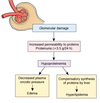
Damage to the glomeruli causes increased permeability of the basement membrane. causes hypoproteinemia which leads to

  1. increased oncotic pressure causing edema
  2. compensatory syntehsis of lipids by the liver causing hyperlipidemia
86
Q

Why does hyperlipidemia occur with nephrotic syndrome?

A

When proteins decrease, the liver compensates by producing more lipids to maintain hydrostatic pressure. Increase lipids = increased lipids in urine

87
Q

What are clinical features of nephrotic syndrome?

A

proteinuria, hypoalbumemia, edema, hyperlipidemia, lipiduria

88
Q

What causes kidney cystic disease? how do cysts differ from tumors?

A

Kidney cysts are normal. Result from a blocked tubule and the resulting pressure creating a cysts.

Cysts - uniform density and thin wall, tumor = abnormal shape, full cells

89
Q

What is adult dominant polycystic kidney disease?

A

Inheriteddisase with an autosomal dominant pattern. Cysts not present at birth, but develop slowly over time, overtaking the whole kidney medulla.

Causes the kidney to grow (T12- L5)

Can lead to kidney failure - gen in mid to later adult life. No cure. Treated with dialysis and transplant

90
Q

What is kidney failure?

A

Onset can be acute or insidious.

When kidneys aren’t removing enough wastes, more wastes build up in the blood (increased BUN + creatinine)

91
Q

What are the different types of acute renal failure?

A
  1. pre-renal: decreased blood supply (dt shock, dehydration of vasoconstriction)
  2. Post renal: Urine flow is blocked (dt stones, tumors, enlarged prostate)
  3. Intrinsic: kidney tubule function is decreased (dt ischemia, toxins, intratubular obstruction)
92
Q

What causes pre-renal failure?

A

Shock, trauma, sepsis, hemorrhage, dehydration

93
Q

What causes intrarenal and post renal ARF?

A

Intra: nephrotoxins

Post: Benign prostatic hypertrophy, stones

94
Q

What are the stages of acute renal failure?

A

Pre-renal: decreased blood flow causes pre-renal oliguria

Acute tubular necrosis (caused by hypoxia) enters the oliguric phase (small amounts of urine dt blockage because of the casts)

When pressure increases, is causes the cast blockages to pass, entering the diuretic phase (sudden increase of urine).

Recovery phase (healing of the tubular cells, takes anywhere from 2 weeks to 6 mos to heal)

95
Q

What is chronic renal failure? What is it characterized by?

A

Loss of functional nephrons. remaining nephrons must filter more leading to hyperfusion and hypertrophy

kidneys are thin and atrophic

Indicated by increase in Cr and BUN

96
Q

When does renal failure become symptomatic

A

losss of 75% of nephrons

97
Q

Who is at risk for chronic renal failure?

A
  • glomerulonephritis
  • Diabetic nephropathy
  • pyelonephritis
  • polycystic kidney disease
  • nephrotoxic exposure
  • obstructive nephropathy
  • Chronic HTN
98
Q

What are the stages of Renal failure and the S&S which accompany them

A
  1. 75% nephrons loss - decreased renal reserve. Asymptomatic. BUN and CR normal
  2. 75-90% loss = renal insufficiency. Polyuria and nocturia. Slight increase in BUN and Cr
  3. >90% loss = end stage failure = uremia
99
Q

WHat is the development of CRF

A

Decreased urine reserve leads to renal insufficiency (bc remaining nephrons are already working as hard as possible). Since nephrons can no longer regulate urine density, body enters renal failure as BUN and CR rise. Leads to end stage renal failure

100
Q

What is end stage renal disease? How is it treated?

A

End stage of renal disorders. Kidneys have atrophied. Associated with high levels of BUN and CR.

Tx with kidney transplant or dialysis

101
Q

What are some clinical consequences of ESRD?

A
  1. Decreased renal filtration causes altered fluid and electrolyte balance (acidosis, hyperkalemia, salt wasting and HTN)
  2. Waste build up in blood - toxins damage RBCS, CNS and platelets
  3. Kidney metabolic fxn decreases - decreases erythropoetin and vit D activation
102
Q

How does ESRD affect bone density? How does it affect CaPO4 salts

A

Decreased amounts of Vit D causes decreased serum calcium (bc calcium isn’t reabsorbed from GI tract). This stimulates the parathyrod hormone which causes bone reabsorption, which increases serum calcium, but decreases density of bones and forms calcium phosphate salts

Decreased GFR decreases phosphate excretion, which contributes to the formation of CaPO4 salts.

These individuals are at risk for kidney stone formation

103
Q

What are the secondary consequences of CRF?

A

Anemia, heart failure, osteoporosis

104
Q

What are the cardiovascular consequences of CRF?

A

Decreased erythropoeitin production decreases RBC production, which causes anemia

Decreased RBCs causes decreased blood viscosity which increases blood flow rate, which increases HR and therefore the workload on the left side of the heart which can cause lt sided ventricular hypertrophy and dilation. Not enough O2 is received by the Lt side to support contractions, which can lead to ischemia, angina or LHF

105
Q

Where would you find tumors or the urothelium?

Renal cell?

A

Urothelium: kidney pelvis, ureter, urinary bladder, urethra

Renal Cell: kidney medulla and cortex

106
Q

What are renal cell carcinomas? Etiology and survival rate? S&S?

A

:slow growing, but potentially massive tumors found in the kidney parenchyma. Common tumor

Etio: no identified risk factors, but 5% of ESRD will result in renal carcinomas. Gen happen in older adults (>50yo)

Survival: 50% survive after 5 years

S&S: flank pain, mass effect, hemturia

107
Q

What is mass effect?

A

compression of surrounding structures

108
Q

What is Wilms tumor?

Etiology?

Prognosis?

S&S?

A

: cancer of the lobules in children composed of immature cells resembling renal blastema

:related to genetic defects on chromosone 11 (some associated with changes of the tumor suppressor gene WT-1)

Good prognosis when surgery combined with chemo

Abdominal enlargement,

109
Q

What are carcinomas of the urethelium?

Prognosis?

S&S

A

: Most are transitional cell carcinomas, but some may be squamous or adenoca

Variable prognosis, dependant on grade

Hematuria, neoplastic cells in urine, urgency and frequency (decreased bladder volume), groin pain, hydronephrosis (aggressive cases)

110
Q
A