patho exam 3 Flashcards
bacteria strategies for evading or surviving host defense systems.
Man and animals have evolved many strategies for defending themselves against bacterial invation. Battle of evolution, as bacteria develop methods to overcome these defenses.
Most of these strategies can be thought of as those aslo suited to a natural enviroments.
i.e. properties such as adherence can be just as useful in staying close to a nutrient source as bacteria sicking to teeth or heart valves ect.
Defenses against protozoa are akin to avoid phagocytosis involved in the immune system.
virulence factors?
the properties (gene products) that enable a microorganism to establish itself on or within a host and enhance its potential to cause disease.
samonella
gram negative
pathogencic interations
- enter the body, attach to host cells for colonization
- evade the host innate and adaptive immune systems and persist in the immuno-evasion or immuno-suppression.
- Obtain nutrients and other requirements susch as iron
- Disseminate (sprad) within the host, replication and travel to other hosts.
- these may result in symptoms from the bacteria/ bacterial products ( i.e. toxins)
Preinfection
factors to consider?
Survival in the External Enviroment Reservoir?
Must be able to survive in the enviroment but also adapt to rapid changes?
- production of endospores to survive harsh enviroments
- Gram +ve walls to help reduce dehydration and protein oxidation
* Animal—animal—human—-human-human
* Enviroment to human
- Gene expression of new proteins
- Production of secondary metbolites - acids, antibiotics, bacterocins, antimicroial products, biofils
Most frequent Entry Portals
- skin
- Intestinal tract
- Respiratory tract
- Genitourinary tract
and many others
medical and dental related entry portals
preferred portals of entry
- many microbs have to enter in a specific manner and in a specific place to cause disease.
- ex vibrio cholerae infects the GI tract therefore, rubbing and infected oyster on a wound of the skin will not caused cholera.
- cross referance modes of transmission in section 2
Penetration of Skin or Epithelial Layer
- Skin -normal does a good job of preventing entry but can be breached
- surgery, catheters, trama, burns
- Biting arthropods?
- Tick,mouse,deer interations
- Borrelia burgdoferi- lyme disease
-
pareternal infections
-by some rout other than through the alimentary canal, such as by subcutaneeous, intramuscular or intravenous injection.
Microorganisms are depositied into the tissues below the skin or mucous membranes
- Puntures
- Injections
- bites
- scratches
- surgery
- splitting of skin due to swelling or dryness.
skin
when intact - it is normally impentrable
- when integrity lost then it becomes portal for entry
- hair folicles and Sweat gland ducts ( many have antimicrobial oils for production)
colonization and invation of host
it has been proposed that the reason the skin and immune systems are so effectiv e for the great majority of organisums is that the host-microbe relationship maybe viewed as somewhat short( evolutionary time).
No bacterium can borrow or penetrate skin alone with so form of breach.
-But there aer many virulence factors that have evolved that promote colonization and survival.
first steps
- penetration of skin
- penetration of the Epithelial and Mucin Layers
Rickettsial Diseases
Rickettsias are small bacteria,
- gram negative
- non-sporeforming
- strictily obligate
- intracellular
- in vertebrates - associated with/ fleas, lice or ticks.
- not cultured in laboratory media
– only in lab animals, mammalian tissue culture and yolk sac of chick embryos.
Rickettsia prowazekii-
common body or head louse, causes
- Typhus
- 3 million death in WW1 among troops, unsanitary conditions, caused more deaths than combate.
Human Louse
Rickettia prowazekii-typus
Borrelia recurrents- relapsing fever
Bartonella quintana-trench fever
Pool feeding
bite of louse puntures skin
- makes a trough
- blood pools and the bacterium enters from louse fecal matter which is aided by scratching
- Bacteria multiply in the cells lining blood vessles that eventually lyse resulting in the skin rash.
Penetration of Epithelial or Mucin Layers
Respiratory tract
- easiest portal to enter and most commonly infected
- Many microbs travel in aerosols which we breath
- ex. cold, TB, influenza,smallpox,Pneumonia
Penetration of the Epithelial or Mucin Layers.
Gastrointestianl tract
- Through food, water and dirty fingers
- Must overcome low pH of the gut
- Ex. Amobic dysentary, Hepatitis, Shigellosis
Penetration of the Epitelial or Mucin Layers
Genitourinary tract
- for STDs
- Broken (parentral route) or unbroken membranes ( depends on the microb
- Ex. HIV, Genital warts, Herpes, Syphillis
Epithelial Layer
-Nesseria gonorrhoeae
-STD attaches to urogenital epithelia more tightly than other tissue via
–Protiein Opa (opacity associated protein).
- Opa binds specifically to host cells processing the protein
- CD66- only found on human epithelial cells.
epithelial Layer
-Evolutionary pressure have made this organism host specific
- behavior of host- transmission, reproductive process?
- procreation (reproduction) vs Recreation
what is Mucin?
Network of protein and polysaccharide
what does Mucin do?
general and specialized functions
- prevent bacteria reaching mucosal cells
- Gi and vaginal tracts act as lubricats
- Goblet cells
- Respiratory tract- ciliated cells expel bacteria caught in mucin
- secoundary infections with colds and flu?
- Mucus is expelled in long streams that forms a network of tangled strands
- however pathogens have sought out weaknesses.
cholera outbreak in Yemen
worlds worst cholera outbreak
2000 people have died, a quarter of them children
more than half a million suspected cases
some bacteria can cross the Mucosal layer?
this is an active system in the bodies immune system.
Antibodies are produced against ingested organisums some of which are termed Secretory and released back into the Lumen t attach incoming organisms.
Preemptive strike!
Mycobacterium tuberculosis is an intercellular pathogen that infects macrophages.
Scanning EM of Mouse Peyers’ Patches
pic a-m- cell
pic b- infected by Y. pseudotuberculosis.
once organisums penetrate the host what are important virulence fcactors?
motility and methods to stick to cells
Motility, chemotaxis and Adherence
mouth, small intestin, bladder?
- mucsal surfaces are washed by fluids. Bacteria capable of adhearing to a surface have a selective advantage.
- many pathogens have mechanisms of attaching themselves to host epithelial cells.
- Specific receptors , diffrent parts of the body infected. Also host specific human strains of the particular bacterial species may not adhere to epithelial cells of an animal and vice-versa.
motility
addvantage to be able to move towards a nutrient source or away from an unfavorable enviroment.
- Flagella- ae specialized structures that provide motility
- Fimbrie/pill- can be involved in motility
chemotaxis
Directe movement of an organism toward ( positive) or away from ( negative ) a chemical gradient.
flagellum
- same basic structure shared between Bacteria and Archaea.
- flagella genes are highly conserved across these 2 domains sugetting motility has Depp evolutionary Roots.
phylogenetic Tree of life
Bacteria- Green filamentous bacteria
gram positives
spirochetes
proteobacteria
cyanobacteria
Planctomyces
Bacteroides Cytophaga
Themotoga
Aqyifex
-Archaea
halophiles
methanosarcina
methanobacterium
methanococcus
T.celer
Themoproteus
Pyrodicticum
-Eukaryota
Entamoebae
slime molds
animals
fungi
plants
ciliates
flagellates
Trichomonads
Microsporidia
Diplomonads
flagella arrangments
polar
lophotrichous
peritrichous
speed of bacteria
- some can move at 60 cell length /sec
- cheetah only 25 lengths /sec
- Polar flagellated organisms -rapid , spinning motion
- Peritrichously and lophotrichous flagellated organisms move in a straight line
-
Approx. 40 genes are involed in flagella
- Regulation genes- for synthesis and control
- structural proteins- make up flagella itself
- Chaperones proteins- mediate export of flagella proteins through cytoplasmic membrane to the outside of the cell to construct the flagellum itself.
the flagella consists of several components
- Base and Filament
gram- ve flagella
several “rings” make the basal body
-an outer L in LPS, P in Peptidoglycan, MS and C rings ar3e located in cytoplasmic Membrane and Cytoplasm.
Gram +ve
lack outer membrane and so no outer L Ring
flagella pic
flagella base
1000-1200 protons/ revolution
- protons flowing through channels exert electrostatic forces on helically arranged charges on the rotor proteins.
- Attractions between + and -ve ions cause the base to rotate 1200 protons/revolution
flagella structre
- at the base the motor that rotates the flagella is anchored in the cytoplasmic membrane and cell wall.
- Acchored int eh cytoplasm membrane ar ethe Mot and Fil proteins acting as the motor and switching ( reverse) mechanisms.
-Motion from te basal body- Proton Motive Force,- protons move across the cytoplasmic membrane through the Mot complex (1000-1200 ions per revolution)
articles on flagella
many belive the flagella is not evolution but intelligent design
chemotaxis
prokaryotes are too small to sense a gradient along the single cell insted sense what they passed through a few secounds before movement /direction therefor apper a little erratic
- without a gradient, cells move in a random fashion
- -Smooth “runs” swimming forward and ” tumbles” where cell stops and jigs about, changes direction then “runs” in a new direction.
flagella attraction
- in gradient, fewer tublems and more runs and bacteria moves towards attractant or way from a repellant.
- Anticlockwise forward motion, clockwise the bundle of flagella pushes apart, foward motion cease and cells tumble.
- Polar flagellated cells, reverse direction of rotation results in backward motion.
chemoreceptores
sensory proteins in the cytoplasmic membran sense the chemical gradients
- changes in the enviroment are sensed at both genetic and biochemical levels.
- Interact with cytoplasmic proteins to affect flagellar motor direction.
Adhesion
surface structures?
surface structures
- Fibriae
- Pilli
- Capsules
- Slime layers
- Specialized proteins
Fimbriae and Pili
Rod shaped protein structures, adhesins that diffre in thickness and length ( often on Gr -ve organisms)
-Fimbriae- shorter, thinner structures
-Pili- longer, thicker structures
-orderly array of single subunits called Pilin packed in a helical way to form a cylindrical structure.
Pili Adhesins
-many pathogens have a two- step process of the pili making initial contact. The pile retract and “pull” the bacteria close to the host cell where the adhesins take over and the pili depolymerize to make a much more strong bond.
Signal transduction-
-once thought that pili were metabolically inert. Now it appers that the attachment of Pili envokes a whole range of “gene switching” of virulence genes. The action may take the form of conformational changes in the pili tips upon binding (proteins E,F,G).
flagella
fimbriae
flagella- long tails
fimbriae- smaller hairs
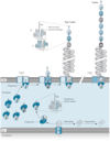
assembly of Pili is undertaken in the periplasmic space.
- additional proteins -ushers interact with chapreons and guide the subunits to the cell surface.
- present in the periplasm , prevent the pili from folding into their final configuration until ready to be extruded from the out membrane
- this is called the Chaperone Usher system
- finally a periplamic Protein H signals the end of the extrusion process.
how the bacteria mesures the length of decided what the final should be is not yet known.
chaperone- usher sytem
steps
- secrection and Specialized tip proteins (E,F,G) cross the inner membrane and into the periplasmic space.
- in addition to the actual pilus proteins there are a number of proteins that help in construction of the pilus.
- Usher
-scaffold structure C
-Chaperones
-the main shaft is assembled from Protein (A)
classes of fimbriae /pili are known.
functions:
Twitching motility. - rapid extension/ reaction enables the cell to “crawl” along the surface.
Key colonization factor-in Nesseria menigitidis ( bacterial meningitis) and Vibrio Cholerae ( cholera)
Avoid host defenses . -Pili break easily when bound by host aantibodies can break away (E,F and G protiens). Also Once the body recognizes the pili another type of pili with a diffrent tip is produced by the bacteria.
Pili (fimbriae) of Gram+ve bacteria
-oral strains able to adhere and form biofilms
strep, salvivarius, Strep, gordonii, Strep. oralis, Strep. Parasaguis and Actinomyces naeslundii.
- short, thin, rod- like fimbriae.
- covalent attachment of sbunits to each other and to peptidoglycan cell wall.
- Biofils can be thought of as a virulence factor.
biofilms
are matrices of organisms where gradients of nutrients exist.
-homoserine lactones-involves communication between cells- Quorum sensing
biofils
quorum sensing
properties and benifits
- reduced metabolic levels
- resistance to antibiotics
- reduction to predation
- pseudomonas aeruginosa
biofilms
exapmles:
- important in pre-infection and establishing colonization/ disease.
- Pseudomonas aeruginosa - in cystic fibrosis
-Legionella pneumophillia in Legionnaire’s disease
-Streptococcus, Bacteroides, Fusobacterium in dental plaque (endocarditis)
-S.epidermidis, Enterococcus facalis, E. coli, Proteus mirablis, P. aeruginosa
-K. pneumoniae- medical catheters.
during _________ organisums must evade or have counter measures to the host immune system.
colonization organisms.
complement system
- is part of the immune system that helps (complements) the ability of antibotics and phagocytic cells to clear microbes and damaged cells from the host organisum Promotes inflamation
- 3 patheays involved but all lead to the formation of the Membrane Attack Complex (MAC)
calassical pathway
antibody controlled
-host may produce antibodies that bind capsule providing sites both for phagocyte binding and activation of complement.
classic pathway
antibody controlled
-IgG and IgM antibodies bind antigens that become cross-linkage by multiprotein C1.
- this antiody -antigen complex leads to C2 and C4 binding at an adjacent membrane.
-
alternative Pathway
- controlled by serum protein
- Serum protein Properdin binds to bacterial cell surface.
- this membrane bound complex binds C3 and protein factor B
- catalyzes the formation of C5-9MAC leading to cell destruction as demonstrated in the classical pathway.
the 3rd pathway is initiated by?
Mannose Binding Lectins present on the cell walls of bacteria.
pore formation
- membrane damage and cell lysis
- recruits the C6-C9 proteins
capsule and slime layers
- many prokaryotes secrete a loose, unstructured network of polysaccharide/protein-carbohydrate.
- may be rigd (capsule) or flexible (slime) thick or thin.
- Evade complement and phagocyte- mediated killing.
types of slime and capsules
- enteropathic (disease of the intestine) e. coli
- extracellular polysaccharide
- Mucoid colonies
functions of capsules and slime layers
- protect from host inflammatory response (complement activation and phagocyte-mediated killing)
- Interferes in “alternative pathway”?
- can prevent binding of the B protein and activation of the C3 complement and the MAC ( membrane Attack Complex)
Alternative pathway
- Serum protein -Properdin binds to cell surface. This membrane bound complex binds C3 and Protein factor B
- -catalyzes the formation of C5-9MAC leading to cell destruction as demonstrated in the classical pathway.
Bacteria not killed by MAC are called?
Serum resistant
- although some C3b (opsonizing) can attach under the loose capsule, the capsule can prevent contact with phagocyte receptors.
- MAC may form too far way from membrane due to capsule that forms a physical barrier and therefore no bactericidal effect.
capsular material
- in some instances, proteins can diffuse through the capsule layer and still cause complement activation
- so just having a capsule or slime layer dosent protect pathogens ompletely.
- can be used for vaccines and have been very successful against a number of pathogens.
ex. Haemophilus influenzae type b
Bacterial meningitis
meningitis is an infection of the fluid of a persons spinal cord and the fluid that surrounds the brain.
- Viral meningitis is generally less severe and resolves without specific treatment, while bacterial meningitis can be quite severe and may result in brain damage, hearing loss, learning disabilities and death.
- Before the 1990’s Haemophilus influenzae type b was the leading cause of bacterial meningitis, but new vaccins being given to all children as part of thier routine immunizations have reudced the occurrence of invasive disease due to H. influenzae.
-leading causes are Streptococcus pneumoniae and Neisseria meningitis are leading causes of bacterial meningitis.
bacteria can produce _____ that resemble or mimicks host polysaccharides.
-non immunogenic or autoimmune problems
some organisum can attach host substances to thier cell walls to evade host immune system.
S.pyogenes- Hyaluronic acid (mammalian polysaccharide)
Neisseria meningitdis-Sialic acid (SA) (mammalin glycoprotein)
Attachment of sialic acid ( and othr substances) to LPS O-antigen prevents formation of C3 convertase.
-SA- can also hide mannose antigens on the surface of host cells or bacteria from mannose- binding lectian ( 3rd pathway of complement system) of host immune cells.
______ prevent effective MAC formation.
changes in the length of the LPS-O antigen side chains
this mechanism is not well understood.
what Bacteria can coat themselves with antibotics using immune cells receptors on thier cell surfaces.
s.aureus-protein A
Step. pyogenes- protein G
optimization-
making the bacteria more sticky.
so phagocytes get trapped and kill the cell
IGg
singal monomer
complement binding site
phagocyte receptor binging site
binds to antigen on forgin materia
IGM
5 monomers=10 binding sites
more efficent
cause clumping action-easier for macro phages to degrade bacteria
S. aureus
IgG
clumping factor binds fibrogen
collegen binding protein
attachment to extracellular matrix proteins
fibrogen binding protein
capsule
Phagocytic process is ______ and a further ______
enhanced 10-fold by antibody binding
10-fold by c3b fixation.
the body can be dampened down the bodys response by
produce enzymes that specifically degrads C5a found in gram +ve’s
-produce toxins that kill ohagocytes inhibit migration or reduce the oxidative burst
,odification of LPS so the immune response is not activated
-H. Pylori
pyogenic
pus forming bacteria
dead phagocytes make up large part of pus
Sty. aureus
produces pigmented compounds called Carotenoids neutralized the “oxidative burst”
Mycobacterium tuberculosis
intercellular pathogen uses cell wall Glycolipids to scavenge toxic oxygen compounds, remove hydroxyl radicals and superoxide anions.
Leukocidins
cytotoxin
produced by some intracellular pathogens such as Stap.aureus and Strep. pyogenes
-organisums are ingested but then released as phagocytes are killed
dead phagocytes make up a large part of pus
capsule
polysaccharide/protein coating
-Encapsulated bacteria aqre often highly resistat to phagocytosis
prevents adherence of phagocyte to the bacteial cell killing action of the MAC
Strep pnemoniae
capsule
10 cells can kill a mouse in a few days
non- encapulated strains are a virulent.
PMN’s release lysosomal granules that causes more tissue
damage as well as killing themselves, Pus is mainly
composed of DNA from these two sources.
•Close control of this system is essential
cytokines /chemokines and complement guide the
phagocytes to the infection
-prime these cells for activation only at the site of infection.
118
cytokines
involved in cell signaling
cytokines -include chemokines, interferons, interleukins, lymphokines, tumor necrosis factor.
-produced by a broad range of cells including immune cells like macrophages, B lymphocytes, Tlymphocytes and mast cells as well as endothelial cells and fibroblast.
Nitric oxide (NO)
free radical
during infection cytokines trigger monocytes and macrophages to produce Nitric Oxide.
- attacks bacterial metalloenzymes, proteins and DNA
- NO also combines withsuperoxide the synergy is more reactivve =Peroxynitrite - that oxidizes amino acids causing protiens to denature
NO- is toxic to both bacteria and human cells
Inflammation
- a non specific reaction to noxious stimuli such as toxins and pathogens -redness, sweling and pain
- mediated by Cytokines
-an bnormal upward deviation from 37C
- may be caused by externally induced pyrogenic agents from bacteria (LPS) (fever inducing/heat generating) or Endogenous Pyrogens-released from leukocytes
Interlukins
are a group of cytokines
interlukins lL-1,IL-6, Tumor Necrosis Factor (TNF-a)
pryogenic
fever/heat generating
types of fever
continuous fever
inter mittent fever
relapsing fever
countinuous fever
body temperature remains elevated over 24 hour period; <1 C variation
intermittent fever
body temperature shifts between normal and raised throught 24 hour period; period characteristic of most infectious diseases
relapsing fever
fever returning after an infection is reestablished.
signs of inflammation
swelling
redness
pain
heat
loss of function
these signs are due to the changes in blood vessel function
goal of inflammation
to bring phagocytes to the injered area to
isolate destroy inactivate the invader
remove debris
prepare subsequent healing