Path Pics Flashcards

1
Q

Name cells that white arrows point to.

A

Type II pneumocyte

2
Q

Name cells at black arrows.

A

club cells or clara cells

3
Q

What type of cell is this?

What do the arrows point to?

A

type II pneumocyte

lamellar bodies

4
Q

Name the cell.

A

macrophage

5
Q

Name the cell.

A

alveolar macrophage

6
Q

Name the cell.

Why does this cell look this way?

A

alveolar macrophage: crack cocaine user

7
Q
A

resorption atelectasis:

tumor obstructing right bronchus, markedly reduced right lung volume and right mediastinal shift

8
Q
A

compression atelectasis:

left pneumothorax from chest wall trauma, left lung collapse and right mediastinal shift

9
Q

What is at the arrow in this liver?

What disease does it indicate?

What lung disease will this person have?

A

pink PAS positive hyaline globules

alpha 1 antitrypsin deficiency

panacinar emphysema

10
Q

What type of emphysema effects this part of the acinus?

A

centriacinar

11
Q
A

emphysema: hyperinflation

12
Q
A

hyperinflation of lungs: emphysema

13
Q

What is at the arrow?

What disease is this?

A

centriacinar emphysema: enlarged airspaces with tissue destruction at APEX

14
Q

What is at the arrows?

What disease is this?

A

central part of acinus has abnormally large airspace with club-shaped alveolar septa that appear to be free floating

centriacinar emphysema

15
Q

What type of emphysema effects this part of the acinus?

A

panacinar emphysema: whole acinus is affected

16
Q
A

destruction with airspace dilation is most severe in lower lobe

Panacinar emphysema

17
Q
A

relatively uniform dilation of all parts of acini

panacinar emphysema

18
Q

What type of emphysema effects this part of the acinus?

A

paraseptal emphysema: distal acini adjacent to interlobular septa and pleura affected

19
Q
A

paraseptal emphysema

bullae: markedly enlarged subpleural airspaces

20
Q
A

paraseptal emphysema: distal airspaces immediatley beneath the pleural space have marked dilation

21
Q
A

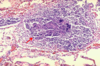
chronic bronchitis: submucosal gland hypertrophy

22
Q
A

chronic bronchitis: lymphocytes (arrow) surround the bronchial epithelium

23
Q
A

chronic bronchitis: goblet cells (arrows) increased

24
Q
A

mucous plug

asthma

25
Q
A

asthma: thick BM with eosinophilic inflammation

26
Q
A

Asthma: Charcot-Leyden (disintegrated eosinophils) and eosinophils

27
Q
A

asthma: Curschmann spirals (mucous tight coils)

28
Q
A

bronchiectasis: dilated airways exted almost to pleura

29
Q
A

bronchiectasis: saccular dilation of airways with surrounding fibrosis

30
Q
A

bronchiectasis: airway dilation accompanied by surrounding fibrosis and luminal pus

31
Q
A

allergic rhinitis: type I hypersensitivity

arrow: Charcot-Leyden crystals
star: inflammation

32
Q
A

nasal polyp with stromal edema and eosinophils

33
Q
A

angiofibroma: large blood vessels and fibrous tissue

34
Q
A

subpleural “net-like” reticulations (arrow)

idiopathic pulmonary fibrosis

35
Q
A

patchy interstitial fibrosis (white arrow) that is most severe subpleurally with interveneing normal tissure

idiopathic pulmonary fibrosis

36
Q
A

usual interstitial pneumonia (UIP): patchy interstitial fibrosis most severe subpleurally

indicitive of IPF or collagen vascular associated lung disease

37
Q
A

usual interstitial pneumonia (UIP): localized areas of fibroblastic proliferation (arrows: fibroblast foci)

lymphocytes

38
Q
A

usual interstitial pneumonia (UIP)

arrows: densely collagenized fibrosis
asterik: honeycomb cysts lined with metaplastic bronchiolar epithelium

39
Q
A

usual interstitial pneumonia (UIP)

seen in IPF and collagen vascular disease associated lung disease

40
Q
A

cryptogenic organizing pneumonia (COP)

intra-alveolar plugs of loose fibroconnective tissue (arrows)

41
Q
A

organizing pneumonia following adult respiratory distress syndrome: alveolar duct plugged by loose fibroconnective tissue (arrows)

similar to COP: need clinical information to differentiate

42
Q
A

simple coal workers pneumoconiosis

arrow: coal dust macule with dilated adjacent airspaces (localized emphysema)

43
Q
A

complicated coal workers pneumoconiosis

BLACK LUNG

44
Q
A

silicosis: dense collagenized nodule with surrounding lymphocytes

45
Q
A

silicosis: birefringent silica particles under polarized light

46
Q
A

asbestos

right: straight stiff: more pathogenic
left: curly flexible

47
Q
A

pleural plaques (dense collagen): arise on parietal pleura and diaphragmatic domes

seen in asbestos inhalation

48
Q
A

ASBESTOS bodies: iron coated asbestos

49
Q
A

ASBESTOS bodies: iron coated on Prussian blue

50
Q
A

asbestosis: diffuse interstitial fibrosis (looks like UIP) with asbestos bodies

51
Q
A

Drug-induced pneumonitis: mild interstitial fibrosis with lymphocytes and type II pneumocyte hyperplasia (arrows)

52
Q
A

sarcoidosis

gramuloma (arrow) alond bronchovascular bundles and interlobular septa

53
Q
A

sarcoidosis

granuloma surrounding bronchiole

54
Q
A

sarcoidosis: non-caseating granulomas with giant cells

55
Q
A

asteroid bodies

sarcoidosis and other granulomatous diseases

56
Q
A

Schaumann bodies (calcification)

sarcoidosis and other granulomatous diseases

57
Q
A

hypersensitivity pneumonitis: lymphocytic interstitial inflammation and fibrosis is centered on bronchiole (arrow)

58
Q
A

hypersensitivity pneumonitis

granuloma with giant cells surrouned by lymphocytes

59
Q
A

hypersensitivity pneumonitis

plug of organizing pneumonia with lymphocytes

60
Q
A

end stage hypersensitivity pneumonitis

HONEYCOMB LUNG: end stage of many interstitial fibrosisng lung diseases

61
Q
A

desquamative interstitial pneumonia or respiratory bronchioloiti: filling of small airways with macrphages with yellow-brown pigment, widening of alveolar walls, mild fibrosis, type II cell hyperplasia

62
Q
A

pulmonary langerhans cell histiocytosis

positive for: langerin, S-100, CD1a

63
Q
A

pulmonary alveolar proteinosis: alveoli are filled with dense, amorphous, protein-lipid granular precipitate (alveolar walls are normal)

64
Q
A

penumocystis jirovecii

Gomori methenamine silver stain

65
Q
A

saddle pulmonary embolism: lodges at bifurcatio nof pulmonary arteries

sudden death due to acute cor pulmonale

66
Q
A

arrows: pulmonary embolism
line: wedge shaped hemorrhagic infarct

67
Q
A

pulmonary embolism with early infarction with alveolar hemorrhage (arrow) and coagulative necrosis of the alveolar septa (asterik)

68
Q
A

resolving pulmonary infarct: after 48 hours

becomes paler red-brown (arrows) due to conversion of hemorrhage into hemosiderin by macrophages (will eventually scar)

69
Q
A

non-thrombotic pulmonary embolism

foreign material (IV drug use)

70
Q
A

pulmonary vascular thickening contricts lumen

pulmonary HTN

71
Q
A

plexogenic lesion: network of capillary formations span lumen of pulmonary arteries

severe primary pulmonary HTN and congenital heart disease with L to R shunt

72
Q
A

diffuse pulmonary hemorrhage: lungs are heavy and show diffuse dark red consolidation

goodpasture, idiopathic pulmonary hemosiderosis, lupus, wegener granulomatosis

73
Q
A

necrotizing hemorrhagic interstitial pneumonitis due to autoimmune alveolar BM destruction: arrow necrosis of alveolar walls with intra-alveolar hemorrhage

goodpasture syndrome

74
Q
A

pulmonary edema

75
Q
A

long standing pulmonary edema with alveolar microhemorrhages and hemosiderin-laden macrophages (arrow= heart failure cells)

76
Q
A

protein rich fluid and necrotic epithlial cells: hyaline membranes

Diffuse alveloar damage: acute stage

77
Q
A

diffuse alveolar damage: organizing stage

hyaline membranes have been resorbed and replaced with plugs of intra-alveolar organizing fibroconnective tissue (arrow)

right: trichrome stain

78
Q
A

bronchopneumonia: patchy pale areas of consolication in upper lobe

79
Q
A

lobar pneumonia: lower lobe is consolidated

80
Q
A

red hepatization: pneumonia

81
Q
A

gray hepatization: pneumonia

82
Q
A

S. pneumoniae

right: alveoli with neutrophils and proteinaceous exudate
left: gram positive lancet shaped diplocci in neutrophil rich exudate

83
Q
A

pulmonary abscess: necrotizing pneumonia

84
Q
A

pulmonary abscess: lots of neutrophils, alveolar destruction

85
Q
A

atypical pneumonia: lymphocytic inflammation of the interstitium (red arrows) and pneumocyte hyperplasia (yellow arrows)

86
Q
A

Ghon complex (black arrow) and caseous hilar nodes (red arrow)

primary TB

87
Q
A

TB granulomas with central necrosis

88
Q
A

granulomatous inflammation with central necrosis (caseation) rimmed by macrophages

TB

89
Q
A

TB: red rods on acid fast stain

90
Q
A

confluent areas of caseous necrosis and cavitation (arrow) on upper lobe (highest O2)

secondary TB

91
Q
A

miliary TB: lymphohematogenous dissemination

multiple foci of granulomatous inflammation

92
Q
A

granulomatous inflammation, necrotizing

histoplasmosis

93
Q
A

histoplasmosis: macrophages with tiny yeast

94
Q
A

blastomycosis

BROAD BASED BUD: snowman

95
Q
A

right: H and E: lighlty basophilic budding yeast surrounded by a clear space (capsule)
left: stain that highlights capsule

Cryptococcus neoformans

96
Q
A

thick walled non-budding SPHERULES filled with ENDOSPORES

Coccidiomycosis

97
Q
A

CMV pneumonia

infected cell: enlarged with cytoplasmic and intranuclear viral inclusions (owl eyes)

98
Q
A

pneumocystis pneumonia

right: central umbilications on SILVER STAIN
left: organism in granular alveloar exudate

99
Q
A

aspergillosis

right: ACUTE ANGLE branching, SEPTATE HYPHAE
left: angioinvasion allows aspergillus to cause hemorrhagic destrction that crosses fissure

100
Q
A

Murcormycosis

angioinvasive

BROAD WIDE ANGLE BRANCHING, NON-SEPTATE HYPHAE

101
Q
A

bronchogenic cysts

102
Q
A

pulmonaory sequestration

103
Q
A

meconium aspiration syndrome

arrows: amniotic fluid/squamous debris and meconium (red)

104
Q

infant

A

hyaline membrane disease: neonatal RDS

105
Q
A

circumscribed coin lesion; peripheral solitary

hamartoma

106
Q
A

grey-white glistening nodule

hamartoma

107
Q
A

lobules of mature cartilage entrapped in benign respiratory epithelium

pulmonary hamartoma

108
Q
A

peripheral spiculated mass

adneocarcinoma

109
Q
A

peripheral tumor

adenocarcinoma

110
Q

cancer

A

pneumonia like consolidation of entire lobe

adenocarcinoma

111
Q
A

lung adenocarcinoma

mucin produing glands

112
Q
A

lepidic pattern shows growth along alveloar septa (arrows)

lung adenocarcinoma

113
Q
A

large centrally located mass

squamous cell lung carcinoma

114
Q
A

large tumor that obliterates entire central portion of lung and shows cavitation

squamous cell lung carcinoma

115
Q
A

keratin pearl

squamous cell lung carcinoma

116
Q
A

large cells with prominent nucleoli

large cell lung carcinoma

117
Q
A

central tumor (arrows) with infiltrative growth into surrounding lung

small cell lung carcinoma

118
Q
A

small cells with scant cytoplasm and crush artifact (arrow), mitoses

small cell lung carcinoma

119
Q
A

endobronchial circumscribed tumor

carcinoid tumor

120
Q
A

nests of bland cells with granular chromatin

carcionoid tumor

121
Q
A

dense core granules on EM are evidence of neuroendocrine differentiation

carcinoid tumor

122
Q
A

pancoast tumor

123
Q
A

metastatic lung cancer: cannonball appearance

124
Q
A

circumscribed nodule of metastatic colon adenocarcinoma with normal surrounding parenchyma

125
Q
A

diffusely thickened pleura with rind like encasement of lung

Malignant pleural meosthelioma

126
Q

mesothelioma type?

A

epithelioid type of mesothelioma

tubules: arrow that mimic adenocarcinoma

127
Q
A

CALRETININ: positive brown stain

mesothelioma

128
Q

mesothelioma or adenocarcinoma?

A

long, slender microvilli

mesothelioma

129
Q

mesothelioma or adenocarcinoma?

A

adenocarcinoma

stubby microvillous rootlets

130
Q
A

nasopharyngeal carcinoma

poorly differentiated squamoous cell carcinoma in background of lymphocytes

131
Q
A

laryngeal papilloma

132
Q
A

laryngeal carcinoma

squamous cell carcinoma of vocal cord