Panoramic Tomography 1 (DPT) Flashcards
What is a tomogram?
- Radiograph showing a slice or section of tissue in focus
- tissues either side of slice are exposed to radiation but are not clearly seen on resulting film
- tissues seen are within focal trough or plane
- multiple tomograms of sequential planes would build up to give 3D image (e.g. CT)
- DPT is a form type of tomography that allows us to bring the teeth and supporting structures into focus.
What is a DPT (Dental Panoramic Tomogram)?
- Dental arch elliptical shaped
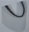
- Focal trough is horseshoe shaped
- Complex rotation – usually with two centres of rotation
- X-ray tube rotates behind patient
- Sensor or cassette move in front of patient synchronised with tubehead
What is a Focal trough?
- Approximately horseshoe shaped
- Narrow anteriorly
- Wider posteriorly
- Corresponds to shape of dental arch

In Panoramic Radiography what is the conventional film?
- Film within cassette
- Film sensitive to light
- Within cassette are intensifying screens
- Screens absorb X-rays and produce light
- Light interacts with film producing image
What are the Disadvantage of intensifying screens?
- Light is emitted in all directions
- Light affects larger area of film than a single photon
- Image quality (fine detail) is not as good as direct action film
In digital what are Indirect action film and intensifying screen replaced by?
Phosphor plates or Solid state sensor/CCD
Why is a bite block used in a DPT?
To bring mandible into same focal trough as maxilla
What are some advantages of DPT?
- Images teeth and facial bones with minimal discomfort
- Shows both sides on one radiograph allowing comparison
- Shows vertical height of mandible and inferior dental canal
- Shows maxillary sinus walls
- Dose is reduced compared to full mouth of intra-oral radiographs and actual time to take is less
What can a DPT show?
- Lesion not completely visible on intra-oral
- Gross dental disease/neglect
- Symptomatic third molars
- Orthodontic assessment
- Mandibular fractures
- Degenerate disease of TMJ
- Implant planning or review
What are the Disadvantages of DPT?
- Lack of fine detail
- Superimposition of other soft and hard tissues, air shadows
- Patient must be correctly positioned for optimal image quality
- Exposure time up to 16 seconds
- Patient co-operation required
- Magnification of image due to object/receptor distance
How should the patient be positioned for a DPT?
- Remove metal jewellery, glasses etc
- Patient stands with spine straight holding handles
- Patient bites incisors edge to edge in groove on bite block
- Light beam markers
- Head immobilised
- Tongue to roof of mouth
- Stand still
What is the relationship with the patient and the Focal trough?
We try and position the patient so that their teeth are in the middle of the focal trough – with the patient biting into the groove of the bite block. The patient brings their mandible forwards into an edge to edge incisal relationship – which is impossible to do if you have a Class III incisal relationship!
Is a DPT good for caries diagnosis?
- Not the “Gold Standard”
- Frequently requested when strong gag reflex
- Poor fine detail
- Overlap, particularly in premolar regions
- Superimposition of anatomy/air (lip shadow)
- Some have suggested DPT may be better for occlusal caries diagnosis (especially in molars)


