Paediatrics Haematology Flashcards
What is haemoglobin made from?
4 protein subunits
Fetal haemoglobin (HbF) = two alpha and two gamma
Adult haemoglobin (HbA) = two alpha and two beta
Draw the oxygen dissociation curve for fetal and adult Hb?
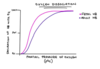
When does the production of HbF decrease?
From 32 to 36 weeks gestation (HbA is produced in greater quantities)
What is the problem in sickle cell disease?
Genetic abnormality for the beta subunit responsible for causing the sickle shape of the RBCs (doesn’t happen in fetal haemoglobin)
What is the treatment in sickle cell disease?
Hydroxycarbamide to increase production of fetal Hb
What is anaemia?
Low level of Hb in the blood (not a disease - the result of one)
What is haemoglobin?
Protein found in RBCs (iron is needed to make it)
What are the causes of anaemia in infancy?
Physiologic anaemia of infancy
Anaemia of prematurity
Blood loss
Haemolysis
Twin-twin transfusion
What are the causes of haemolysis in a neonate?
Haemolytic disease of the newborn
Hereditary spherocytosis
G6PD deficiency
When does physiologic anaemia of infancy occur and why?
At six to nine weeks of age in healthy term babies - high oxygen delivery to tissues caused by high Hb levels at birth cause negative feedback - production of erythropoietin by the kidneys is suppressed and there is reduced production of Hb by the bone marrow
Why do premature neonates become anaemic?
Less time in utero receiving iron from mother
RBC creation can’t keep up with rapid growth in first few weeks
Reduced erythropoietin levels
Blood tests remove a significant proportion of circulating volume
How to test for haemolytic disease of the newborn?
direct Coombs test (DCT)
What are the 2 main causes of anaemia in older children?
Iron deficiency anaemia secondary to dietary insufficiency
Blood loss from menstruation in older girls
What are the rarer causes of anaemia in children?
Sickle cell anaemia
Thalassaemia
Leukaemia
Hereditary spherocytosis
Hereditary eliptocytosis
Sideroblastic anaemia
What is a common cause of chronic anaemia and iron deficiency worldwide?
Helminth infection with roundworms, hookworms or whipworms
Can be very common in developing countries
What is the treatment of helminth infection?
Albendazole
Mebendazole
What are the causes of microcytic anaemia?
T – Thalassaemia
A – Anaemia of chronic disease
I – Iron deficiency anaemia
L – Lead poisoning
S – Sideroblastic anaemia
What are the causes of normocytic anaemia?
A – Acute blood loss
A – Anaemia of Chronic Disease
A – Aplastic Anaemia
H – Haemolytic Anaemia
H – Hypothyroidism
What are the two types of macrocytic anaemia?
Megaloblastic (result of impaired DNA synthesis preventing cell from dividing normally - vitamin deficiency)
Normoblastic
What are the causes of megaloblastic anaemia?
B12 deficiency
Folate deficiency
What is normoblastic macrocytic anaemia caused by?
Alcohol
Reticulocytosis (usually from haemolytic anaemia or blood loss)
Hypothyroidism
Liver disease
Drugs e.g. azathioprine
What are the symptoms of anaemia?
Tiredness
Shortness of breath
Headaches
Dizziness
Palpitations
Worsening of other conditions
What symptoms are specific to iron deficiency anaemia?
Pica - dietary cravings for abnormal things e.g. dirt
Hair loss - iron deficiency anaemia
What are some generic signs of anaemia?
Pale skin
Conjunctival pallor
Tachycardia
Raised resp rate
What are the specific signs of iron deficiency anaemia?
Koilonychia (spoon shaped nails)
Angular chelitis
Atrophic glossitis (smooth tongue due to atrophy of papillae)
Brittle hair and nails
What sign is specific to haemolytic anaemia? and Thalassaemia?
Haemolytic = Jaundice
Thalassaemia = bone deformities
What are the initial investigations for anaemia?
Full blood count for haemoglobin and MCV
Blood film
Reticulocyte count
Ferritin (low iron deficiency)
B12 and folate
Bilirubin (raised in haemolysis)
Direct Coombs test (autoimmune haemolytic anaemia)
Haemoglobin electrophoresis (haemoglobinopathies)
What does a high level of reticulocytes indicate in anaemia?
Active production of RBCs to replace lost cells - anaemia is due to haemolysis or blood loss
What is the management of anaemia?
Establish underlying cause and direct treatment accordingly
If severe then may need blood transfusions
What are the causes of iron deficiency?
Dietary insufficiency - most common
Loss of iron e.g. in heavy menstruation
Inadequate absorption e.g. in Crohn’s
Where is iron mainly absorbed?
Duodenum and jejunum
In what form is iron mainly absorbed? What can interfere with this?
In the soluble ferrous (Fe2+) form - when less acid in stomach it changes to the insoluble ferric (Fe3+ form)
PPIs can interfere and coeliacs / Crohn’s disease
How does iron travel around the blood?
As ferric ions bound to carrier proteins called transferrin
What is the total iron binding capacity (TIBC)?
Total space on the transferrin molecule for the iron to bind
What is the transferrin saturation?
Serum iron / TIBC
What is ferritin?
Form that iron takes when it is stored in cells (extra is released when there is inflammation e.g. with infection or cancer)
What can cause low / high ferritin?
Low = iron deficiency (could still be normal especially if infection)
High = difficult to interpret and means inflammation usually
Is serum iron a useful measure?
No - significantly varies throughout the day - higher levels in the morning and after eatin iron containing meals
On its own its not a useful measure
What can increase the levels of serum ferritin, serum iron and TIBC?
Supplementation with iron
Acute liver damage (lots of iron is stored in the liver
What is the management of anaemia?
Treat underlying cause
Iron supplemented with ferrous sulphate or ferrous fumarate - slowly corrects the iron deficiency (causes constipation and black coloured stools - unsuitable for malabsorption)
Blood transfusions are very rarely necessary
What is leukaemia?
Cancer of a particular line of the stem cells in the bone marrow - causing unregulated production of certain types of blood types (chronic is slow and acute is fast) cell line myeloid or lymphoid
Draw out cell lineage?

What are the types of leukaemia?
Acute lymphoblastic leukaemia (ALL)
Acute myeloid leukaemia (AML)
Chronic myeloid leukaemia (CML) is rare
What age does ALL and AML peak?
ALL peaks aged 2-3 years
AML peaks aged under 2 years
What is the result of the excessive production of a single type of cell?
Pancytopaenia:
Low RBCs (anaemia)
Low WBCs (leukopenia)
Platelets (thrombocytopaenia)
What is the main environmental risk factor for leukaemia?
Radiation exposure
Which conditions are predisposing to leukaemia?
Down’s syndrome
Kleinfelter syndrome
Noonan syndrome
Faconi’s anaemia
Which conditions are predisposing to leukaemia?
Down’s syndrome
Kleinfelter syndrome
Noonan syndrome
Faconi’s anaemia
How does leukaemia present in children?
Persistent fatigue
Unexplained fever
Failure to thrive
Weight loss
Night sweats
Pallor (anaemia)
Petechiae and abnormal bruising (thrombocytopania)
Abdo pain
Generalised lymphadenopathy
Unexplained or persistent bone or joint pain
Hepatosplenomegaly
What is the first step if leukaemia is suspected?
FBC within 48 hours
What investigations to establish the diagnosis of leukaemia?
FBC for anaemia, leukopenia, thrombocytopenia and high numbers of WBCs
Blood film for blast cells
Bone marrow biopsy
Lymph node biopsy
What testing can be done to stage leukaemia?
Chest X-Ray
CT scan
Lumbar puncture
Genetic analysis and immunophenotyping of abnormal cells
What is the treatment of leukaemia?
Chemotherapy
Radiotherapy
Bone marrow transplant
Surgery
What are the complications of chemotherapy?
- Failure to treat leukaemia
- Stunted growth and development
- Immunodeficiency and infections
- Neurotoxicity
- Infertility
- Secondary malignancy
- Cardiotoxicity
What is the prognosis for leukaemia?
Overall cure rate for ALL is 80% but prognosis depends on individual factors
Outcomes less positive for AML
What is idiopathic thrombocytopenic purpura?
Idiopathic (spontaneous) thrombocytopenia (low platelet count) causing a purpuric rash (non-blanching rash)
What is ITP caused by?
Type II hypersensitivity reaction - antibodies which target and destroy platelets - can be spontaneous or triggered by something e.g. viral infection
How does ITP present?
Children under 10
Recent viral illness
Onset over 24-48 hours
Bleeding e.g. gums, epistaxis, menorrhagia
Bruising
Petechial or purpuric rash caused by bleeding under skin
What are petechiae, purpura and ecchymoses?
NON BLANCHING REGIONS
Petechiae = pin-prick spots (around 1mm) of bleeding under skin
Purpura = larger (3-10mm) spots of bleeding
Ecchymoses = large area of blood (more than 10mm)
How is ITP diagnosed?
Urgent FBC for the platelet count (other values in FBC should be normal)
Other causes excluded e.g. heparin induced thrombocytopenia and leukaemia
What is the treatment of ITP?
Only if patient is actively bleeding or severe thrombocytopenia (platelets below 10)
Prednisolone
IV immunoglobulins
Blood transfusions if required
Platelet transfusions only work temporarily
Why do platelet transfusions only work temporarily?
Antibodies against platelets will begin destroying the transfused platelets as soon as infused
What advice for patients with ITP?
Avoid contact sports
Avoid IM injections
Avoid NSAIDs, aspirin and blood thinners
Advice on managing nosebleeds
Seek help after any injury which may cause internal bleeding e.g. car accidents or head injuries
What are some complications of ITP?
Chronic ITP
Anaemia
Intracranial and subarachnoid haemorrhage
Gastrointestinal bleeding
What is sickle cell anaemia?
Genetic condition causing sickle (crescent) shaped RBCs - making them fragile and more easily destroyed leading to haemolytic anaemia
What is the pathophysiology of sickle cell anaemia?
Patients with sickle cell disease have an abnormal variant called haemoglobin S (HbS) - causing the RBCs to be “sickle” shaped
What is the inheritance for sickle cell anaemia? What does it affect?
Autosomal recessive condition where there is an abnormal gene for beta-globin on chromosome 11 (one copy results in sickle-cell trait - usually asymptomatic)
What is the relation of sickle cell disease to malaria?
Sickle cell disease is more common in patients from areas affected by malaria e.g. africa, india, middle east, carribean. Having one copy of gene reduces the severity of malaria - more likely to survive and pass on their genes - there is selective advantage to having the gene
How is sickle cell disease diagnosed?
Pregnant women at risk of being carrier as offered testing during pregnancy
Also tested for on newborn screening heel prick test at 5 days of age
What are the complications of sickle cell disease?
Anaemia
Increased risk of infection
Stroke
Avascular necrosis in large joints e.g. hips
Pulmonary hypertension
Painful and persistent penile erection (priapism)
Chronic kidney disease
Sickle cell crises
Acute chest syndrome
What is the general management of sickle cell disease?
Avoid dehydration and other triggers of crises
Ensure vaccines are up to date
Abx prophylaxis to protect against infection usually with pen V (phenoxymethypenicillin)
Hydroxycarbamide used to stimulate production of HbF (protective against sickle cell crises and acute chest syndrome)
Bone transfusion for severe anaemia
Bone marrow transplant can be curative
What is a sickle cell crisis?
Umbrella term for a spectrum of acute crises related to the condition - range from mild to life threatening
Spontaneous or triggered by stresses e.g. infection, dehydration, cold or significant life events
How are sickle cell crises managed?
Low threshold for admission to hospital
Treat any infection
Keep warm
Keep well hydrated with IV fluids
Simple analgesia e.g. paracetamol and ibuprofen (NSAIDs avoided in renal impairment)
Penile aspiration for priapism
What is a vaso-occlusive crises?
Sickle shaped blood cells clog capillaries causing distal ischaemia
Associated with dehydration and raised haematocrit
What are the symptoms of a vaso occlusive crisis?
Pain, fever and symptoms of infection (if present)
How is priapism caused by vaso-occlusive crisis treated?
Aspiration of blood from the penis
What is a splenic sequestration crisis?
RBCs block blood flow within the spleen - causing it to be acutely enlarged and painful
What can pooling of blood in spleen cause?
Severe anaemia
Circulatory collapse (hypovolaemic shock)
What is the management of splenic sequestration crisis?
Emergency treatment
Management is supportive
Blood transfusions
Fluid resusfor anaemia and shock
What is the management of recurrent splenic sequestration?
Splenectomy
(recurrence can cause splenic infarction = susceptibility to infections)
What is aplastic crisis?
Temporary loss of the creation of new blood cells commonly caused by infection with parvovirus B19
Leads to significant anaemia - management is supportive with blood transfusions if necessary
Usually resolves spontaneously within a week
How is acute chest syndrome diagnosed?
Fever or respiratory symptoms with new infiltrates seen on CXR
What can cause acute chest syndrome?
Infection e.g. pneumonia or bronchiolitis
Non-infective causes (e.g. pulmonary vaso-occlusion or fat emboli)
What is the management of acute chest syndrome?
Abx or antivirals for infections
Blood transfusions for anaemia
Incentive spirometry using a machine which encourages effective and deep breathing
Artifical ventilation with NIV or intubation
What is thalassaemia?
Genetic defect in the protein chain that makes up haemoglobin
Defect in alpha globin chain = alpha thalassaemia
Defect in beta globin chain = beta thalassaemia
Both conditions are autosomal recessive = varying degrees of anaemia
Why does thalassaemia lead to splenomegaly?
RBCs are more fragile and break down more easily - spleen collects all destroyed RBCs
Why are patients with thalassaemia prone to fractures and prominent features e.g. pronounced forehead and malar eminences (cheek bones)?
Bone marrow expands to produce extra RBCs
What are the signs and symptoms of thalassaemia?
Microcytic anaemia (low mean corpuscular volume)
Fatigue
Pallor
Jaundice
Gallstones
Splenomegaly
Poor growth and development
Pronounced forehead and malar eminences
How is thalassaemia diagnosed?
FBC for microcytic anaemia
Haemoglobn electrophoresis to diagnose globin abnormalities
DNA testing to look for genetic abnormality
Pregnant women are offered screening test for thalassaemia at booking
Why does iron overload occur in thalassaemia?
Faulty creation of RBCs
Recurrent tranfusions
Increased iron absorption in gut in response to anaemia
How are patients with thalassaemia monitored for iron overload?
Serum ferritin monitored
How is iron overload managed?
Limiting transfusions
Performing iron chelation
How does iron overload present?
Fatigue
Liver cirrhosis
Infertility
Impotence
Heart failure
Arthritis
Diabetes
Osteoporosis and joint pain
Where is the genetic defect for alpha-thalassaamia?
Protein on chromosome 16
What is the management of alpha-thalassaemia?
Monitoring FBC
Monitoring for complications
Blood transfusions
Splenectomy performed
Bone marrow transplant can be curative
Where is the defect for beta-thalassaemia?
Chromosome 11 (can be abnormal copy or deletion genes where there is no function in beta globin protein)
What are the three types of thalassaemia?
Thalassaemia minor / intermedia / major
What are the features of thalassaemia minor?
Patients are carriers of an abnormally functioning beta globin gene (one abnormal and one normal gene)
What is the management of thalassaemia minor?
Mild microcytic anaemia usually patients require only monitoring
What are the features of thalassaemia intermedia?
Two abnormal copies of beta globin gene (two defective or one defective and one deletion gene)
Causes more significant microcytic anaemia
What is the treatment of thalassaemia intermedia?
Require monitoring and occasional blood transfusions (maybe also iron chelation to prevent iron overload)
What is the genetic defect in thalassaemia major?
Homozygous for the deletion gene - no functioning betal globin genes at all - most severe = severe anaemia and failure to thrive in early childhood
What are the features of thalassaemia major?
Severe microcytic anaemia
Splenomegaly
Bone deformities
What is the management of thalassaemia major?
Regular transfusions
Iron chelation
Splenectomy
Bone marrow transplant can potentially be curative
What is hereditary spherocytosis?
Condition where RBCs are sphere shaped making them fragile and easily destroyed
What is the most common inherited haemolytic anaemia in northern europe?
Hereditary spherocytosis
What is the inheritance of hereditary spherocytosis?
Autosomal dominant
How does hereditary spherocytosis present?
Jaundice
Anaemia
Gallstones
Splenomegaly
What is a haemolytic crisis in hereditary spherocytosis?
Haemolysis, anaemia and jaundice is more significant (often triggered by infections)
What is an aplastic crisis?
Increased anaemia, haemolysis and jaundice without normal response from bone marrow of creating new red blood cells (no reticulocyte response often triggered by infection with parvovirus)
How is hereditary spherocytosis diagnosed?
Family history and clinical features along with spherocytes on the blood film
What is the abnormality on FBC for hereditary spherocytosis?
Increased mean corpuscular haemoglobin concentration (MCHC)
Increased reticulocytes
What is the management of hereditary spherocytosis?
Folate supplementation and splenectomy
Cholecystectomy (removal of gallbladder) if gallstones a problems
Transfusions during acute crises
What are the features of hereditary elliptocytosis?
Similar to hereditary spherocytosis except RBC are ellipse shaped
Autosomal dominant
Presentation and management is similar to hereditary spherocytosis
What is G6PD deficiency?
Condition where there is a defect in the G6PD enzyme found in all cells in the body
Who is G6PD deficiency most common in?
Mediterranean
Middle eastern
African patients
What is the pattern of inheritance for G6PD deficiency?
Inherited in an X linked recessive pattern - usually only affects males as they have a single copy of the gene
What does G6PD deficiency cause?
Crises that are triggered by infections, medications or fava beans (broad beans)
What is the function of the G6PD enzyme?
Protects cells from damage by reactive oxygen species - molecule which contain oxygen produced during normal cell metabolism and during stress - v important in RBCs - a deficiency makes cells more vulnerable to ROS leading to haemolysis in RBCs - causing acute haemolytic anaemia
How does G6PD present?
Neonatal jaundice
What are the other features of G6PD?
Anaemia
Intermittent jaundice (particularly in response to triggers)
Gallstones
Splenomegaly
What may be seen on blood film for G6PD deficiency?
Heinz bodies - blobs of denatured haemoglobin (“inclusions”) seen within the RBCs
How can G6PD deficiency be diagnosed?
G6PD enzyme assay
What is the management of G6PD deficiency?
Avoid triggers (including fava beans and certain medications)
Which medications trigger haemolysis and should be avoided?
Primaquine (an antimalarial)
Ciprofloxacin
Nitrofurantoin
Trimethoprim
Sulfonylureas (e.g gliclazide)
Sulfasalazine and other sulphonamide drugs
