OSCE: Cervical Spine Treatments Flashcards
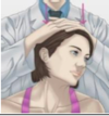
Compression Test
How do you perform this test?
What does a positive test indicate?
Test:
- Physician position: standing behind seated patient.
- Physician applies an axial compression of the head in neutral.
Positive test: Pain down the arm in the nerve root distribution
Indicates: Nerve root compression (cervical radiculopathy)

Spurling’s Maneuver
How do you perform this test?
What does a positive test indicate?
Physician position: standing behind seated patient.
The test is done in three stages, each of which is more provocative. If symptoms are produced, the test is considered positive and there is no need to proceed to the next stage.
- Compression of the head in neutral.
- Compression of the head with head in extension.
- Side bend away from the affected side then toward the affected side and add compression.
Positive test: pain down the arm in the nerve root distribution
Indicates: nerve root compression (cervical radiculopathy)

OA (C0-C1 Articulation) MET
Physician position: sit or stand at the head of the table with the patient supine.
- The patient is supine, and the physician sits at the head of the table.
- Place one hand under the patient’s occiput with the pads of the fingers contacting the sub occipital musculature. The index and middle fingers of the physician’s opposite hand is placed on the patient’s chin beneath the lower lip.
- Gently flex and side bend the patient’s occiput to the right to the restrictive barrier, isolating motion to the OA articulation. Add rotation left.
- Instruct the patient to gently lift the chin up into the physician’s fingers against an equal counterforce for 3 to 5 seconds, and then the patient is instructed to stop and relax.
- Flex the patient’s occiput to the edge of the new restrictive barrier by pulling cephalad on the patient’s occiput and pressing gently downward with the fingers on the patient’s chin.
- Steps 4 & 5 are repeated 3-5 times or until motion is maximally improved at the dysfunctional segment.
- Reassess
Example:
OA E RrSl
- Initial treatment position: flexion, left rotation, right side bending
- Patient’s activating force: extension, right rotation, left side bending
AA (C1-C2 Articulation) MET
Physician position: sit or stand at the head of the table with the patient supine.
- Doctor supports the back of the head with palms
- Fingers are gently placed between the descending ramus of the mandible and the mastoid process
- FULLY FLEX THE CERVICAL SPINE
- This locks out the rotation of the typical cervical vertebra isolating the atlas on the axis
- Rotate the head to the barrier through the atlas
- Have patient rotate the head to the barrier against your resistance for 3-5 seconds
- NOTE: In acute painful dysfunctions, the patient can very gently rotate or look to the right (reciprocal inhibition, oculocervical)
- Have the patient relax/stop and then engage the new barrier by increasing rotation
- Repeat this process 3-5 times or until no new barriers are reached and motion is restored
- Reassess
Example:
AA Rr
- Initial treatment position: left rotation
- Patient’s activating force: right rotation

Typical Cervical Vertebrate MET
Physician position: sit or stand at the head of the table with the patient supine.
- Patient supine with the physician seated at the head of the table
- Physician’s 2nd metacarpal-phalangeal joint placed on the articular pillar to aide in inducing side bending to the RB.
- The physician cradles the patient’s occiput within their hands.
- Flex the patient’s c-spine until dysfunction segment is isolated. Then, extend the c-spine to engage the dysfunctional segment extension RB. Induce side bending to engage the side bending RB and then rotation to engage that RB. (The RB is engaged in all three planes)
- The physician instructs the patient to side bend or rotate their head away from the RB, while meeting the patient’s force with equal and opposite counterforce (resulting in isometric contraction)
- After 3-5 seconds the patient is instructed “stop” their force. After 1-2 seconds of relaxation take up the slack to engage the new RBs in all 3 planes.
- Repeat steps 4-6 for 3-5 repetitions or until there is no further RBs engaged and then reassess for 2-4 TART findings.
Example:
C4 E RrSr
- Type 2 SD
- Initial treatment position = flexion, left rotation, left side bending through C4
- Patient’s activating force = push right ear toward the right shoulder (E RrSr)
OA (C0-C1 Articulation) ART
Physician position: sit or stand at the head of the table with the patient supine.
- Doctor contacts:
- One hand under the occiput (pads of the fingers contact the sub-occipital musculature)
- Opposite hand’s index and middle finger are placed on the patient’s chin beneath the lower lip
- Flex or Extend the head through the occiput
- Rotate the head to the barrier through the occiput
- Side bend the head to the barrier through the occiput
- Apply an articulatory force into the barrier
- Hold for 1-2 seconds and return to neutral
- Repeat this process until no new barriers are reached
- Reassess
Example:
OA E RrSl
Initial treatment position: flexion, left rotation, right side bending
AA (C1-C2 Articulation) ART
Physician position: sit or stand at the head of the table with the patient supine.
- Doctor supports the back of the head with palms
- Fingers are gently placed between the descending ramus of the mandible and the mastoid process
- FULLY FLEX THE CERVICAL SPINE
- This locks out the rotation of the typical cervical vertebra isolating the atlas on the axis
- Rotate the head to the barrier through the atlas
- Apply an articulatory force into the barrier
- Hold for 1-2 seconds and return to neutral
- Repeat this process until no new barriers are reached
- Reassess
Example:
AA Rr
Initial treatment position: left rotation
Typical Cervical Vertebrate ART
Physician position: sit or stand at the head of the table with the patient supine.
- Doctor contacts the articular pillar of the affected segment with 1st MCP joint
- Flex/Extend the cervical spine through the affected segment
- Rotate the cervical spine to the barrier through the affected segment
- Side bend the cervical spine to the barrier through the affected segment
- Apply an articulatory force into the barrier
- Hold for 1-2 seconds and return to neutral
- Repeat this process until no new barriers are reached
- Reassess
Example:
C4 E RrSr
- Type 2 SD
- Initial treatment position = flexion, left rotation, left side bending through C4
Bilateral Forearm Fulcrum ST/MFR
Physician position: sit or stand at the head of the table with the patient supine.
- Arms are crossed under patient’s head and hands placed palm down on patient’s shoulders
- Repetitively flex patients neck, giving a longitudinal stretch of the paravertebral muscles
- Repeat for 2-3 minutes or until desired effect is achieved
- Re-evaluate for TART improvement

Bilateral Forearm Fulcrum ART
Physician position: sit or stand at the head of the table with the patient supine.
- Arms are crossed under patient’s head and hands placed palm down on patient’s shoulders
- Repetitively flex patient’s neck (giving a longitudinal stretch of the paravertebral muscles)
- Repeat for 2-3 minutes or until desired effect is achieved
- Reassess

Bilateral Forearm Fulcrum MET
Physician position: sit or stand at the head of the table with the patient supine.
- The patient lies supine, and the physician sits at the head of the table
- The physician gently flexes the patient’s neck to the edge of the restrictive barrier
- Instruct the patient to extend (backward bend) the head and neck while the physician applies an equal counterforce
- Maintain isometric contraction for 3 to 5 seconds, and then the patient is instructed to stop and relax
- Once the patient has completely relaxed, the physician gently flexes the neck to the edge of the new restrictive barrier
- Repeat Steps 3 to 5 3-5x or until motion is maximally improved
- The same sequence is repeated for left and right side bending and rotation
- Reassess

Oculocephalogyric Reflex
Direct MET
Physician position: sit or stand at the head of the table with the patient supine.
- Engage restrictive barrier with eye motion
- Patient holds for 3-5 seconds while physician is monitoring the SD
- Relax (patient closes eyes)
- Doctor further engages restrictive barrier using the head and neck
- Continue until no new barriers are met
- Reassess SD and TART findings
Examples:
- Extension SD –> instruct the patient to look down towards feet (induces F)
- Flexion SD –> instruct the patient to look up toward top of head (induces E)
- R Sidebending SD –> instruct the patient to look up and to the left (induces SBl)
- L Sidebending SD –> instruct the patient to look up and to the right (incudes SBr)
- R Rotation SD –> instruct the patient to look directly to the left (induces Rl)
- L Rotation SD –> instruct the patient to look directly to the right (induces Rr)


