Ortho Radiology 2 Flashcards
Midforearm Fractures
- Which parts of the forarm are unstable?
Isolated ulnar fractures
- If displaced less than 50% bone diameter and less than 10 degrees angulation what should we do?
3 YO that stops weight bearing = R/O what?
1.
- Radius
- ulna,
- midshaft are unstable
2. Short arm cast 2 weeks then functional splint 2-6 weeks
septic arthritis
What will an xray of a Frykman I AP view show?
3
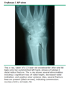
- What percent of all distal radial fractures are treated?
- What are the normal distal radius findings on XR? 3
- 1/6 all fractures treated
- XRs
- Radial inclination
- Radial height- 1 cm
- Volar tilt- normal 10 degrees
- What is this?
- What signs will you find to support this Dx? 2

- scaphoid fracture- proximal part becomes avascular
2.
- anatomical snuff box tenderness and
- proximal part dies- scaphoid fracture
Scaphoid fractures
- XR of wrist: which kinds?
- Look for widening of what? at what value?
- High risk for what? Greatest risk of this where?
- What may be a reason that you initially miss this?
- XRs of wrist
- PA,
- lateral
- scaphoid view (AP with 30 degress of supination and ulnar deviation) - Look for widening of scapholunate distance (>3 mm)
- High risk of nonunion: Greatest at proximal pole
- XRs may be normal initially
- What is scapholunate dissociation?
- Signs? 3
- scaphoid ligament lunate is torn
2.
- tenderness to palapation on dorsal radial side of wrist
- could have click with movement
- Terry Thomas sign
Scaphoid Fractures
- Repeat the Xray when?
- Other imaging? 3
- Repeat XR in 10-14 days
2.
- Bone Scan
- MRI
- CT
- Benefits of a bone scan with scaphoid fractures? 2
- What would a CT help with? 2
1.
- More cost effective than MRI
- Can show uptake in 72 hours
2. Help see fracture line and displacement
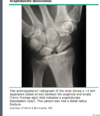
What is this?

Buckle fracture- pinching out on the outside like a water bottle
- What does this show?
- What is this fracture characterized by? 2

- Each of these anteroposterior radiographs shows an intrarticular fractures of the 1st metacarpal.
- the Type I or “Bennett’s” fracture of the proximal 1st metacarpal illustrated here is characterized by
- its articular involvement and
- the persistent attachment of the a volar fragment to the trapezium
- Where is the fragment that is dislocated in a Bennet’s fx?
- by what?
- Management?
Intra-articular fx through base of first metacarpal bone
- Large distal fragment dislocated radially and dorsally by
- the abductor pollicus longus muscle
- ORIF
Metacarpal Fractures
Thumb
- Bennett’s is a fracture combined with what? 2
- Whats a Rolando fracture?
- If extra-articular fracture how can we manage?
- What are acceptable limits for thumb for angulation and rotation?
- Bennett’s- fracture combined with
- subluxation or
- dislocation of metacarpal joint - Rolando fracture- T or Y shaped fracture involving joint surface (comminuted)
- If extra-articular fracture- can consider closed reduction and thumb spica cast for 4-6 weeks
- Less than 25 degrees angulation and rotation are acceptable limits for thumb
Would you fix this?
How should you check this?

You have to check rotation
If rotation if affected you need to fix if not you dont
make a fist and pinky dives underneath or sticks out
- What is this?
- How can you tell?
- Describe how this injury can progress with further trauma?
- If only one is broken how can you treat?

- Boxer’s fracture
- Nondisplaced and minimally angulated, neck
- boxers fracture- break 5th metacarpel and the next time they punch it they break the 4th. if they keep going they will keep breaking metacarpals
- with one you can probably just treat in a cast
What is this?

Shaft, proximal transverse fracture
What does this show?

This anteroposterior radiograph shows a displaced fracture of the base of the 2nd metacarpal.
Metacarpal Fractures
- What should we examine for?
- How should we instruct the pt to do this?
- XRs?
- Examine for rotational malalignment
- Pt to make a fist
- XRs
- 3 view
Transverse fractures of the proximal phalanx are best seen on what kind of radiographs?
Lateral

How would you describe this fracture?
3

- transverse fracture of the proximal phalanx (white arrow).
- slight angulation of the two major fragments and
- the diastasis on the side adjacent to the arrow.
What kind of fracture is this?

This anteroposterior radiograph shows an oblique fracture of the proximal phalanx (white arrow). Oblique fractures may be stable if the periosteal sleeve is intact
What will heal faster: a transverse or oblique fx?
oblique: more surface area
What does this show?

The radiographs reveal a nondisplaced, nonangulated fracture (white arrow) of the proximal phalanx that does not appear to involve the proximal interphalangeal joint.
What kind of fx is this?
This AP x-ray shows a condylar fracture at the head of the proximal phalanx. These fractures most often involve one condyle and are unstable.
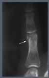
What is this?

The radiographs above show a fracture of the cancellous bone at the tip of the distal phalanx, often referred to as a tuft fracture. These fractures are generally stable and managed conservatively.
Have a subunguil hematoma- poke it? maybe
Distal Phalanx Fracture
- Defined as?
- Stable or unstable?
- Tx?
- If its the PIP?
- Closed with minimal to no displacement- tuft or shaft
- Stable
- Aluminum splint over tip of finger for 3-4 weeks
- Leave PIP free
What are the distal phalanx fracture types?
6

What do you see in this?
2

DDD in lumbar spine
Sclerotic and White/Arthritis
What is this showing?
2

- Arthritis
- Femoral neck is broken/rotates in thats how you know its broken
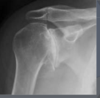
What is this?

Acetabulum/ impingement
Head is rounded off
Pistal grip deformity
What is this?

Arthritis. medially (medial is way more common)
Everything medial is more common
Bone on bone medial compartment arthririts
Knock kneed girls = lateral compartment arthritis
What are you worrying about with this specific fracture?
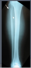
This anteroposterior radiograph shows an oblique proximal fibular fracture (arrows). When this results from an ankle injury with concomitant rupture of the tibiofibular syndesmosis, it is referred to as a Maisonneuve fracture.
ankle mortis is a little wide so you press up and it hurts. dont do anything up there but may put a few screws in
This anteroposterior radiograph of the ankle shows widening of the ankle mortise. This appearance strongly suggests what?

a tear of the distal tibiofibular syndesmosis, an injury that is referred to as a syndesmotic sprain or, in layman’s terms, as a high ankle sprain.
Care must be taken to exclude additional injuries (eg, proximal fibular fracture) commonly found with fractures of the what?

of the medial or posterior malleoli.
- What is a jones fracture?
- Describe Type 1?
- Management?
- In all types you should consider what?
- Proximal shaft of 5th metatarsal
- Type I- nondisplaced or displaced less than 2mm
- Short leg cast with strict non-weight bearing for 6-8 weeks/May take longer
- All types (I-III) consider surgical fixation
This fracture demonstrates typical extension of the fracture line towards the intermetatarsal joint. Note the characteristics of an acute fracture:
3
- narrow fracture line,
- normal thin cortex adjacent to the fracture,
- normal intramedullary canal
What is this?

Gout
What is this?
Dont show them the Xray
plantar fascitis is why they have the pain and the cause of the bone spur
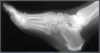
What is this?
lipoma
What is this?

old stress fracture that has healed
could be cancer but unlikely- ask about night pain- could feel heat over it
What is this?

most common benign bone tumor
-osteochondroma- going to be in metaphysis- tibia
What is this?

Scaphoid fracture
What is this?
Arthritis/Nodes
What is this?

Osteoarthritis lateral compartment probably a female
Calcification in the medal compartment of the other knee. pseudogout probably
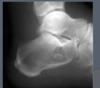
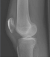
What is this?
2

Was either AVN or an osteocondryle defect
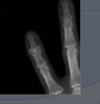
What is this?

fibular fracture. rolled the ankle
What is this?

distal phalanx fx
How would you describe this?
most likely it was AVN and the head collapsed
head elevated flat could be old AVN
Osteophytes present
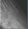
What is this?

physis are open, buckle fracture
What is this?

Boxer fracture
Oblique fracture- see if the rotation is off. probably treat conservatively
What is this?

Bennet’s fracture on the left thumb
How would you manage this?
cast em and immobilize
What is this?

MM
lytic lesions
