Organelles and cell function Flashcards
4 Types of organic molecules
- Carbs - Glycogen is a polysacharide
- Proteins
- Lipids
- Nuclei Acid
Carbohydrates
Function
Organisation
Example:
- Main source of Energy
- Mono, Di, Polyscaharides
- Glucose / Sucrose / Glycogen
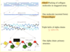
Dehydration Sythesis forms bonds between sugars
Proteins(Amino acids)
Function
Organisation
Example:
- Major componet of most Tissues
- • Primary: Amino acid sequence • Secondary: Conformation (Alpha helices, B-sheets) • Tertiary: Folding to shapes (Ionic bonds) • Quaternary: 2 or more subunits
- Heamoglobin (Quaternary)
Lipids (Glycerol and fatty acids)
Function
Organisation
Example
- Cell Membranes / Energy reserves
- • Fatty Acids – Energy (glycerol backbone and hydrocarbon chain) • Phospholipids/Glycolipids – Cell membrane bilayer, hydrophobic= fluid and impermeable to polar substances (charged polar head, glycerol backbone, fatty acid chain) • Steroids – Hormones (4 bonded C rings)
- Fatty Acid→Triacylglycerol Phospholipids→Phosphatidylcholine Steroid→testosterone
Nucleic Acid (Nucleotides)
Function
Organisation
Example
- Carry genetic info
- • DNA Adenine←→Thymine Guanine←→ Cytosine • RNA Uracil instead of Thymine
- DNA RNA
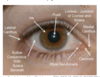
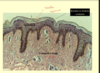
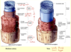
Glycocalyx
Carb enriched coating covering outside of eukaryote cells
Found on apical portion of microvilli (BRUSH BORDER) in digestive tract

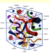
Cells
Charactaristics
All living things composed of cells
Basic structural and fuctional unit of life
All cells from pre-existing cells
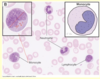
Red blood cells only cells without nucleus
Cell membrane Functions
- Respond to stimuli via receptors
- Fluid Mosaic model of plasma membrane – Phospholipid bilayer – Made of phosphatidylcholine – Can fuse with other membranes – No movement of anything large or polar across membrane (except lipids) – Membrane proteins travel throughout surface
Cell membrane proteins:
Integral Proteins - Penetrate bi-layer or not completely
Peripheral Proteins - Inside or Outside cell
Lipid anchored proteins

Passive transport through Cell Membrane
Passive
Diffusion: High to low con gradient ( Non polar O2 / Co2)
↑speed = ↑ Temperature, ↑ energy state (gass), ↓ size, ↑ conc. gradient
Osmosis: H2o From high to low concentration (Hyper/Hypo/Iso)
Facilitated Diffusion: Much faster than simple diffusion Requires a membrane-bound carrier/ Reaches endpoint
Aqauporins: Integral proteins allow passive movement of water even through it is polar
Filtration: Only in the Kidney tubeoles

Active transport through cell membrane
Requires energy / against gradient /includes pumps / carrier proteins
PRIMARY
Maintains specific gradient ↑NA+ outside, ↑K+ intside – uses energy from ATP and Phosphorylation
SECONDARY
Energy stored in electrochemical gradient of another solute (usually sodium/potassium) – uses NA/K pump to go against gradient

Endocytosis
Pinocytosis
Phagocytosis
Pinocytosis: extracellular fluids, solutes Receptors not required
Endocytosis: requires membrane receptor to bind & internalize
Phagocytosis: Large molecules and foreign particles
Cytoskeleton function (4)
- Structure and Support
- Intracellular transport
- Contractility and Motility
- Spatial Organization
Microtubules
Size 24nm
Protein: TUBULIN
Hollow structure / MAP hold mictrotubles in place
Types of Microbubules
- Axonal transport: highway that motor proteins carry organelles on
- Cilia: Help transport fluid and materials / 9 - 2 cofiguration
- Basilar body Anchor cilia / 9 triples config
- Centrioles : Appear at poles during mitosis, attach to mitotic spindle separating DNA into daughter cells 9 triples config

Microfilaments + function
6nm
Protein: ACTIN
Functions: Wound healing
movement of axons
vesicles
phagocytosis
cytokinesis
Intermediate filaments
10nm
Most stable
Provide shape and structure to cell
Protein (various)
Cytokeratin - Epithelium

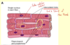
Rough Endoplasmic Reticulum
Synthesis/processing of secretory proteins
Continuous with nuclear envelope
Labyrinth of flattened sacs
Ribosomes on outside (Rough appearance)

Golgi Apparatus
Modifications of secretory proteins
• Adding sugars, folding etc.
− Stacks of flattened disklike membranous cisternae
− Usually less than 8 in a stack
− Vesicles bud from sides
Smooth endoplasmic Reticulum
Synthesis of lipids
o Liver: detoxification of drugs, toxins
o Liver: Release of glucose from glycogen when required
o Muscle cells: sequestering of Ca and regulated release
- Most remain membrane bound
- more “tubular” than the rough ER
- some like hormones secreted
Lysosomes
Fuses with vacuoles for digestion of cellular organelles, bacteria, macromolecules
− membrane-bound bags of enzymes
− Peroxisomes (special type) destroy peroxides

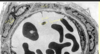
Nucleus
Contains genetic information (DNA)
− Pores in envelope allow for mRNA and protein movement
− Usually 1 per cell but some special cells can have 2
− Easiest structure to see under microscope
Nucleolus
rNA synthesis
(2 types of chromatin)
- Heterochromatin: (Inactive, clumps)
- Euchromatin: (dispersed) Active in RNA synthesis

Mitochondria
Energy production through cellular respiration
− 1 glucose = 36 ATP − Glycolysis (anaerobic) in cytoplasm, 2 pyruvic acid into mitochondria →2 ATP
− Oxidative phosphorylation in mitochondria matrix →34 ATP