Oral Medicine Flashcards
(39 cards)
What is Lichen Planus?
What are the 6 main oral types?
Lichen Planus = Chronic, mucocutaneous + premalignant disease that can be oral, cutaneous or genital and appear with a wide spectrum of presentation.
(Purple People Break And Enter Raves)
- Papular (small white spots)
- Plaque-like (white patch)
- Bullous (RARE - blood-filled blisters)
- Atrophic (Erythematous)
- Erosive (Ulcerative)
- Reticular
What is the aetiology and epidemiology (3) of Lichen Planus?
Aetiology:
Unknown (may be AI)
Epidemiology:
- Age (4th-8th decade)
- Gender (60% female)
- Europeans and Indians
What are the 4 histological signs seen from biopsy sample to diagnose Lichen Planus?
- +/- Hyperkeratosis
- “Saw tooth” rete ridges in epidermis
- (T) Lymphocyte cells dominant sub-epithelial band
- Basal cell degeneration and liquifaction
Clinical features of Oral Lichen Planus?
(THINK: Sites, Symptoms & Types)
- Symmetrical/Bilateral - Often posterior buccal mucosa, labial mucosa, tongue or ginigva
- Gingival involvement may = Desquamative gingivitis (differentials = LP, PV or MMP)*
- Koebner Phenomenon - Often present in sites of increased friction (e.g. ill-fitting denture)
- Asymptomatic or discomfort on heating spicy/acidic foo
6 Oral Presentations:
- Papular
- Plaque like
- Bullous (rare)
- Atrophic (Erythematous)
- Erosive (Ulcerative)
- Retricular
Clinical features of Cutaneous Lichen Planus?
- Symmetrical/Bilateral
- Koebner’s Phenomenon (e.g. knees/shins)
- Purple Polygonal Pruretic (itchy!) Papules (flat-topped and few mm diameter)
- Wickham’s Striae (surface network of fine white striations)
Other than Oral and Cutaneous, what are 3 other sites Lichen Planus may present?
(And how does it present?)
1) GENITAL
- More common in females
- Vulvo-Vaginal Gingival Syndrome → Scarring
2) NAILS
- Dystrophic (poorly formed and discoloured)
- Longitudinal grooving and pitting
- Possible loss of nail
3) HAIR
- Scarring alopecia
What is the difference in oral and cutaneous lichen planus in terms of longevity and quantity of patients who have both?
Oral (MORE COMMON)
- Chronic (4-25 years)
- 10-30% have cutaneous lesions
Cutaneous
- Transitional (last average 18 months)
- 70-77% have oral lesions
What are 3 differentials for Desquamative Gingivitis?
Which is most common?
- Lichen Planus (most common)
- Pemphigus Vulgaris
- Mucous Membrane Pemphigoid
What is the malignant transformation % of Lichen Planus?
What advice should be given to patients? (3)
~0.5-2%
(“Transformation = independent of smoking and alcohol”)
Patients should be warned of malignant potential and adviced:
- Regular reviews
- Smoking cessation
- Good OH
What are 9 differentials for a white patch lesion in the mouth?
- Candidal Infection
- Epithelial Dysplasia
- Hyper-keratosis
- Lichen Planus
- Lichenoid Reaction
- Discoid Lupus Erythematous (DLE)
- Graft vs. Host Disease
- Leukoplakia
- Squamous Cell Carcinoma
How would you manage a patient with symptomatic oral Lichen Planus?
- Educate pt: No cure, will manage symptoms but low risk of malignant change (so regular reviews and reduce risk factors e.g. smoking)
- Eliminate provoking factors (e.g. sharp cusps or ill-fitting denture causing trauma)
- Avoid chemical irritation (e.g. use Sensodyne SLS-free toothpaste and avoid spicy foods)
Treatment options:
1st line = Topical Corticosteroids (e.g. Hydrocortisone/Betamethasone0
2nd line = Topical Immunosuppressants (Calcineurin Inhibitors)
Severe = Systemic Corticosteroids or Immunosuppressants
Lichen Planus has no cure. Other than elimination of provoking factors and advice to reduce malignant risk factors, what 3 pharmaceutical interventions can be used?
- Topical Corticosteroids (1st Line)
* E.g. Hydrocortisone, Betamethasone or Benzydamine (Difflam)* - Topical Immunusuppressants (Calcineurin Inhibitors) (2nd line)
* E.g. Ciclosporin, Tacrolimus or Pimecrolimus* - SEVERE = Systemic Corticosteroids or Immunosuppressants
* (E.g. above, Azathioprine, Dapsone, Mycophenolate mofetil or Thalidomide)*
What are 4 issues concerning long-term corticosteroid use?
- Adrenal gland suppression → Adrenal Insufficiency (ME)
- Skin thinning, easy bruising and slowed wound healing
- Increased risk of infection
- Hypertension
Topical Calcineurin Inhibitors are 2nd line treatment for Lichen Planus, how do they work, how long to they take to work and what are 3 examples?
Calcineurin = Protein involved in T-lymphocyte activation
Inhibition = Immunosuppression
(Use causes initial “burning sensation” followed by lesion resolution within 8-12 weeks)
- Ciclosporin
- Tacrolimus
- Pimecrolimus
What are 10 drugs that can cause drug-induced Lichenoid Reaction?
(HINT: Harry Potter is A BAD MAN G)
- Hypoglycaemics (oral)
- Penicillamine (RA)
- ACE inhibitors (“prils”)
- Beta blockers (e.g. Furosemide, Indapamine or Mannitol)
- Allopurinol (Gout)
- Diuretics
- Methyl-dopa (Hypertension)
- Anti-malarials
- NSAIDs
- Gold salts (RA)
How is the:
- Aetiology
- Presentation
- Histology
- Management
of Lichenoid Reaction different to Lichen Planus?
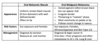
1) Aetiology
LP = Unknown (may be AI)
LR = Drug-induced or Dental-material related (resins or metals, e.g. amalgam)
2) Presentation
Similar, but LR:
- Mostly erosive (ulcerative) type→ Soreness
- Can affect palate
- May be unilateral (e.g. if DM related)
3) Histology
Similar, but LR:
- Deeper and less defined infiltrate
- Lots of plasma and eosinophils
4) Management
- Patch test to see if local (DM) cause - remove
- If possible, withdraw drug and monitor
- No pre-malignant risk
- Otherwise, manage as with LP
What is Graft vs. Host Disease?
What is the aetiology and risk factors (2)?
Immune reaction of graft lymphocytes against host cells
Aetiology: Following allogenic bone transplant (within first 6-24 months)
Risk Factors:
- Poorly-matched graft
- Old age doner or recipient
N.B. GvHD = Increased risk of OSCC
What are 3 oral clinical features of Graft vs. Host Disease?
- Asymptomatic or Burning sesation (Erosive/Ulcerative or Reticular)
- Oral dryness → Superficial palatal and labial mucoceles
- Sclerotic GvHD → Trismus (reduced opening)
How is Graft vs.Host Disease managed? (4)
- Topical analgesia (e.g. LA)
- Topical Corticosteroids (e.g. Betamethasone M/W)
- Topical Immunsuppressants (e.g. Tacrolimus ointment)
- Regular review (as GvHD = Increased OSCC risk)
What is Lupus Erythematous?
What is the aetiology?
What are the 2 main types?
Are they more common in males or females?
Lupus Erythematous = Chronic, indolent (little/no pain) oral and cutaneous disease
Aetiology = AI (may be precipitated by drugs, virus’ or environment)
- Systemic LE (Multi-systemic + affects vascular and CT)
- Discoid LE
Both = More common in females (esp. child bearing age)
In Discoid Lupus Erythematous, what are the clinical features of:
- Oral lesions? (seen in 20-50%)
- Cutaneous lesions?
1. ORAL LESIONS
- Similar site to LP - Bilateral buccal/labial mucosa
BUT also: Palate + Vermillion border - Less-well demarcated erythematous areas, surrounded by border of fine white striae
- May ulcerate - Sign of active lesion or progression to SLE
2. CUTANEOUS LESIONS
- Scaley, atrophic plaques on sun-exposed skin
- Oval keratin-plug plaques (may appear on skin, hair, genitals or orally)
- Increased blood vessel formation (telangiectasia)
- Scarring alopecia or scalp pigmentation
Main method of DLE diagnosis is biopsy (through in 25% cases autoantibodies are visible). What 6 histological features of DLE would you observe?
- Para or Ortho-keratosis
- Chronic inflammatory cell infiltrate in sub-epithelial CT
- Hyalinisation of sub-epithelial CT
- Basal cell liquefaction and degeneration
- Irregular pattern of acanthosis
- MAY SEE: Keratin plugs and Civatte bodies
How are the following lesions treated/managed in Discoid Lupus Erythematous:
- Oral lesions? (hint: same as LP)
- Cutaneous lesions?
1. Oral Lesions
- 1st line = Topical Corticosteroids
- E.g. Hydrocortisone, Betamethasone or Benzydamine (Difflam)*
- 2nd line = Topical Immunsuppressants (Calcineurin inhibitors)
- Ciclosporin, Tacrolimus or Pimecrolimus*
- Severe = Systemic Corticosteroids or Immunosuppressants
2. Cutaneous Lesions
- Suncream (SPF 50)
- Chloroquine (anti-malarial)
- Potent steroids
What are 10 causes of LOCALISED oral pigmentation changes?
- Diet/Beverages
- Ecchymosis (Bruising)
- Varices
- Black Hairy Tongue
- Amalgam Tattoo (Focal Agyrosis)
- Graphite Tattoo
- Ethnobotanical Tattoo
- Metal Salt deposits
- Oral Melanotic Macules
- Oral (Malignant) Melanoma