Normal RAD EXAM 2 Flashcards
Name the line of mensuration and give the maximum measurements for the child and the adult:
Atlantodental Interspace Child: 3mm Adult: 5mm
Name the line of mensuration and explain what a normal finding:
George’s Line Normal: Smooth vertical alignment of each posterior body corner
Name the line of mensuration and explain what a normal finding:
Posterior Cervical Line Normal: When each spinolaminar junction point is joined, a smooth arc-like curve results. At the C2 level, the spinolaminar junction line in children should not be > 2 mm anterior to this line
Name the line of mensuration: Canal stenosis may be present when the measurement is less than _______
Sagittal dimension of the cervical spinal canal Stenosis: <12mm
Name the line of mensuration and explain what a normal finding:
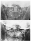
Atlantoaxial alignment Normal: The lateral margins of the atlas lateral masses are compared to the opposing lateral corner of the axis articular surface should be in vertical alignment.
Name the line of mensuration and explain what a normal finding:
Cervical gravity line Normal: A vertical line is drawn through the apex of the odontoid process should pass through the C7 body
Name the line of mensuration and list the minimum and maximum degrees for the angle:
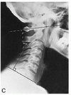
Cervical lordosis i.e. Angle of the cervical curve Minimum: 35deg Maximum: 45deg
Name the line of mensuration and explain what a normal finding:
Stress lines of the cervical spine Normal Flexion: These lines normally should intersect at the level of the C5-C6 disc or facet joints Normal Extension: These lines normally should intersect at the level of the C4-C5 disc or facet joints
Name the line of mensuration and list the normal values for the neutral position:
Prevertebral soft tissues C-1: 10mm C-2: 5mm C-3: 7mm C-4: 7mm C-5: 20mm C-6: 20mm C-7: 20mm
Name the view and identify the numbered structures 1-6:
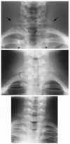
A-P Lower Cervical 1. C7 spinous process 2. C7 lamina 3. C7 pedicle 4. C7 transverse process 5. C6 articular pillar 6. C5-C6 von Luschka joint
Name the view and identify the numbered structures 7-12:
A-P Lower Cervical 7. T1 spinous process 8. T1 lamina 9. T1 pedicle 10. T1 transverse process 11. 1st costotransverse joint 12. 1st rib
Name the view and identify the numbered structures 13-19:
A-P Lower Cervical 13. 2nd costotransverse joint 14. Medial clavicle 15. Trachea 16. Mastoid process 17. Angle of mandible 18. C5 intervertebral foramen 19. Lung apex
Name the view and identify the numbered structures 1-5:
A-P Open Mouth 1. Atlas lateral mass 2. Atlas anterior arch 3. Atlas posterior arch 4. Atlas transverse foramen 5. Atlas transverse process
Name the view and identify the numbered structures 1-7:
Cervical Oblique 1. C6 vertebral body 2. C5 transverse process 3. C6 pedicle 4. C5 lamina 5. C6 articular pillar 6. C6 spinous process 7. C6-C7 intervertebral foramen
Name the view and identify the numbered structures 11-16:
(C) A-P Open Mouth 11. Axis spinous process 12. Axis transverse foramen 13. Axis transverse process 14. Mandible 15. Tongue 16. Styloid process
Name the view and identify the numbered structures 6-10:
A-P Open Mouth 6. Atlanto-occipital joint 7. Mastoid process 8. Odontoid process 9. Axis pedicle 10. Axis lamina
Name the view and identify the numbered structures 8-14:
Cervical Oblique 8. C5-C6 von Luschka joint 9. C4 pedicle 10. C3 pedicle 11. C6 transverse process 12. First rib 13. Trachea 14. Mandible
Name this cervical view?
Right Anterior Cervical Oblique (RACO)
Name this cervical view?
Left Anterior Cervical Oblique (LAO)
Name this cervical view?
Right Posterior Cervical Oblique (RPO)
Name this cervical view?
Left Posterior Cervical Oblique (LPO)
Name the view of image C on the left and identify the numbered structures 1-6:
A-P Thoracic 1. Rib 2. Transverse process 3. Costotransverse joint 4. Costovertebral joint 5. Pedicle 6. Spinous process
Name the view of image C on the left and identify the numbered structures 7-13:
A-P Thoracic 7. Inferior endplate 8. Intervertebral disc space 9. Clavicle 10. Diaphragm 11. Trachea 12. Paraspinal line (arrowheads) 13. Aorta (arrows)
Name the view of image C on the left and identify the numbered structures 8-16:
(C) Lateral Thoracic 9. Rib head 10. Posterior rib 11. Lateral rib 12. Diaphragm 13. Posterior costophrenic sulcus 14. Heart 15. Lung hilus 16. Trachea
Name the view of image C on the left and identify the numbered structures 1-8:
(C) Lateral Thoracic 1. Vertebral body 2. Endplate (arrowhead) 3. Intervertebral disc 4. Pedicle 5. Intervertebral foramen 6. Apophyseal joint 7. Spinous process 8. Axillary margin, scapula (arrow)
Name the line of mensuration and explain how it is measured:
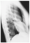
Thoracic kyphosis A line is drawn parallel to and through the superior endplate of the T1 body. A similar line is drawn through the inferior endplate of the T12 body. Perpendicular lines to these endplate lines are then constructed, and the resultant angle is measured at the intersection of the lines.
Name the line of mensuration and list the minimum and maximum values for the male and female in addition to the minimum and maximum values for the male and female with Straight Back Syndrome”:”
Thoracic cage dimention Normal Male: Min 11mm Max 18cm* Normal Female: Min 9mm Max 15cm* SB Male: Min 9mm Max 13cm* SB Female: Min 8mm Max 11cm*
Name the view:
A-P Full Spine
Identify the numbered structures 1-5:
Rib 1. Anterior rib 2. Posterior rib 3. Rib tubercle 4. Costotransverse joint 5. Rib head
Name the line of mensuration and list the minimum and maximum angles then explain the difference between hyper/hypolordsis:
Lumbar lordosis Minimum 50deg - <50deg = Hypolordosis Maximum 60deg - >60deg = Hyperlordosis
Name the line of mensuration and list the average measurement for each level:
Lumbar intervertebral disc angles L-1: 8 deg L-2: 10 deg L-3: 12 deg L-4: 14 deg L-5: 14 deg
Name the line of mensuration and explain what the normal measured tolerance is on an upright radiograph:
Lumbosacral angle Normal: 41deg +/- 7deg
Name the line of mensuration and list what the normal tolerance is:
Lumbosacral disc angle Normal: 10-15deg
Identify the static vertebral malpostition in each picture A-D:
A: Extension B: Lateral flexion C: Laterolisthesis and rotation D: Anterolisthesis and retrolisthesis
Name the line of mensuration and explain what is classified as anterior versus posterior weight bearing:
If the line falls >1cm posterior to the anterior tip of the sacrum there is “posterior weight bearing””
Name the line of mensuration and explain what is classified as normal or abnormal:
McNab’s line Normal: The line should lie above the tip of the adjacent superior articular process Abnormal: If the line intersects the superior articulating process, subluxation may be present
Name the line of mensuration and explain what is classified as normal or abnormal:
Ulmann’s line Normal: The L5 body should lie posterior to or just contact this perpendicular line Abnormal: If the anterior margin of the L5 body crosses the perpendicular line, then anterolisthesis may be present
Name the line of mensuration and what its used to detect:
Meyerding’s grading scale The degree of anterolisthesis of the affected vertebral body can be categorized according to the division in which the posterior-inferior corner of the body lies
Name the line of mensuration and list the minimum measurements for the cervical, thoracic, and lumbar spine:
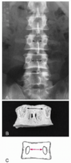
Interpediculate distance Cervical: 24mm Thoracic: 14mm Lumbar: 21mm
Name the line of mensuration and explain how an abnormal finding is detected:
Eisenstein’s method A canal measurement < 15 mm may indicate the presence of spinal stenosis
Name the view and identify the numbered structures 1-4:
(C) Lumbar Oblique 1. Pedicle 2. Superior articular process 3. Pars interarticularis (isthmus) 4. Lamina
Name the view and identify the numbered structures 5-9:
(C) Lumbar Oblique 5. Inferior articular process 6. Transverse process 7. Spinous process 8. Intervertebral disc 9. Interlaminar space
Name the view of image C on the left and identify the numbered structures 1-7:
(C) A-P Lumbar aka Lumbopelvic 1. Spinous process 2. Pedicle 3. Superior articular process 4. Transverse process 5. Inferior articular process 6. Lamina 7. Pars interarticularis (isthmus)
Name the view of image C on the left and identify the numbered structures 8-13:
(C) A-P Lumbar aka Lumbopelvic 8. Twelfth rib 9. Sacral ala 10. First sacral tubercle 11. Sacroiliac joint 12. Descending colon 13. Psoas muscle
Name the Anomaly:
Agenesis of the lumbar articular process
Name the Anomaly:
Agenesis of the odontoid process
Name the Anomaly:
Agenesis of the posterior arch
Name the anomaly:
Occipitalization of the atlas
Name the anomaly:
Complete agenesis of the posterior arch of the Atlas
Name the anomaly:
Partial agenesis of the posterior arch of the Atlas
Name the anomaly:
Spina Bifida of the Atlas
Name the anomaly:
Spina Bifida of the Axis
Name the anomaly:
Posterior Ponticle
Name the anomaly:
Os odontoideum with instability
Name the anomaly:
Os odontoideum on a lateral view
Name the anomaly:
Hypoplastic odontoid process
Name the anomaly:
Congenital Block Vertebra
Name the anomaly:
Cervical ribs
Name the anomaly:
TVP Hyperplasia
Name the anomaly:
Lumbar block vertebra
Name the anomaly:
Butterfly vertebra
Name the anomaly:
Agenesis of the odontoid process with instability
Name the anomaly:
Schmorl’s Nodes
Name the anomaly:
Limbus Bone
Name the anomaly:
Lateral Hemivertebra
Name the anomaly:
Nuclear Impressions
Name the anomaly:
Spina Bifida Oculta
Name the anomaly:
Lumbosacral transitional segment
Name the anomaly:
Agenesis of a lumbar articular process
Name the anomaly: https://o.quizlet.com/B0fQ4xi4S6MoRO505IhnSw_m.png
Clasped Knife Syndrome type I
Name the anomaly: https://o.quizlet.com/VWtp9Lud4v1L1aseEy-TuA_m.png
Clasped Knife Syndrome types II and III
Name the anomaly: https://o.quizlet.com/wD9DQw2.Q.2Dshz-6IO1jA_m.png
Congenital Hip Dysplasia
Name the anomaly: https://o.quizlet.com/eCvn8.TW18oJBfYf-yXfNA_m.png
(A) Bipartite Patella (B) Tripartite Patella