neurospine2 Flashcards
(39 cards)
- A 65-year-old man presents 6 months after a motor vehide collision complaining of severe disabling neck pain. Cervical CT reveals a non-united type II odontold fraclure. The MOST appropriate management is:
A. Posterior C1-C2 arthrodesis
B. B. Halo immobilization
C. Hard cervical collar
D. Anterior odontoid screw fixation
A. Posterior C1-C2 arthrodesis
Posterior C1-C2 fixation/arthrodesis immobilizes the entire C1-C2 complex, and is the treatment of choice in this condition. Generally, a fracture older than 3 months Is considered a chronic fracture. Acute type II odontoid fractures do not respond well to external orthrosis (hard collar or halo), and healing rates are even poorer for chronic fractures. Published clinical series have reported <50% healing rates for anterior odontoid screw fixation in chronic fractures. Low healing rates are generally ascribed to pannus formation at the fracture site.
- A 65-year-old woman presents with severe, progressive back pain is found to have degenerative, thoracolumbar scoliosis as depicted in the pigures. Which radiographic parameter has the greatest impact on physical function and disability?
A. Abnormal coronal balance
B. Presence of apical rotation
C. Scoliosis curbe magnitude
D. Positive sagittal balance
E. Presence of single curve

D. Positive sagittal balance
of the choices provided, the radiographic parameter that has been shown to have the greatest impact on health status measures is positive sagittal balance (measured from C7 to the posterior margin of the sacrum). Glassman and colleagues assessed the impact of multiple radiographic parameters on health status measures based on a large population of adult deformity patients. Patients with a positive sagittal balance had significantly greater pain, decreased physical function, worse self image, and poorer social function. This underscores the important of achieving sagittal balance when surgically addressing spinal deformity. This also underscores the importance of ensuring that an adequate degree of lordosis is provide when performing instrumented lumbar fusions in order to prevent development of “flat back”
- A 66-years-old white male present after a witnessed fall down six steps complaining of pain and weakness, worse in arms than legs. Plain radiographs, CT and MRI of the cervical spine are shown in the pigures. What underlying medical condition does this patient have:
A. Severe Cervical Spondylosis
B. Ankylosing Spondylitis
C. Osteoporosis
D. Diffuse Idiopathic Skeletal Hyperostosis

D. Diffuse Idiopathic Skeletal Hyperostosis
the answer is Diffuse Idiopathic Skeletal Hyperostosis (DISH). DISH is a common condition characterized by abundant ossification throughout the body. It occurs most often in the spine with the anterior longitudinal ligament being most often affected. It affect males predominantly and may occur in 20% of the population. For the diagnosis, there should be flowing calcification or ossification of at least four condiguous vertebral bodies in addition to preservation of disc height in the involved areas and absence of sclerosis of fusion of the SI joints. Other differentiating factors from ankylosing spondylitis (AS) include syndesmophytes and calcification of the annulus fibrosis and nucleus pulposus in AS. Among others complications, patiens with DISH are predisposed to spine injures from minor otherwise trivial mechanisms. Most fractures are of the spine seen in DISH are characterized as distraction extension injuries. Often there is a transverse shift of the fractured segment as opposed to the compression fracture, probably due to limited or absent dissipations.

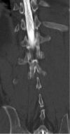
- A 68 years-old male with a history of prostatic cancer present with low-grade fever and severe low back pain progressing to include lower extremity numbness. Thoracic CT shows extensive destruction of the T11 and T12 vertebral bodies with relative sparing of the T11-12 disc space, as well as a large paraspinous abscess with calcification. Thoracic MRI shows a kyphotic deformity with enhancing soft tissue and bone extending into the anterior spinal canal and resulting in moderate stenosis and spinal cord compresion. The MOST likely pathologic process is:
A. Osteoporotic Compression Fracture
B. Discitis – Staph Aureus
C. Pott’s Disease – Tubercolosis
D. Pathologic Fracture – Metastatic Tumor
E. Discitis – Staph Apidermidis
C. Pott’s Disease – Tubercolosis
the answer is Pott’s Disease, resulting from tuberculosis. The radiographic description is classic for Pott’s Disease. Staphylococcal infection is unlikely due to the fact that discitis centered on the disc space and generally spares the vertebral bodies (opposite to the imaging appearance described in this case). Pathological fracture due to metastasis is would not explain the presence of a paraspinous abscess. Osteoporotic compression fracture would not presence with extensive, enhancing epidural soft tissue extension.
- A 68 years-old woman presents with progressive myelopathy with MRI shown. An isolated posterior approach is contraindicated in this patient in the presence of:
A. Fixed kyphotic deformity
B. Posterior ligamentous hypertrophy
C. Subluxation at C3/4 and C4/5
D. Ventral compressive pathology
E. Compressive pathology across multiple levels

A. Fixed kyphotic deformity
an isolated posterior approach is contraindicated due the fixed kyphotic deformity. Among the listed factors, appropriate assessment of the cervical alignment is most important when determining if a posterior approach is appropriate. The posterior approach is ideally suited for patiens demonstrating a degree of lordosis. The presence of a straight or kyphotic alignment will limit dorsal migration of the spinal cord and potentially cause the spinal cord and nerve roots to drape across ventral pathology, leading to further neurologic compromise
- A 70 years-old male with a type II dens fracture has non-union of the dens despite 6 month of halo fixation. His new cervical CT scan demonstrates non-union of the dens with 6 mm of posterior displacement. Cervical x-rays reveal that anatomic reduction of the fracture is not possibble. No other associated fractures are identified. He is otherwise neurologically intact and is in good medical condition. What is the most appropriate treatment option at this time?
A. Continued halo management for an additional 3 months
B. Management in a hard cervical collar for 3 month
C. Removal of halo fixation and observation
D. Anterior odontoid screw fixation
E. Posterior C1-2 instrumented arthrodesis
E. Posterior C1-2 instrumented arthrodesis
the most appropriate management would be to perform posterior C1-2 instrumented arthrosis. The reported rate of chronic nonunion fractures of the dens after halo fixation in the elderly is around 28%. A posterior fixation would provide the best rate of fusion as compared to an anterior odontoid screw fixation in the setting of chronic nonunion of dens. The reported rate of fusion from a posterior C1-2 arthrodesis is approximately 85%. Contraindications to anterior odontoid screw fixation include: disruption of the transverse ligament, concomitant atlantoaxial joint injuries, fracture line parallel to screw trajectory, cervical kyphosis, barrel chest habitus, obesity that would make the screw trajectory not possible. Anterior screw fixation should not be used in patient with nonunion fractures in which fractures healing and/or fixation will be impaired. Furthermore, the 6 mm of posterior displacement would make the posterior approach superior to an odontoid screw.
- A 72 years old woman has been diagnosed with osteoporosis, with a T-score of -2.6 on a recent central DXA scan. She has recently been started on supplemental calcium (1500 mg daily) and bisphosphonate. Which one of the following would be the most appropriate next step in management of her osteoporosis?
a. Screening urinary calcium levels
b. Doubling the calcium supplement
c. Repeating bone density testing in 2 years
d. Measuring hormonal levels
e. Obtaining x-rays of the lumbar spine
c. Repeating bone density testing in 2 years
in asymptomatic postmenopausal women with osteoporosis, many physicians monitor changes in bone mineral density (BMD) by central DXA every year or two during pharmacologic therapy for osteoporosis. It is important to note that drugs may decrease a patient’s risk of fracture even when there in no apparent increase in BMD. As with most test, BMD has some precision error. Therefore, changes of less than 2 to 4% in the vertebra and 3 to 6% at the hip from test to test can be due to the precision error of the method. Repeat bone density measurements for monitoring disease progression in response to therapy should generally be performed after 1 or 2 years. Two years is consistent with guidlines developed by the Centers for Medicare & Medicaid Services (CMS).
- A C1-2 transarticular screw is most likely to result in injury to the vertebral artery when misdirected:
A. Medially
B. Caudally
C. Laterally
D. Anteriorly
E. Cranially
B. Caudally
the vertebral artery is most vulnerable to injury during C1-2 transarticular screw placement due to drill misdirection caudally. The vertebral foramen lies caudal to the pars of C2, and anatomic variations of the foramen can place the vertebral artery at risk during placement of a transarticular screw. Cranial misdirection of the screw could possibly affect the occiput-C1 joint altough this occurrence is extremely uncummon. Medial placement of a transarticular screw could result in dural laceration, with or without spinal cord injury. Placement of a transarticular screw too far anteriorly has been reported to cause hypoglosal nerve dysfunction in few case reports.
- A 54 years old male presents with a three day history of bladder problems. He states that he does not feel his bladder filling, and that urine sometimes leaks out. Neurologic exam is normal with the exception of hypoactive reflexes. His CT myelogram is displayed below. What is the most appropriate treatment for this patient?
A. Endovascular therapy with particulate embolic agents
B. A ventral approach, carpectomy and recection of the lesion
C. A posterior approach, laminectomy, durotomy, and ligation of draining vein
D. Continued observation, physical therapy and lumbal strenghtening
E. Endovascular embolization followed by radiosurgery
C. A posterior approach, laminectomy, durotomy, and ligation of draining vein
the patient has a Type I spinal malformation, or a spinal arteriovenous fistula. The fistula lies within the dura and is an abnormal connection between the radicular vein and artery lying dorsally on the spinal cord making a posterior approach preferable. His problem is caused by venous engorgement in the spinal cord from backflow of venous blood through the radicular draining vein into the coronal venous system on the dorsal aspect of the spinal cord. An arterial steal phenomenon may also be contributing to the problem, altough this is likely less significant

- A normal intraoperative electromyographic study during lumbar fusion surgery has been shown to the BEST corelate with:
A. Improved fusion rates
B. Improved patient outcomes
C. Lack of a neurological injury
D. Pedicle fractures
C. Lack of a neurological injury
intraoperative electrophysiological monitoring during pedicle screw placement is useful as a diagnostic test, as a normal evoked EMG response is highly predictive of intrapedicular screw placement in the pedicle adjacent to the monitored root. The use of such monitoring has, however, never been shown to devrease the incidence of pedicle breach during screw placement, decrease the incidence of neurological injury, or improve patient outcomes. An abnormal EMG response, moreover, is not highly predictive of a neurological injury (because of a high false-positive rate of EMG abnormalities of this setting)
- A patient awakens from left-sided costotransversectomy at T10 for resection of a ventral metastatic tumor with complete paraplegia and loss of pain and temperature sensation. His sensation to light touch in the lower extremities is spared. Post operative MRI reveals no evidence of spinal cord compression or hematoma. What the most likely diagnosis?
A. Posterior cord syndrome
B. Anterior cord syndrome
C. Central cord syndrome
D. Brown-Sequard Syndrome
B. Anterior cord syndrome
the most likely diagnosis is anterior cord syndrome. The sacrifice of the left T10 nerve root likely compromised the artery of Adamkiewicz. This artery supplies the anterior 2/the most likely diagnosis is anterior cord syndrome. The sacrifice of the left T10 nerve root likely compromised the artery of Adamkiewicz. This artery supplies the anterior 2/3 of the spinal cord. It commonly arises at T10 on the left but may arise anywhere from T7-L4. It may be seen on the right in 17% of patients. When planning o posterolateral approach for ventral access to the spinal cord, a spinal angiogram may be helpful in identifying the artery of Adamkiewicz to prevent a vascular insult to the cord when sacrifying a nerve root.
- A patient present with a classic Type II odontoid fracture. A MRI and Flexion-Extension Dynamic Radiographs of the cervical spine are consistent with disruption of the transverse ligament. The MOST appropriate treatment is:
A. Anterior odontoid screw fixation
B. Halo immobilization
C. Occipital-cervical fusion
D. Hard cervical collar
E. Posterior C1-2 fixation
E. Posterior C1-2 fixation
Disruption of the transverse ligament indicates an unstable C1-2 complex, requiring C1-2 fixation. Because the instability is ligamentous is nature. Treatment must be based on functionaly fusing the unstable segment. Hard cullar and halo treatment immobilize bony fractures for healing but are inadequate to address ligamentous instability. Anterior odontoid screw fixation simply reapproximates the fractured odontoid peg to the C2 body and is inadequate to address an unstable C1-2 segment. Occipital-cervical fusion sacrifies adjacent motion segments needlessly, including the occiput-C1 motion segment which is functionally critical.
- A patient present with bilateral nondisplaced fractures through the C2 pars interarticularis (hangman’s fracture). Flexion-extension dynamic cervical radiographs show < 2mm motion and no significant deformity. The most appropriate treatment modality is:
A. Anterior C2-3 discectomy and fusion
B. Posterior C1-2 fixation
C. Posterior C1-3 fixation
D. External immobilization
E. Occipito-cervical fixation
D. External immobilization
the majority of hangman’s fractures are considered stable fractures. The literature consistently reports exellent healing rates with external orthosis, both hard collar and halo immobilization. Operative fixation is reserved for unstable fractures (i.e > 5 mm displacement or gross malalignment)
- A spinal cord injured patient’s examination reveals the presence of voluntary but non-functional strenght in the lower extremities (more than half of the muscles have less than antigravity strenght). Sensation to pain, temperature, and proprioception is markedly diminished distal to the injury. The patient’s american spinal injury association (ASIA) impairment scale grade is:
A. E
B. C
C. A
D. D
E. B

B. C
the ASIA grading system was developed to standardizet the reporting of spinal cord injuries. It is a modification of the Frankle scale. Grades range from A to E. A lessions are characterized by no motor or sensory function detected below the level of the lession, including the sacral segment. Grade B is characterized by no motor function detected below the level of the lession and some preserved sensory function below the level of the lession. Grade C patients characteristically have some voluntary motor function preserved below the level of the lession, but this function is too weak to serve any useful purpose (define as more than half of key muscle having a strenght grade of 3 or less). Sensation may or may not be preserved. Grade D patients have functionally useful voluntary motor function below the level of the injury. While grade E patients have normal strength and sensation, altough abnormal reflexes may persist based upon the above grading scheme, this patiens injury would be catagorized as grade C
- A thirty six years old female presents to the emergency room with a two day history of difficulty with urination, anterior tibialis weakness and diminished rectal tone. Her MRI is attached. What is the most important surgical goal?
A. Instrumented stabilization and fusion
B. Generous posterior decompression
C. Lysis of adhesions in the cauda equina
D. Resection of the mass
E. Sectioning of the filum terminale
E. Sectioning of the filum terminale
the patient has a terminal lipoma of the conus and filum terminale that is tethering the spinal cord. The lession is not malignant, nor is it likely to grow significantly. The patient’s symptoms are most likely related to spinal cord tethering. Her best chance of improvement and prevention of further neurological deterioration is to completely untether the cord, including section the filum terminale and detachment of the lipoma from adjacent dura. Some of the lipoma may need to be removed to accomplish an untethering, but it does not require resection. Attempts at comprete resection puts the patient at risk for neurologic deficit as it is likely adherent to the neural elements.
- A twenty four years old woman present with right arm and leg weakness and left cranial nerve VI palsy following a high-speed motor vehicle accident. Her lateral cervical spine x-ray shows upper cervical prevertebral soft tissue swelling and a basion-dental interval of 16 mm. Her head CT is negative for intracranial hemorrhage. What is the recommended treatment of this injury?
A. In situ arthrodesis and immobilization in a 4-poster brace
B. Traction followed by immobilization in a halo
C. Immobilization in a hard collar
D. Internal fixation and arthrodesis
E. Emmobilization in a halo
D. Internal fixation and arthrodesis
the most appropriate treatment option is internal fixation and arthrodesis. Traumatic atlanto-occipital dislocation frequently results in death at the time of injury. Surviving patients may present with a normal neurological exam, but often present with hemiparesis or quadriparesis. Cranial nerve palsies may be seen. This injury is extremely unstable and patients frequently deteroriate without timely occipitocervical instrumentation.
- According to the BEST medical evidence available regarding lumbar fusion, the addition of pedicle screw fixation to a single or double level posterolateral fusion performed for chronic low back pain due to degenerative disease without deformity or neurological deficit is associated with which of the following outcomes:
A. Unchanged complication rate
B. Higher fusion rate
C. Lower overall cost
D. Shorter hospital stay
E. Improved patient outcomes
B. Higher fusion rate
according to the best medical evidence available, the placement of pedicle screws as an adjunct to posterolateral fusion performed for low back pain in patiens without deformity or radiographic instability improves fusion rates, but does not improve patient outcomes. There is compelling evidence that the addition of pedicle screws increase cost and complications.
- According to the guidlines for the Performance of Fusion Procedures for Degenerative Disease of the Lumbar Spine, the literature supports the use of fusion in lumbar decompression surgery for degenerative stenosis when associated with:
A. Conjoined nerve root
B. Focal disc herniation
C. Spondylolisthesis
D. Spina bifida
E. Congenital stenosis
C. Spondylolisthesis
medical evidence does not support the addition of fusion to decompressive surgery in patient with degenerative lumbar stenosis unless there are additional structural problems such as spondylolithesis, for which there is Class I evidece. Some Class III evidence supports the addition of fusion in patients with no deformity but with instability demonstrated on flexion and extention x-rays and on patiens undergoing wide laminectomies (with significant facet disruption). There is no substansial evidence available on the addition of fusion to decompression for patients with stenosis and congenital stenosis, spina bifida, conjoined root, or focal disc herniation.
- According to the guidlines for surgical management of degenerative spinal disease, the short and long term economic impact of lumbar fusion surgery for degeneratve spinal disease is:
A. Positive in the short term and negative in the long term
B. Negligible in the short term and positive in the long term
C. Negative in the short term and negative in the long term
D. Negative in the short term but positive in the long term
E. Positive in the short term and positive in the long term
D. Negative in the short term but positive in the long term
the economic impact of lumbar fusion surgery is negative in the short-term, partiqularly if instrumentation is involved. In the long term, however, there are beneficial economic effects, partiqularly with respect to return to employment data. The impact is not negligible. It is quite costly up front but often has a significant long term beneficial impact. In a carefully selected patient pool the initial negative impact of fusion surgery is outwighed by the beneficial effects over time.
- After a type II odontoid fracture, the function of which ligament/membrane most strongly influences treatment options:
A. Alar
B. Tectorial
C. Apical
D. Transverse
E. Anterior longitudinal
D. Transverse
rupture of the transverse ligament (also known as the cruciate ligament) allows translation of C1 on C2, evidenced by an increase in the anterior dental interval. Incompetence of this ligament is a contraindication to odontoid screw fixation and is associated with delayed instability. Incompetence may be diagnosed based on either increase in the anterior dental interval (>3mm in adults is considered abnormal) or MRI findings. The other ligaments (alar, apical, interspinous and anterior longitudinal) are important in determining the degree of stability, need for bracing or internal fixation, prognosis for healing and other factors, but individually are less determinative of the spesific operative approach, in particular the need for C1-2 bony fusion.
- An eleven years old boy is brought to clinic by his parents with a one week history of neck pain. On questioning, the boy denies any falls or other accidents. He has no significant past medical history. A review of system is non-contributory. On physical exam, he is well-develoved, well-nourishd young male with a normal posture. He is slightly tender to midline palpation of the servical spine. Cervical range of motion is somewhat limited. His neurological exam is normal. His vital signs are normal, and he is afebrile. An MRI of the servical spine was obtaine by the boy’s pediatrician. The lateral view is displayed. What is the most likely diagnosis?
A. Paget’s disease
B. Metastatic cancer
C. Hurler’s syndrome
D. Pott’s disease
E. Eosinophilic granuloma

E. Eosinophilic granuloma
Eosinophilic Granuloma is the spine is a benign, non-infectious disease process which typically present as a solitary lession, restricted to the vertebral body without involvement of the disc space and/or posterior elements. A systemic workup of such patients is generally advisable.
- Based on published medical evidence review, discography may BEST be used as a diagnostic test for which clinical purpose?
A. Functional Imaging relevant to patient selection for lumbar fusion
B. Repair of annular tears
C. Visualization of clinically relevant disc space abnormalities
D. Prediction of outcome following lumbar interbody fusion
E. Diagnosis of discogenic low back pain
A. Functional Imaging relevant to patient selection for lumbar fusion
discography does not have adequate sensitivity or spesificity as an independent predictor of outcome following lumbar fusion surgery. Discography does not allow visualization of clinically relevant disc abnormalities not visualized by non-invasive axial imaging techniques. When employed using concordant pain response as an outcome measure, however discography may serve as a functional study of the lumbar spine relevant to the identification of pain arising from individual degenerative discs.
- Class I medical evidence supports the use of lumbar fusion in patients with degenerative low back pain, without stenosis or spondylolisthesis, under which of the following circumstances:
- Acute severe axial back pain
- Protracted pain responding to medical management
- Protracted pain arising from 3 or more levels
- Acute pain refractory to epidural injections
- Protracted pain refractory to multi-modality medical management
- Protracted pain refractory to multi-modality medical management
Class I medical evidence supports the use of fusion surgery in the treatment of patients with degenerative spine disease who have been carefully selected and who have had pain that has been refractory to the best medical management. Such patients do better with surgery than do patients treated with conservative therapy. Stenosis or spondylolisthesis need not be a component of the disorder. Precise definition of selection criteria remains elusive. Degenerative disc disease is ubiquitous. Multi-level changes make designation of the pain-generator problematic. Patients may respond to conservative measures despite profound multi-level degenerative changes on radiographic studies.
- During a retroperitoneal approach to the lumbar spine, what structure runs along the medial aspect ot the psoas muscle ang lateral aspect of the spine?
- Ilioinguinal nerve
- Ureter
- Sympathetic trunk
- Genitofemoral nerve
- Aorta
- Sympathetic trunk
the sympathetic trunk runs medially along the medial border of the psoas. The ilioinguinal nerve emerges along the upper lateral border of the psoas to the quadratus lumborum. The genitofemoral nerve travels more laterally along the psoas. The ureter is usually adherent to the posterior peritoneum and generally falls away from the psoas and spine during the dissection and exposure, as does the aorta.


