Neuropathology 1 cerebrovascular disease (pie) Flashcards
What are the different causes of nervous system injury ?
- Hypoxia
- Trauma
- Toxic insult
- Metabolic abnormalities
- Nutritional deficiencies
- Infections
- Genetic abnormalities
- Ageing
What are the 2 main cellular responses to injury that damage to nerve cells &/or their processes can lead to ?
- Rapid necrosis with sudden acute functional failure (as seen in “stroke”)
- Slow atrophy with gradually increasing dysfunction (as seen in age-related cerebral atrophy)
What is the typical appearance of acute neuronal injury and what is this commonly due to?
Occurs in the context of hypoxia / ischaemia and is typically visible 12-24 hours after an irreversible “insult” to the cell ==> Results in neuronal cell death
Appearance of neuron:
- Shrinking and angulation of nuclei
- Loss of the nucleolus
- Intensely red cytoplasm
Pic shows the appearance of a normal neuron compared to one which has undergone acute neuronal injury

What are some of the other responses which can occur in neurons in response to insult?
Axonal reactions:
- Increased protein synthesis -> cell body swelling, enlarged nucleolus
- Chromatolysis – margination and loss of Nissl granules
- Degeneration of axon and myelin sheath distal to injury “Wallerian degeneration”
Simple neuronal atrophy (chronic degeneration):
- Shrunken, angulated and lost neurons, small dark nuclei, lipofuscin pigment, reactive gliosis
Sub-cellular alterations – inclusions:
- Common in neurodegenerative conditions, e.g. neurofibrillary tangles in Alzheimer’s disease
- Inclusions appear to accumulate with ageing
- Also get inclusion in viral infections affecting the brain

Define what is meant by the term gliosis
- Gliosis is a nonspecific reactive change of glial cells in response to damage to the central nervous system (CNS).
- In most cases, gliosis involves the proliferation or hypertrophy of several different types of glial cells, including astrocytes, microglia, and oligodendrocytes.
What is the typical response seen in astrocytes in response to CNS injury of any cause ?
In early gliosis:
- Astrocytes undergo hyperplasia and hypertrophy i.e. they grow in number and size
- They get enlarged vesicular nuclei and prominent nucleoli
- Cytoplasmic expansion with extension of ramifying processes
In older gliosis:
- In old lesions, such as at the edge of an old infarction – nuclei become small and dark and lie in a dense net of processes (glial fibrils)

What is the most important histopathological indicator of CNS injury, regardless of cause?
Astrocytic Reponse
Describe the response of oligodendrocytes to injury
They are less sensitive than neurones so often have a limited reaction to injury, due to having low anti-oxidant reserves and high intracellular iron, they are sensitive to oxidative stress, and will die in response to significant hypoxic injury.
What is oligodendorycte damage a feature of ?
Demyelinating disorders
What is the response of microglia cells in response to CNS injury ?
Recall the function as the macrophage system in the CNS
Response to injury:
- Microglia proliferate
- Recruited through inflammatory mediators
- Form aggregates
- Around areas of necrotic and damaged tissues
What is the most vulnerable CNS cell to hypoxic injury and why ?
Neurons because they are so metabolically dependent on oxidative phosphorylation
What are some of the causes of hypoxic brain injury ?
- Cerebral ischaemia, infarct, haemorrhages, trauma, cardiac arrest, cerebral palsy
- After onset of ischaemia, mitochondrial inhibition of ATP synthesis leads to ATP reserves being consumed within a few minutes ==> neurons damaged
What happens when the autoregulatory mechanisms of the brain which help maintain blood flow at a constant rate by dilatation and constriction of cerebral blood vessels ?
Results in hypoxic brain injury in ischemia leading to infarction.

Define cerebrovascular disease and what are the common causes of it ?
Any abnormality of brain caused by a pathological process of blood vessels
This includes:
- Brain ischaemia and infarction
- Haemorrhages
- Vascular malformations
- Aneurysms
What is the commonest cause of disability in Scotland ?
Cerebrovascular disease
What are the 2 processes that cerebrovascular disease involves ?
- Hypoxia, ischaemia and infarction resulting from impairment of blood supply and oxygenation of tissue
- Haemorrage resulting from rupture of CNS vessels, in this case a SAH resulting from a ruptured berry aneurysm
What can cerebral ischaemia be classified into and what are the causes ?
Can be classified as global or focal
Global hypoxic ischaemic damage - when the systemic compromise to circulation that cannot be compensated for by CNS auto-regulatory mechanisms
- Generalised reduction in blood flow / oxygenation
- Cardiac Arrest
- Severe hypotension, e.g. trauma with hypovolaemic shock
Focal - restriction of blood to a certain area of brain
- Vascular obstruction
When does global ischaemia occur to the brain ?
- If there is a generalised reduction of cerebral perfusion Mean arterial pressure below 50mmHg and autoregulatory mechanisms cannot sufficiently compensate
- Watershed areas at the periphery of a vascular territories contain neurons particularly sensitive to hypoxia, because they are the most distant from the heart and least well supplied
- Neurons more sensitive than glial cells, and some neurons are more sensitive than others - Neocortex and hippocampus areas particularly sensitive
- Severe ischemia leads to pan-necrosis
Go over the pathological stages following a cerebral infarction

At 48hrs following a cerebral infarction what change in cell type is the main feature ?
Neutrophil infiltration drops off after 48 hours and microglia gradually become the predominant cell type present
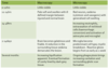
After a week following a cerebral infarction what histological process starts to occur ?
Reactive gliosis occurs in which astrocytes increase in number and size
After a few weeks following a cerebral infarction what begins to form ?
- A cavity begins to form which is lined by a gliotic scar characterised by astrocytes with abundant fine cytoplasmic processes.
- Eventually, even the gliotic scar desists and a cystic gap remains as a permanent marker of the site of an old infarction
What are the changes HTN causes in the brain ?
- Accelerated atherosclerosis - contributes to thromboembolism
- Lacunes - Lacunae are by definition lake like infarcts of less than 15mm maximum diameter. They occur when there is occlusion of a small penetrating vessel such as is seen due to occlusion of part of a lenticulostriate artery
- Hyaline arteriolosclerosis - results in thinning and weakening of small vessel walls making them more prone to occlusion and to rupture.
- Micro-aneurysms (Charcot-Bouchard)
What are the consequences of the changes HTN causes on the brain ?
- Lacunar infarcts - Atheroma, embolism small penetrating vessels leads to occlusionBasal ganglia
- Multi-infarct dementia
- Ruptured aneurysms and intra-cerebral haemorrhage
- Hypertensive encephalopathy - PM: global cerebral oedema, tentorial and tonsillar herniation, petechiae and arteriolar fibrinoid necrosis
