Neurology Flashcards

1
Q

Define the different layers of organisationa and classification of the nervous system

A
2
Q

What are the gross regions of the brain and what are their functions?

A

Frontal lobe: personality

pariental = sensory

Occipital –> vision processing

Temporal: personality (fear and anxiety)

Cerebelllum: fine motor skills

3
Q

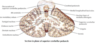
What are Gyri and Sulci in the brain?

What is their function?

A

folds in brain –> larger suface area

4
Q

List the Cranial nerves

A
5
Q

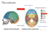
What is the function of the meningines?

Describe their organisation

A

The meningines protect the brain

6
Q

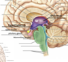
What are other important structures in the brin, than just the major regions?

A
7
Q

How is the spinal cord arranged? (inkl. grey matter, white matter, dorsal route, ventral route)

A
8
Q

How can the PNS regenerate in comparison to the CNS?

A

PNS : regeneration possible (phagocytes remove inhibitory cells)

CNS: barely any regeneration –> glia cells inhibit and form scaring

9
Q

Dorsal-comumn-medial lemniscus pathway

What is its function?

Which route does it take?

A

Sensory pathway

Crossing over in medulla oblangata

10
Q

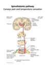
Spinothalamic pathway

What is its function?

Which route does it take?

A

Sensory pathway

Crosses over while entering the spinal cord

11
Q

List two examples of motor pathways

What is their function and which route do they take?

A

Lateral corticospinal tract:

–> e.g. arm movine –> crosses sides in medulla oblangata

Vesibulospinal tract

–> ear and balance

–> ipsilateral side all the time

12
Q

Ipsilateral

A

Same side

13
Q

Contralateral

A

entgegengesetze Körperseite oder hälfte

14
Q

Define a neuron

A

structional and functional unit of nervous system

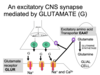
conduction of electrical siganals and communication via chemical synapse

15
Q

Describe the structure of a neuron

A
16
Q

What is the function of an axo-dendritic synapse?

How abundant is it?

A

“normal” synapse, most abundant

usually exitation

17
Q

Explain the organisation of the Cytoskeleton in Neurons

A

very abundant to ensure size and shape

intermediate fillaments: stability

Microtubule: transport of vesicles along axon

18
Q

How abundant is a Axo-somatic synapse?

What is its usual function?

A

rarer, usually inhibitory

19
Q

Describe the structure of a axo-axonic synapse

A

end at next cells axon

20
Q

Explain the function of Astroglia (Astocytes)

A

most abundant, gap junctions suggest astroglia-astrogial signaling

–> Barrier function (form Brain-Blood Barrier, signaling between blood and neurons)

–> Remove Neurotransmitter

fibrous and protoplasmic

21
Q

What is the function of Oligodendrocytes?

What are their characteristics?

A

Form Myelin and retian for lifetime (CNS)

highly metabolic active (ER, Golgi, Mitochondria)

22
Q

What is the origin of Microglia cells?

What is their function?

A
  • derive from bone marrow,

immune function –> Macrophage activation

23
Q

What is the function Schwann cells?

Where are they found?

A

Astrocytes and Oligodendrocytes function in PNS

–> myelinate one axon (wrap around one axon as a cell)

24
Q

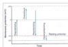
Define Electrochemical equilibrium

and Equilibrium potential

A

Electrical force balances diffustion force

Potential at which equilibrium is achieved

25
What are the Normal intracellular and extracelluar concentration for Na+ and K+ in Neurons
K+ Ex: 5, In 150 Na+ : Ex: 150, In: 10 mMol
26
Why has potassium a stronger effect on resting membrane potential than sodium?
Membranes are more permeable to potassium than to sodium (finite permeability to sodium)
27
Why do you use the GHK equation? What do you use it for? How is it calculated? Goldmann equation
More accurate to calculate potential than Nernst: more Ions+ takes into account permeability of membrane
28
Name the differnet Phases of a neural action potential
29
What is Graded potential ?
Potentials in membrane potential as response to a stimulus --\> **responses determined by time, distance, strength of** **stimulus** **often at synapse + sensory receptors** **--\>** No action potential (all or nothing), contribute to action potential e.g. weak stimulus = weak response, response gets weaker over time or distance etc.
30
Explain action potential and ion movement in neurons
31
What is Saltatory conduction?
Faster conduction: AP jumps to nodes of Ranvier --\> very rich in Na+ channels --\> new AP at each node triggered
32
Explain the structure and function of a Meutorneuron
Neuromuscular junction (yellow = examples for disease) Contraction by inducing AP in muscle cell by binding to ACh receptors One Motorneuron many muscle fibers, one muscle fiber only one motor neuron
33
Three disorders of NMJ
Botulism --\> Botulinum toxin: irreversible inhibition of release of ACh --\> paralysis Myasthenia Gravis: autoimmune: antibodies against ACh receptors --\> paralysis Lambert-Eaton myasthenic syndrome --\> inactivation of Ca2+ channels in presynaptic --\> no release of ACh possible
34
Define the components required for neurotransmitter release
quick 1. **Synapse** is required (presynaptic nerve ending, gap, postsynaptic membrane + **receptors**(ion channel linked + G-protein linked receptors) 2. **-Ca2+** is required (200 micromols) 3. **Synaptic vesicles + neurotransmitters required** 4. **AP** for release required --\> depolarisation for opening of Calcium channels --\> calcium influx in cell 5. **Proteins complex required for release** --\> rapid response due to interaction of proteins at vesicles and synaptic membrane protein --\> ca2+ is recognized by proteins at vesicles causes conformational change 6. **ATP and recycling needed**
35
Explain the difference between excitatory and inhibitory transmission
**Inhibition:** e.g. GABAR receptor + GlyR--\> influx of Cl- causes hyperpolarisation --\> Negative charge in; often at soma, further away of threshold **Excitatio**n: Positive charge in, causing depolarization closer to threshold
36
Identify mechanisms of termination of neurotransmitter action at the synapse
1. **rapid reuptake** by the transporter in neuronal cells and Glial cells for glutamate (excitatory amino acid Transporter EAAT) 2. **Enzymatic break down** of neurotransmitter
37
Twitch
zucken, one stimulus, al little response to it
38
Wha is a Summation in muscle innervation/contraction?
Several stimuli adding up and separating but increasing different movements
39
How would you describe a Fused tetanic contraction?
Many single contractions which can't be separated anymore --\> contraction of maximum force
40
Explain the structure of a Peripheral Nerve
fascicles = bundles of axons endoneurium (loose collagen fibers) Perineurium (gives main structure and strength to nerve Epineurium (collagen) which also contains blood supply and some fat tissue
41
Conduction: list the factors that affect conduction velocity of peripheral axons
**Myelin** (myelinated axons = quicker) **Diameter** (bigger = faster because of less resistance) **Resistance** Can be influenced by **local anesthesia**
42
What is a dermatome? What does damage lead to?
Area of skin supplied by a single spinal nerve route --\> Damage = weakness of sensation because skin is still innervated by other spinal nerves
43
WHat is a Myotome? What does damage of a myotome lead to?
Muscle supplied by a single spinal nerve route--\> Damage = muscle weakness because muscle is still innervated by other spinal nerves
44
What is a Ramus? Explain the organisation of a rami route
Abzweigung eines Nerves --\> Ventral + dorsal route form Mixed spinal nerves , together (ventral + dorsal) = roots and ramus are splitting of this mixed spinal nerve nerves (e.g. dorsal ramus or ventral ramus)
45
What is a plexus? How is it organised?
**Many spinal nerves** together form **one plexus** which recombines them to a single other nerves which then **e.g. innervates one muscle** ## Footnote E.g. phrenic nerve from cervical plexus
46
What is Mono and what is Polyneuropathy?
Mononeuropathy = only affects one nerve Polyneuropathy = affects many nerves
47
Classify the different levels of PNS nerve injury and its chance for recovery
1. full recovery possible 2. demyelination + axon loss, but recovery possible (epi + perineurium still intact) 3. No recovery possible, severe damage, epineurium damaged
48
List diagnostic techniques for PNS pathology
Electromyography EMG * distinction between neural or muscular cause for weakness Nerve conduction study: * Speed of conduction (demyelination or axon damage?) Somatosensory Evoked Potential * ( Peripheral or central lesion?)
49
Spinal Levels of upper and lower limb innervation
**Upper Limb:** Brachial Plexus: C5,C6,C7,C8, T1 **Lower Limbs** Lumbar Plexus:T12-L5 Sacral Plexus : L4-S4
50
Spinal Cord: What are the different segments of the spinal cord? How many nerves does each segment have, where do they emerge from?
C1-C7 emerge superior to vertebra, C8 emerges inferior (because there are 7 C vertebra but 8 spinal nerves emerging from them) Other than that:
51
Parasympathetic anatomy (Where do the fibres synapse, Which nerves do they emerge from? )
Emerge from Cranial Nerves + Sacral spinal cord: * III Oculomotor – pupil constriction * VII Facial nerve – Salivation * IX Glossopharyngeal – Salivation * X Vagus – bradycardia, gastric motility, digestion * And S2-S4 Level Nerves go directly to a target organ, ganglia in the target organ (long pre-synaptic, short post-synaptic branches)
52
Sympathetic Nervous system (Where so the fibres synapsing, where do they emerge?)
T1-L3 First synapse in sympathetic chain --\> Short pre-ganglionic fibre, lond post-ganglionic fibre
53
What effects dos the Parasympathetic NS have on the body and differnt organ systems?
54
What effects does the SNS have on different organ systems?
Fight and Flight: Dilation of pupils, sweating, increasing blood glucose levels, increased Heart rate and breathing, dilation of bronchi
55
WHat is the main Neurotransmitter in PSN? What other transmitter does it also use?
Acetylcholine (ACh) (Glutamate, Glu for down of Spinal cord to S2-S4) but afterward also ACh)
56
Neurotransmitters in SNS
Many different ones for different targets, main transmitter : Noradrenaline
57
Summarise the biosynthesis of Acetylcholine
58
Summarise the biosynthesis of Noradrenaline
59
In which structure of the spinal cord do the autonomic neurons emerge? In which structure the sympathetic neurons?
Autonomic in general: dorsal horn Sympathetic: Intermediolateral cell column
60
Examples of regulation of the autonomic NS
1. **Heart rate:** Baroreflex (regulation via BP) --\> Baroreceptors in Aorta+ Carotis sense change --\> change in innervation of Vagus nerve on Heartbeat 2. **GI tract:** * cephalic response (insulin secretion + gastric acid production even before tasting food) * Mechanorecpeptors show dialation of stomach, Chemoreceptors secrete satiety Hormones 3. **Respiration:** Chemoreceptors (peripheral in Aorta+ Carotis, central in central respiratory center changes in O2, pH, CO2) up respiratory rate, Mechanoreceptors in lungs down(to prevent hyperinflation) --\> signals to respiratory muscles
61
What are the Receptors of Sympathetic NS? Where are they located?
1. All Nicotonic in the sympathetic chain 2. differ, but all G-Protein linked Adrenergic receptors
62
What are the Receptors of the PNS? What kind of receptors are they? Where are they located?
Nicotinic = ion chanel Muscarinic= G Protein-coupled
63
Orthostatic hypotension
Drop of BP after standing up
64
What can abnormalities in ANS lead to? | (Name 3 disorders)
1. Primary hypertension: Hypertension = exaggerated sympathetic nerve activity to blood vessels and heart---\> increased circulating volume and vascular tone 2. heart failure 3. Parkinsons Disease
65
Explain the regulation of the Baroreceptor reflex
66
Cerebrum, Cerebellum, Diencephalon, Brainstem, Medulla oblangata brief location + function
67
Spinal cord: Vertebra + spinal nerve pairs (and where they dissent from vertebra)
30 Vertebra, but 31 pairs of spinal nerves 1-7 above vertebra 8-31 below vertebra
68
Explain the Anatomy of spinal cord (cross section)
69
What are the function of the spinal cord?
1. Connects the PNS and ANS to the brain 2. Carries sensory signals to the brain 3. Carries motor signals to the muscles 4. Coordinates reflexes
70
Explain the organisation and function of the Diencephalon
Hypothalamus: * integration hub: * Regulates temperature, hunger, thirst, hormone (connected with pituitary)+ autonomic function **Thalamus** * integration center for somatic and special senses information and projection to cortex. * Involved in emotional status, consciousness, appropriate motor response
71
What is a Basal Ganglia? Where is it located? What is its function?
Different structures in Cortex --\> accumulation of Cell bodies •Control of movement: facilitating voluntary movement, inhibiting unwanted or inappropriate movements, “fine-tuning”
72
What are the function of the Limbic system?
It is involved in: * motivation, * instinctive behavior, * emotion, * memory
73
Describe the anatomy, and function of the Cerebellum
Anatomy: * two hemispheres divided in lobes, Grey cortex with folds and deeper white matter vestibular system for balance * •spinal cord and muscles of locomotion, posture; muscle tone * •Motor cortex and thalamus for learned movements (egmanual skills, trajectory, timing,, speed and force)
74
What is the Corpus callosum? What is its function?
Connects the two hemispheres of the cerebral cortex --\> information exchange and processing between two hemispheres
75
What is the Ventricular system? Which cells are important for its function?
System which produces and transports Cerebrospinal fluid Produced by **choroid plexus**
76
Explain the route of the CVF fluid from production into spinal cord
77
Composition and function of Cerebrospinal fluid
Composition similar to plasma (high Na+, low K+) but: * Lower glucose (2/3) * Much lower protein (200x) * Lower Ca2+, K+ * Higher Cl-, Mg * Slightly lower pH (7.33) Function: * cushioning * nutrient supply * removal of waste products * immune defence
78
Name and describe the different layers of meninges
79
Where can you find fibrous astrocytes? What are their characteristics?
* white matter * have relatively few organelles * and exhibit long unbranched cellular processes
80
Where are protoplasmic astrocytes found? What are their characteristics?
* most prevalent * and are found in grey matter tissue * possess a larger quantity of organelles * and exhibit short and highly branched tertiary processes.
81
Which cells are involved in the damage of MS?
Oligodendrocytes --\> Normally: Myelination of Axons If damaged: demyelination of the CNS
82
Explain the effects of Botulinum toxin in the Nervous system
Botulinum toxin: irreversible inhibition of release of ACh --\> paralysis
83
Explain the mechanism and effect of myastenia Gravis on the Nervous system
Myasthenia Gravis * Autoimmune disease * antibodies bind and block ACh receptors --\> **paralysis**
84
Explain the mechanism and effect of Lambert-Eaton myasthenic syndrome on the Nervous system
Lambert-Eaton myasthenic syndrome --\> inactivation of Ca2+ channels in presynaptic membrane --\> no release of ACh possible --\> **no excitation of muscle possible**
85
What does damage to a nerve supplying a myotome/dermatome lead to? Why?
It leads to a weaker sensation and muscle weakness because area is still supplied by more nerves
86
What does damage to a nerve after the plexus lead to? Why?
Many spinal nerves supply one muscle but form **only one nerve after plexus** --\> damage to the one nerve after plexus leads to **total numbness, paralysis** **etc****.**
87
Which structures belong to the limbic system? Where is it located?
It is located at both sides of the thalamus It has three areas: 1. Cortical area (contains e.g. hippocampus) 2. Sub-cortical area (contains e.g. amygdalla) 3. Diencephalic structures (contains e.g hypothalamus, part of thalamus)
88
What is meant by the term recruitment on muscle contraction?
recruitment means increasing the number of active motor-neurons
89
Which unit does the Equilibrium potential of an ion have?
It is shown in mV (milli volt)
90
What does binding of ACh to the receptors of the post-synaptic membrane of a Neuro-muscular junction lead to?
It leads to the opening of cation-selective channels (Na+, K+)
91
What is neuromodulation? (neuromodulatory synapse)
A neuromodulators synapse is an Axo-axonic synapse, leading to regulation of a neuron it is relatively slow and neurotransmitter often bind to G-protein coupled receptors
92
What does fast-anterograde transport transports?
Anterograde: * is movement of molecules/organelles outward, from the soma to the synapse or cell membrane. Fast anterograde transport includes * synaptic vesicles * transmitters * mitochondria
93
What involves slow anterograde transport?
Anterograd= from cell body to synapse, away from cell body Slow anterograde: delivery of cytoskeletal and cytoplasmic constituents
94
What is retrograde transport?
Transport of things from synaps, periphery back to soma
95
Which substances are transported retrograde?
1. material returns from the terminals to the cell body either for degradation or recycling 2. substances from an extracellular space 3. microtubule-associated ATPase to drive particles along microtubules(?-\> please check again)
96
How is a neurotransmitter inactivated?
* Diffusion out of the cleft * Enzymatic destruction in the synaptic cleft * Re-uptake by the pre-synaptic terminal * Uptake by glial cells
97
Explain the mechanism of function of Benzodiazepines
They enhance GABA function --\> GABA is a special neurotransmitter that has an inhibitory effect on Membrane potential by causing hyperpolarization of the membrane (Cl- influx)
98
Which structures does the central sulcus devide the cerebral hemisphere in?
It divides it into frontal and parietal lobe
99
Compare subdural and epidural hemorrhages (bledding, appearance, position etc.)
100
What does the brainstem consist of?
Pons, medulla and midbrain
101
Explain the role of local currents in NMJ activation
Local currents flow from depolarized region and adjacent region --\> action potential triggered and spreads along surface membrane
102
How is ACh broken down in a NMJ?
It is broken down by the enzyme acetylcholine esterase
103
What does a EMG record? How does it do this?
It superficially records Muscular action potential --\>
104
What are the 2 main types of hydrocephalus and their causes?
**1.** **Communicating****:** * block in CSF absorption * CVS can still flow over all 4 ventricles (--\> communication) * meningitis, head injury, congenital, hemorrhage **2. Non- communicating:** * Obstruction on flow * ventricular, paraventricular tumor, aqueduct stenosis
105
What are the symptoms of hydrocephalus?
In children: cranial swelling Other symptoms: because of increased intercranial swelling: vomiting, sleepiness, irritability, downward deviation of the eyes (also called "sunsetting"), and seizures.
106
How can hydrocephalus be treated?
* remove the cause, e.g. papüiloma * open alternate pathways, * divert CSF
107
Compare grey and white matter
Grey matter: cell bodies (all synapses) White matter: axons
108
109
110
111
112
113
114
What happens during neuropraxia?
* **reversible** conduction block * selective demyelination of the axon sheath * endoneurium and axon still intact - e.g., nerve compression
115
What happens during axonotmesis?
* demyelination and axon loss * epineurium and perineurium remain intact * still some continuity within the nerve * degeneration occurs below and slightly proximal to the site of injury
116
What happens during Neurotmesis?
* most severe form of nerve injury * associated with complete nerve division and disruption * commonly seen after toxic or ischemic injuries * Damage to the epineurium (around the entire nerve) - no nerve growth
117
Explain the process of nerve regeneration in the PNS
Degernation + phagocytosis of parts is calles Wallerian Degeneration
118
What is the most commonly prescribed anti-epileptic drug and what is its target?
Tiagabine --\> is a GABA agonist
119
Which neurotransmitters does the somatic nervous system use?
Preganglionic synapse: Glutamate Postgangionic: ACh
120
What are two cholinergic drugs? What is their target site?
121
What kind of drug is Salbutamol? What is its target site?
Beta2 receptor agonist (relaxes smooth airway muscle)
122
Which enzyme converts testosterone into dihydrotestosterone?
Enzym **5α-Reductase**
123
Which enzyme converts testosterone in estradiol?
Aromatase /Estrogen synthase
124
How can you classify the three types of neurons in the sympathetic nervous system?
1. Neuron (goes down the spinal cord)= **Presympathetic** **/Preautonomic nerve** 2. Neuron (from the spinal cord to sympathetic chain)= **preganglionic nerve** 3. Neuron (from sympathetic chain to target organ) = **Postganglionic nerve**
125
In which "year" do the parasympathetic cranical nerves emerged?
1973 Cranial nerves that provide parasympathetic supply: * X, IX, VII, III
126
Which structures lie above the brainstem?
The diencephalon
127
How do the cerebral hemispheres and the diencephalon relate to one another?
The cerebral hemispheres wrap around the diencephalon
128
What is the function of the brainstem?
* Relay center --\> Contains All fibres that connect cerebrum, cerebellum + spinal cord * Contains nuclei of all cranial nerves * control breathing, heart rate, blood pressure, swallowing balance, etc., sleep * defense reflexes (cough, gag)
129
Which part of the brain degenerated in Parkinson's?
**Midbrain** includes the **substantia** **nigra which** degenerates in Parkinson’s Disease
130
Which structures belong to the basal ganglia?
Caudate nucleus Putamen --\< together corupus striatum Putamen + Globus Pallidus= Lentiform nucleus Amygdulla Substantia Nigra Subthalamic nucleus
131
What is the overall function of the basal ganglia?
To relay and control the **fine-tuning of movement** * facilitating voluntary movement * inhibiting unwanted or inappropriate movements
132
133
134
135
136
137
What does the Striatum consist of?
It consists of the Caudate nucleus and Putamen (both parts of the basal ganglia)
138
What does the lentiform nucleus consist of?
The Putamen and Globus Pallidus
139
How thick is the cerebral cortex? How much of it is exposed to the outside, how much in sulci?
* 2-4 mm thick * ~30% exposed 70% within sulci
140
How do you divide the cortex into forebrain, midbrain, and hindbrain?
**Forebrain** =cerebral hemispheres and diencephalon **Midbrain** **Hindbrain:** Pons, Medulla, Cerebellum --\> classification derives from embriology
141
What is a Astroglia cell?
most abundant, gap junctions suggest astroglia-astroglial signaling --\> Barrier function (form Brain-Blood Barrier, signaling between blood and neurons) --\> Remove Neurotransmitters fibrous and protoplasmic
142
What are the main function of the cerebral hemispheres?
Many functions: * Motor cortex --\> initiation of movement * Motor association area --\< planning of movement * Primary sensory cortex --\> interpretation of sensation * Association area --\> interpreation of sensation (e.g. touch/feel to the shape of an object) * Personality
143
What are commissural fibres of the brain?
Fibres connecting the two parts of the brain (e.g.Corpus callosum )
144
What are the Association fibers?
Fibres, that connect structures on the same side of the brain
145
What are projection fibres?
Fibers that run vertically down (can cross sides) e.g. sensory fibres (run down spinal cord)
146
What is the zona radiata?
Zone in the cerebral cortex, where the projection fibers spread to different structures
147
Which cells produce cerebrospinal fluid?
Ependymal cells in choroid plexi
148
Compare ion channel receptors and G-protein coupled receptors for mediating a neural action potential Name examples in each
Ion channels: fast (ms) (GABA+ ACh) G-protein coupled: slow (s/min) (ACh at muscarenic, dopamine)
149
Which changes might lead to seizures in epilepsy?
A **decrease in** **GABAmediated** **inhibition** or an **increase in glutamate-mediated excitation** in the brain may result in seizure activity
150
How is GABA synthesised?
GABA is synthesised by Glutamic Acid Decarboxylase (GAD) – Known as the Vitamin B6 enzyme.
151
Explain the binding and inactivation of GABA
**Binding** * GABA binds to the receptor and allows entry **Cl- ions which hyper-polarises the cell.** **Inhibition:** * Reuptake of pre-synaptic membrane and glia cells * inactivated by **GABA transaminase,** giving **Succinate semialdehyde**
152
Explain the effect and inactivation of Glutamate as a neurotransmitter
* causes entry of sodium and calcium through the NMDA receptor. * Uptake over pre-synatptic neurone and glia cells via mainly **EAAT2 transporter** --\> inactivated by glutamine synthetase to make glutamine (addition of an amino-acid).
153
What are examples of anti-epileptic drugs? How can they be exploited for epilepsy?
All modulate GABA receptors * e.g. Valproate (weakens GABA transaminase) * Benzodiazepine (enhances GABA action)
154
Whar are areas 4 and 6? Where are they located and what is their function?
Located in the **frontal lobe:** **4: Primary motor complex** * send signals + coordinate movements (initiation of movement) **6: Motor association complex:** * Movements are planned
155
What are the areas 1,2,3 5,7? Where are they found? What is their function?
Found in Parietal lobe: **1,2,3: Primary somesthetic cortex** * interprets sensory information (touch, pain temperature **5,7: Somesthtetic association area** * Associated felt sensation with known+ hand-eye coordination
156
What are the two areas? Where are they located and what is their function?
Mainly occipital lobe **18,19 (green): Visual associaton area** * recognises faces, other familliar things **17 Primary visual cortex** * recieves visual information
157
Located in the **temporal lobe** **Primary auditory cortex** * receives auditory information **Auditory association area** * recognizes e.g. speech
158
Profrontal cortex * judgement, associations --\> personality
159
Broca area: ## Footnote Right-handed on the left side, left hand on the right side (+ other way around) --\> speech
160
Which structures are located in the anterior cranial fossa?
The **frontal cortex** lobes in it
161
Which structures lie in the middle cranial fossa?
The pituitary and the temprotal lobes
162
Which structures are located in the posterior cranial fossa?
The occipital lobe and the brainstem
163
What is latency?
The time difference between a stimulus and a response in a EMG (At picture: Nerve conduction study done at distal + proximal location)
164
How is a nerve conduction study made? Which factors influence latency?
2. Measure the latency of response at both locations (distal + proximal) – i.e. the time difference between stimulation and response. Latency time is affected by: * Activation time. * Conduction delay from cathode to NMJ. * Delay at NMJ. * Conduction delay along muscle fibre to EMG electrodes. 3. Conduction velocity is measured by: 𝐷𝑖𝑠𝑡𝑎𝑛𝑐𝑒 𝑏𝑒𝑡𝑤𝑒𝑒𝑛 𝐸𝐸𝐺 𝑎𝑟𝑒𝑎 */ Dif𝑓*𝑒𝑟𝑒𝑐𝑒 𝑖𝑛 𝑙𝑎𝑡𝑒𝑛𝑐𝑦 𝑖𝑛 𝑏𝑜𝑡ℎ 𝐸𝐸𝐺 𝑎𝑟𝑒𝑎𝑠
165
What is the difference between the ependymal cells and the choroid plexus?
The Ependymal cells line all ventricles whereas the choroid plexi are modified ependymal cells that produce CVS
166
How fast can a nerve in the PNS regernerate per day?
2-5 mm
167
What is a neuroma?
Swelling of Peripheral nerves (or growth), often due to the failed reconnection after nerve damage
168
What is chromatolysis?
It is the dissolvement of the Nissel Body in a nerve (organelles with abundant ER and ribosomes --\> protein synthesis) --\> happens to the cell body in peripheral verve damage
169
Explain the route an efferent postganglionic fiber in the sympathetic Nervous system takes when it goes via the ventral route
1. Enters **ventral route** 2. **Mixed spinal nerve** 3. Via the **White ramus** to the sympathetic chain 4. (Evenutally: goes up and down to other ganlglia) 5. **Synapse** in ganglia 6. Back to spinal nerve (same level, other level) vie the **grey ramus**
170
What is a splanchnic nerve?
Autonomic fibers that travel within their own nerve 1. Synapse in the sympathetic ganglion and have an **own post-ganglionic nerve** (does not leave via grey ramus) 2. Neurons that **enter the Sympathetic gangion but don't synapse there** (synapse in distal ganglion)
171
What is the mechanism of action of Phenobarbital?
It enhances GABA action And Inhibits synaptic exitation (used in all types of epilepsy except absensce seizures)
172
What is the mechanism of action of Valpoate?
It has a weak effect on GABA transaminase + Na+ channels
173
What is the mechanism of action of Vigabatrin?
It inhibits GABA transaminase --\> effective for patients which are resistant to other drugs
174
What is the mechanism of action of Tiagabine?
It inhibits GABA reuptake
175