Neuro Flashcards
Testing tone in upper limb
Supinator catch
flexion and extension of elbow (clasp knife, lead pipe)
dorsiflexion and collar flexion at wrist (cogwheel, reinforced by synkinesis)
Pyramidal pattern of weakness
Testing movements and their roots, and peripheral nerves

How do you test hand function?
grip - C8- long flexors
pincer grip
prayer sign
fine movement (play the piano, general pyramidal function)
Positive prayer sign
RA
scleroderma
diabetes
ulnar nerve palsy
T1 palsy
Dupuytren’s
What does pincer grip test?

Cerebellar signs
DANISH
dysdiadokokinesia
ataxia
nystagmus- 20 dog from centre
intention tremor
scanning (staccato) or slurred speech
hypotonia
Test for cerebellar speech
west register street, edinburgh
baby hippopotomus
british constitution
Management of acute stroke
Contraindications to thrombolysis
high BROW
high BP diastolic >140
bleeding tendancy
recent surgery
over 80
woke with sx
Causes of carpal tunnel
RAPID TTT
RA
acromegaly
pregnancy
idiopathic
diabetes
trauma
tenosynovitis
thyroid (hypo)
4 signs of ulnar nerve palsy
sensory loss over 5th and ulnar 1/2 of 4th finger
wasting and weakness of dorsal interossei
weak pincer grip
partial claw hand (medial lumbricals)
How does a t1 lesion cause claw hand?
Affects the fibres to all lumbricals (ulnar and median)

Lower limb nerve roots
L2- hip flexion
L3- knee extension
L4- inversion and dorsiflexion of the foot
L5- dorsiflexion of the big toe
S1- eversion of the foot
TACI criteria
PACI criteria
2/3:
contralateral hemiparesis
contralateral homonyms is hemianopia
higher cortical dysfunction
POCI criteria
Any 3:
cerebellar signs
contralateral homonyms is hemianopia
Brain stem signs (horners, cranial nerve lesion)
LACI features
No:
contralateral hemiparesis
contralateral homonyms is hemianopia
higher cortical dysfunction
drowsiness
Path of the optic nerve

Visual defect vs site of lesion

Causes of papilloedema
raised ICP due to tumor
essential intercranial hypertension
malignant hypertension
hypercapnoea
How does optic neuritis cause optic atrophy

Signs of optic neuropathy
pale disc
loss of visual acuity
loss of red colour vision (desaturation or red dyschromatopsia)
central scotoma
afferent pupillary defect
Causes of optic neuropathy
demyelination (optic neuritis)
trauma
compression (pituitary tumour or meningioma)
ischaemic (diabetes, cranial arteries, temporal arteritis)
toxic (methanol, ethambutol)
secondary to papilloedema (essential Intercranial HTN)
Bell’s palsy Mx
eye drops
close eyelid at night
high dose steroids if seen <72hrs
consider referral if still weak at 6/12
check BP, sometimes vascular cause
Parkinson’s quadrad
tremor
rigidity
bradykinesia
loss of postural reflexes
Parkinson’s Hx
ADLs
handwriting (micrographia)
buttons and shoelaces
turning over in bed at night
getting in and out of car
Parkinson’s Ex
poverty of facial expression
5Hz tremor
cogwheel rigidity at wrist
thumb finger test
toe tap test
function
exclude Parkinson’s plus
Parkinsonism causes
parkinsons disease
vascular parkinsonism
parkinsons-plus syndromes
other (antidopaminergics, Wilson’s)
Parkinson’s plus syndromes
progressive supernuclear palsy : vertical limitation, axial rigidity
multi-system atrophy- cerebellar signs, autonomic problems
corticobulbar degeneration
lewy body dementia
Features of Parkinson’s gait
loss of arm swing (early)
hesitancy
shuffling
hurrying (festination)
retropulsion (falling backward)
clock face turning
Motor complications in Parkinson’s
drug induced dyskinesias: choreo-athetoid movements
fluctuations: predictable (end of dose) unpredictable (on-off effect)
all commoner with L dopa and less common with DA agonists
Parkinson’s management

Pathophysiology of unilateral inter nuclear opthalmoplegia

Causes of bilateral inter nuclear opthalmoplegia
wernickes encephalopathy
stroke
demyelinating disease
MS
Ix MS
clinical exam and hx may be sufficient
2 separate attacks affecting different parts of the CNS
MRI: periventricular and juxtacorticol plaques
visually evoked potentials: delay in EEG response due to optic neuropathy
somatosensory evoked potentials: delay in EEG response after episode of sensory demyelination
CSF- oligoclonal bands (60% after one attack, 90% after 2)
Treatment of acute MS
may not need treatment if just sensory
IV methylprednisolone for 3 days
reduces length and severity, no impact long term
small risk of psychosis, diabetes, AV necrosis femoral head
high dose oral may be effective, but also increase relapse rate of optic neuritis
Oculomotor palsy features
partial ptosis
eye abducted and depressed
pupil dilated (loss of parasympathetic fibres)
Causes of oculomotor palsy
Diabetes (pupil often spared)
PCA aneurysm (often painful)
raised ICP (false localising)

Why is the pupil spared in diabetes?
parasympathetic fibres have a separate blood supply from the nerve sheath vessels
Features of horners syndrome
slight ptosis
constricted pupil
reduced sweating on forehead
eye is bloodshot early
loss of alpha vasoconstrictor tone
Causes of acquired ptosis

Causes of 6th nerve palsy
Course of 6th nerve
The nucleus is in the lower pons.
The nerve exits anteriorly and travels up the brainstem on either side of the basilar artery, in the subarchnoid space (here it is susceptible to meningitis and rarely basilar aneurysms)
It passes forward over the base of the skull towards the tip of the petrous temporal bone (here it.can be damaged by severe ear infections associated with bone infections- osteomyelitis- as well as by skull fractures and by nasopharyngeal cancer)
It enters the cavernous sinus and then goes through the superior orbital fissure to reach the eye.
Facial nerve palsy assessment algorithms

Causes of a unilateral facial nerve palsy
Brain stem vascular accident (associated contralateral hemiparesis)
acoustic neuroma, within the cerebello-pontine angle
trauma
Ramsay hunt syndrome - Hz affecting geniculate ganglion
bells palsy- in facial canal
pagets disease of bone
parotid trauma, surgery, tumour
mononeuropathy - sarcoid diabetes
Assessment of speech
Look for signs of CVA (hemiparesis) Parkinson’s disease, scleroderma etc;
Then say
Hello, my name is ….., would you mind if I ask you some questions to check your speech and memory?
Check orientation while listening to quality of voice Can you tell me your name?
How old are you?
Do you know what day of the week it is?
And what year this is?
And where are we at the moment?
If you suspect problems with cognitive function after this, proceed with mini mental state examination including counting backwards from 20 etc
Check for dysphasia
Can you lift your left hand and place it on top of your head? (tests receptive dysphasia- important not to move your arm as patient may copy you- you need to check that spoken instruction is understood without any visual clues).
What is this? (tests for nominal dysphasia using keys or pen or newspaper etc)
Check for dysarthria
If the speech is suggestive of dysarthria, ask patient to say “Baby hippopotamus” or “British constitution” or “West Register Street, Edinburgh”.
May be appropriate to check cerebellar signs; eg ask patient to stretch out their arms looking for ataxia of upper limbs.
If pseudobulbar palsy suspected, look in mouth (for spastic tongue) plus check jaw jerk- increased in pseudobulbar palsy.
Ask about symptoms
If no abnormality demonstrated, proceed to listen to a longer segment of speech and check symptoms:
“What symptoms led to your admission to hospital?”
In general
Be prepared to modify your approach according to what you find eg if patient has Parkinson’s, be prepared to demonstrate the signs once you have listened to the voice.
Remember that the examiners will probably point you in the right direction if you are going down a blind alley. The main thing to demonstrate is that you are aware of simple ways to elicit dysphasia, dysarthria and orientation.
Causes of dysarthria
difficulty with mechanism of speech
bulbar palsy= flacid
pseudobulbar palsy = spastic
cerebellar = staccato
myasthenic = weak, quiet, fatigable
Causes of dysphasia
Difficulty with processing of speech
expressive (Broca’s)= knows what they want to say, but can’t say it
Receptive (Wernicke’s)= fluent, effortless, lacks meaning, can’t follow cues written/spoken.
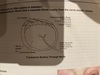
What is a DaT scan?
Dopamine transporter scan
single photon scan with contrast
normal ‘comma’ appearance of basal ganglia
loss of DA neurones in lentiform nucleus
occurs in Parkinson’s and parkinsonism
not essential tremor
Dopamine agonists
parkinsons
less effective then LDopa but cause less effects, so used in younger people
used in addition to LDopa in advanced stages
older ones e.g. pergolide cause pulmonary and cardiac fibrosis —> MR
side effects
sleepiness
hallucinations
impulse control
Overview of Parkinson’s Tx

MS possible mechanisms
Probably autoimmune once triggered
antigens attack myelin and oligodendrocye glycoprotein
initial attack leaves inflammation of the bbb
allows B and T cells to cross
direct damage to axons and indirect damage (demyelination)

Types of MS
Relapsing remitting 80%
primary progressive 10%
benigh 10%
secondary progressive (after about 10 years of relapsing remitting)
MS main presenting symptoms
sensory 40%
optic neuritis 20%
cerebellar/vertigo 20%
internuclear opthalmoplegia 10%
motor (usually spastic paraparesis) 10%
Adult onset spastic paresis differentials
MS
amyotrophic lateral sclerosis
transverse myelitis
spinal cord compression/trauma
intramedullary structural lesions
cervical myelopathy
spinocerebellar degeneration
B12 deficiency
Luetic disease
Indications for DMT in MS
ambulant patients with RRMS
>2 relapses in 2 years
one relapse + radiological progression
