Nervous 3 Flashcards
What kind of fibres pass through the corpus collosum?
Commissure fibres
What are the 4 parts of the corpus collosum?
Rostrum
Genu
Body
Splenium
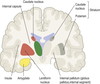
What is A?

Body
What is B?

Genu
What is C?

Rostrum
What is D?

Spleenium
What is the septum pellucidum?
Thin sheet that lies in the mied sagittal plane and seperates the anterior horns of the two lateral ventricles

What is the septum pellicuidum inferior to?
Corpus collosum

What is inferior to the septum pellicudum?
Fornix

What is the fornix?
Bundle of fibres that linkes the hippocampus with the mammilary bodies
What is the connection between the lateral ventricles and III ventricle?
Interventricular foramen
What is E?

Septum pellicidum
What is F?

Fornix
What are the black spaces at each side?

Lateral ventricles
What is the bulbous projection on the floor of the lateral ventricle?
Caudate nucleus
Caudate nucleus is one of what?
The basal nuclei
What is the thalamus?
Sensory relay area made up of smaller masses of gray matter nuclei, each with a different function
What of the general sensory information from the body relays to what nucleus?
Ventro-postero-lateral (VPL) nucleus of the thalamus
What are the functions of the hypothalamus?
Releasing hormone
Regulating temperature
Regulating hunger
Managing sexual behaviour
Regulating emotional response
Regulating thirst
A fold of what immediately overlies the pituitary gland?
Dura mater (called Diaphragma sellae)
What fold overlies the pituitary gland?
Diaphragma sellae
What gland is immediately posterior to the thalamus?
Pineal gland
Where are the colliculi in relation to the pineal gland?
Inferior
The colliculi are a component of what?
Midbrain
What is A?

Third ventricle
What is B?

Intermediate mass of thalamus
What is C?

Hypothalamus
What is D?

Optic chiasma
What is E?

Pituitary gland
What is F?

Mammilary body
What is G?

Pons
What is H?

Medulla oblogata
What is I?

Spinal cord
What is J?

Cerebellum
What is K?
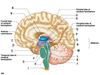
Choroid plexus
What is L?

Fourth ventricle
What is M?

Cerebral peduncle of midbrain
What is N>

Cerebral aquaduct
What is O?

Corpora quadridgemina
What is P?

Pineal body (part of epithalamus)
What is Q?

Thalamus
What is r?

Choroid plexus of third ventricle
What is S?

Corpus collosum
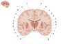
What is A?

Median longitudinal fissure
What is B?

Lateral ventricle
What is C?

Anterior limb of internal capsule
What is D?

Genu of internal capsule
What is E?

Posterior limb of internal capsule
What is F?

Thalamus
What is G?

III ventricle
What is H?

Lentiform nucleus (putamen is medial and globus pallidus is lateral)
What is I?

Caudate nucleus
What structures together constitute the basal ganlia nuclei?
Caudate nucleus, putamen and globus pallidus
What is the internal capsule made up of?
Myelinated axons (white matter)
What is the name given to fibres that connect cerebral hemispheres to other parts of the brain, such as through the internal capsule?
Projection fibres
What is the internal capsule made up of?
Anterior limb, genu and posterior limb
Which cerebral artery through one of its branches supplies the internal capsule?
Middle cerebral artery
What is A?

Superior colliculus
What is B?

Periaquaductal grey matter
What is C?

Oculomotor nucleus
What is D?

Medial lemniscus
What is E?

Red nucleus
What is F?

Substantia nigra
What is G?

Corticospinal fibres
What is H?

Cerebral peduncle
What is I?

Reticular formation
What is J?

Cerebral aquaduct
What group of structures is the substantia nigra apart of?
Basal ganglia
Where would the red nucleus be in relation to the substantia nigra?
Posterior to it
What fibres does the cerebral peduncle contain?
Ascending sensory and descending motor fibres
What part of the brain does this show?

Pons
What is A?

Middle cerebellar peduncle
What is B?

Medial lemniscus
What is C?

Transverse pontine fibres
What is D?
Deep pontine nucleus
What is E?

Corticobulbar and corticospinal tracts
What is F?

Sensory and motor nucleus of CN V
What is G?

IV ventricle
What is H?

Superior cerebellar peduncle
What is I?

Cavity of IV ventricle
What is J?

Superior cerebellar peduncle
What is K?

Main sensory nucleus of CN V
What is L?

Motor nucleus of CN V
What is M?

Middle cerebellar peduncle
What is N?

Medial lemniscus
What is O?

Pontine nucleus
What is P?

Bundles of corticospinal and corticonuclear fibres
What is the medial lemniscus?
Large ascending bundle of heavily myelinated axons that decussate in the brainstem, specifically in the medulla oblongata
What are the pontine nuclei?
Nuclei of the pons involved in motor activity
What forms the medial lemniscus?
Gracile nucleus (medial) - carries sensory information from lower half of body
Cuneate nucleus (lateral) - carries sensory information from upper half of body
both found in junction between medulla and spinal cord
What does this section show?

Medulla
What is A?

Nucleus gracilis
What is B?

Fasciculus gracilis
What is C?

Fasciculus cuneatus
What is D?

Spinal tract (CN V)
What is E?

Spinal nucleus (CN V)
What is F?

Posterior spinocerebellar tract
What is G?

Anterior spinocerebellar tract
What is H?

Decussation of pyramids
What is I?

Spinothalamic tracts
What is J?

Nucleus cuteatus
What is K?

Medial longitutinal fasciculus
What is L?

Spinal tract (CN V)
What is M?

Spinal nucleus (CN V)
What is N?

Fibres of CN X
What is O?

Nucleus ambiguus
What is P?

Posterior spinocerebellar tract
What is Q?

Anterior spinocerebellar tract
What is R?

Deccusation of medial lemniscus
What is S?

Pyramid
What is T?

Inferior olivary nucleus
What is U?

Spinothalamic tracts
What is V?

Reticular formation
What is W?

Central canal
What is X?

Accessory cuneate nucleus
What is Y?

Nucleus cuneatus
What is Z?

Nucleus gracilis
What fibres make up the elevations of the pyramids and olives?
Pyramids - corticospinal and corticobulbar tracts
Olives - contains inferior olivary nucleus
What are the 3 main coronal sections of the brain you need to be aware of?
Anterior - coronal plane passes just anterior to optic chiasma
Middle - coronal plane passes just posterior to optic chiasma
Posterior - coronal plane passes through plenium (posterior part) of corpus callosum

What is A?

Cingulate gyrus
What is B?

Body of fornix
What is C?

Central part of lateral ventricle
What is D?

Body of caudate nucleus
What is E?

Third ventricle
What is F?

Lenticular nucleus
What is G?

Insular lobe
What is H?

External capsule
What is I?

Globus pallidus
What is J?

Tail of the caudate nucleus
What is K?

Optic tract
What is L?

Hippocampus
What is M?

Substantia nigra
What is N?

Mammillary bodies
What is O?

Subthalamic nucleus
What is P?

Temporal horn of the lateral ventricle
What is Q?

Globus pallidus internal segment
What is R?

Claustrum
What is S?

Putamen
What is T?

Extreme capsule
What is U?

Internal capsule
What is V?

Thalamus
What is W?

Thalamus
What is X?

Choroid plexus
What is Y?
Body of corpus collosum
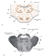
What part of the spinal column is this?

Cervical
What part of the spinal column is this?

Thoracic
What part of the spinal column is this?

Lumbar
What part of the spinal column is this?

Sacral
How do you identify where from the spinal column a section comes from?
Cervical - thin and pointy
Thoracic - thin and pointy, smaller than cervical
Lumbar - thick
Sacral - thickest, more geometric than lumbar

What aspect of the spinal cord receives sensory nerve fibres?
Dorsal route ganglion
From which aspect of the spinal cord do motor nerve fibres project?
Ventral
Why does the size of the ventral grey horn vary along the spinal cord (most notibly the cervical and lumbar regions)?
Due to brachial and lumbosacral plexus
What is the name given to the hole in the middle of the spinal cord and what does it contain?
Central canal, contains CSF
