Myology Flashcards
Sternocleidomastoid
O: Mastoid Process
I:
- Sternal Manubrium
- Sternal 1/3 Clavicle
In: Axillary N.
Fx:
- Head Extension
- Lateral Tilt
- Contralateral Rotation

Trapezius
O:
- External occipital protuberance
- Superior Nuchal Line
- SP C7-T12
I:
- Scapular Spine
- Acromial 1/3 clavicle
In: Accessory N. (CN XI)
Fx:
- Shoulder Elevation
- Scapular ER

Rhomboids
O:
- Major: SP T2-T5
- Minor: SP C7-T1
I: Medial Scapula
In: Dorsal Scapular N.
Fx: Scapular Adduction and Rotation

Levator Scapulae
O: TP C1-C4
I: Superomedial Scapula
In: C3-C4
Fx: Scapular elevation + rotation

Latissimus Dorsi
O:
- SP T6-S5
- Illium
I: Crest of LT
In: Thoracodorsal N.
Fx:
- Adduction Humerus
- Flexion Humerus
- IR Humerus

Pectoralis Major
O:
- Clavicle
- Sternum
- Ribs 1-7
I: Crest of GT
In: Medial & Lateral Pectoral N.
Fx: Shoulder Adduction & IR

Pectoralis Minor
O: Ribs 1-5
I: Coracoid Process (conjoint tendon)
In: Medial Pectoral N.
Fx: Scapular Protraction

Serratus Anterior
O: Ribs 1-12
I: Medial Scapula
In: Long Thoracic N.
Fx: Scapular Retraction

Subclavius
O: 1st rib
I: Inf. Clavicle
In: N. to Subclavius
Fx: Clavicle Stabilization

Deltoid
O:
- Clavicle
- Acromion
- Scapular Spine
I: Deltoid Tuberosity
In: Axillary N.
Fx:
- Shoulder Abduction
- Shoulder Extension
- Shoulder Flexion

Supraspinatus
O: Supraspinous Fossa
I: GT
In: Suprascapular N.
Fx: Shoulder Abduction

Infraspinatus
O: Infraspinous Fossa
I: GT
In: Suprascpular N.
Fx: Shoulder ER

Teres Minor
O: Lateral boarder scapula
I: GT
In: Axillary N.
Fx: Shoulder ER

Teres Major
O: Lateral boarder scapula
I: Crest LT
In: Subscapular N.
Fx:
- Shoulder Adduction
- Shoulder Extension
- Shoulder IR

Subscapularis
O: Subscapular Fossa
I: Lesser Tuberosity
In: Subscapular N.
Fx: Shoulder IR

Coracobrachialis
O: Coracoid Process
I: Proximal, medial 1/3 humerus
In: Musculocutaneous N.
Fx:
- Shoulder Flexion
- Shoulder Adduction

Biceps Brachii
O:
- Long Head: Supraglenoid Tubercle
- Short Head: Coracoid Proess
I: Radial Tuberosity
In: Musculocutaneous N.
Fx:
- Forearm Supination
- Elbow Flexion

Brachialis
O: Anterior Distal 1/2 Humerus
I: Ulnar Tuberosity
In: Musculocutaneous N., Radial N.
Fx: Elbow Flexion

Triceps Brachii
O:
- Long Head: Infraglenoid Tubercle
- Lateral Head: Superolateral Humerus
- Medial Head: Inferomedial Humerus
I: Olecranon
In: Radial N.
Fx: Elbow Extension

Pronator Teres
O: Medial Epicondyle
I: Radial Shaft
In: Median N.
Fx: Forearm Pronation

Flexor Carpi Radialis (FCR)
O: Medial Epicondyle
I: Base of 2nd & 3rd metacarpal
In: Median N.
Fx: Wrist flexion and radial deviation

Flexor Digitorum Superficialis (FDS)
O: Medial epicondyle
I: Volar aspect, middle phalanx 2-5
In: Median N.
Fx:
- PIP Flexion
- MCP Flexion
- Wrist Flexion

Palmaris Longus
O: Medial epicondyle
I: Palmar aponeurosis
In: Median N.
Fx: Anchors palmar fascia/skin

Flexor Carpi Ulnaris (FCU)
O:
- Humeral Head: Medial epicondyle
- Ulnar Head: olecranon
I: Base of 4th & 5th Metacarpals
In: Ulnar N.
Fx: Wrist flexion & ulnar deviation

Flexor Digitorum Profundus (FDP)
O: Proximal 1/3 of anteromedial ulna
I: Volar aspect, base of distal phalanx 2-5
In: 2/3: AIN, 4/5 Ulnar N.
Fx:
- DIP Flexion
- PIP Flexion
- MCP Flexion
- Wrist Flexion

Flexor Pollicis Longus (FPL)
O: Proximal Radius
I: Base Distal Phalanx D1
In: AIN
Fx: Thumb IP Flexion

Pronator Quadratus (PQ)
O: Volar aspect, distal 1/3 ulna
I: Volar aspect, distal 1/3 radius
In: AIN
Fx: Forearm pronation

Brachioradialis
O: Prox 2/3 alteral supracondylar ridge of the humerus
I: Lateral aspect distal radius
In: Radial N.
Fx:
- Elbow Flexion (in mid-pronation)
- Wrist radial deviation

Extensor Carpi Radialis Longus (ECRL)
O: Distal 1/3 lateral supracondylar ridge of humerus
I: Radial aspect 2nd metacarpal
In: Radial N.
Fx: Wrist extension & radial deviation

Extensor Carpi Radialis Brevis (ECRB)
O: Lateral epicondyle
I: Base of 2nd & 3rd metacarpal
In: PIN
Fx: Wrist flexion & ulnar deviation

Extensor Digitorum Communus (EDC)
O: Lateral epicondyle
I: Dorsal expansion 2-5
In: PIN
Fx:
- Extension PIP/DIP (via dorsal expansion)
- Extend MCP

Extensor Digiti Minimi (EDM)
O: Common extensor tendon
I: Dorsal expansion D5
In: PIN
Fx: Extends 5th DIP/PIP/MCP

Extensor Carpi Ulnaris (ECU)
O: Lateral epicondyle
I: Base of 5th metacarpal
In: PIN
Fx: Extension & ulnar deviation of wrist

Anconeus
O: Posterior aspect lateral epicondyle
I: Lateral olecranon & proximal ulnar shaft
In: Radial N.
Fx: Extend elbow with triceps

Abductor Pollicis Longus (APL)
O: Posterior ulnar shaft
I:
- Base of 1st metacarpal
- Trapezium
In: PIN
Fx: Abduct thumb

Extensor Pollicis Longus (EPL)
O: Posterolateral aspect, middle 1/3 ulnar shaft
I: Base of distal phalanx D1
In: PIN
Fx: Extend Thumb

Extensor Pollicis Brevis (EPB)
O: Posterior surface radius
I: Base of Distal Phalanx D1
In: PIN
Fx: Thumb extension

Extensor Indicis (EI)
O: Posterior ulna
I: Extensor Exapansion D2
In: PIN
Fx: Extend 1st digit (DIP, PIP, MCP)

Supinator
O: Proxima 1/3 ulna
I: Latearl aspect, prox 1/3 radius
In: PIN
Fx: Supinate forearm

Abductor Pollicis Brevis
O:
- Scaphoid Tuberosity
- Trapezium Ridge
- Transverse Carpal Ligament
I: Lateral Base proximal 1st phalanx
In: Median N. (Reccurent Branch)
Fx: Abduction of Thumb, CMC & MCP joint

Flexor Pollicis Brevis
O:
- Superficial head: trapezium
- Deep head: trapezoid, capitate and palmar ligaments of distal carpal row
I: Base of proximal 1st phalanx, extnesor expansion
In:
- Superficial: Median N. (recurrent branch)
- Deep: Ulnar N.
Fx: CMC & MCP Flexion of Thumb

Opponens Pollicis
O:
- Trapezium
- Transverse Carpal Ligament
I: Radial Side of 1st Metacarpal Shaft
In: Median N. (recurrent branch)
Fx: Thumb opposition

Adductor Pollicis
O:
- Oblique Head: Capitate, Bases of 2nd and 3rd metacarpals
- Transverse Head: proximal 2/3 of palmar surface of 3rd metacarpal
I: Ulnar side of base of 1st proximal phalanx
In: Ulnar N.
Fx: CMC adduction of thumb

Abductor Digiti Minimi
O: Pisiform
I: Ulnar side, base of 5th proximal phalanx
In: Ulnar N.
Fx: MCP abduction of 5th digit

Flexor Digiti Minimi
O:
- Hamate
- Transverse Carpal Ligament
I: Ulnar Side of proximal 5th phalanx
In: Ulnar N.
Fx: MCP Flexion 5th digit

Opponens Digiti Minimi
O:
- Hook of Hamate
- Transverse carpal ligament
I: Ulnar boarder of entire 5th metacarpal
In: Ulnar N.
Fx: MCP flexion & rotation of 5th digit

Palmar Interossei
PAD 3
O:
- 1st- ulnar side of 2nd MC
- 2nd- radial side of 4th MC
- 3rd - radial side of 5th MC
I: Extensor expansion of 2nd, 4th & 5th digits
In: Ulnar N.
Fx: Adduction of 1st, 2nd, 4th and 5th digits towards midline of hand

Dorsal Interossei
DAB 4 BIPENNATE
O:
- 1st lateral head- ulnar side of 1st MC
- 1st medial head - radial side of 2nd MC
- 2nd, 3rd, 4th spaces between metacarpals
I:
- 1st - radial side 2nd proximal phalanx
- 2nd - radial side of 3rd
- 3rd- ular side of 3rd
- 4th - ulnar side of 4th
In: Ulnar N.
Fx: Abduction 2nd, 3rd and 5th finger from midline

Lumbricals
O: Tendon of FDP
I: Extensor expansion on dorsal and radial side of each digit
In:
- 1st & 2nd: Median N.
- 3rd & 4th: Ulnar N.
Fx:
- MCP flexion digits 2-5
- DIP & PIP extension digits 2-5

Illiacus
O: Illiac Fossa
I: Lesser Trochanter
In: Femoral N.
Fx: Flexes Hip

Pectineus
O: Pectineal Line of Pubis
I: Pectineal Line fo Femur
In:
- Obturator N.
- Femoral N.
Fx:
- Hip Flexion
- Hip Abduction
- Hip Internal Rotation

Psoas
O: Transverse Process L1-L5
I: Lesser Trochanter
In: Femoral N.
Fx:
- Hip Flexion
- Lateral Flexion Trunk

Rectus Femoris
O:
- Straight Head: AIIS
- Reflected Head: groove above acetabulum
I: Quads tendon to Patella
In: Femoral N.
Fx:
- Flexes Hip
- Extends Knee

Sartorius
O: ASIS
I: Gerdy’s Tubercle Medial Tibia
In: Femoral N.
Fx:
- Knee Flexion
- Ext Rotation Hip
- Abduction Hip

Adductor Magnus
O: Inferior Pubic Ramus & Ischial Tuberosity
I: Linea aspera to adductor tubercle
In: Obturator N (Posterior Division) & Sciatic (Tibial Fibres)
Fx: Hip Abduction & Extension

Adductor Brevis
O: Inf Pubic Ramus
I: Linea Aspera/pectineal Line
In: Obturator N. (Posterior)
Fx:
- Hip Adduction
- Assists in Hip Flexion

Adductor Longus
O: Anteiror Pubic Ramus
I: Linea Aspera
In: Obturator (Ant)
Fx:
- Adducts Hip
- Assists in Hip Flexion

Gracilis
O: Inferior pubic symphasis/pubic arch
I: Anteriomedial Aspect of Tibia (Pes)
In: Obturator (A)
Fx:
- Hip Adduction
- Knee Flexion

Gluteus Maximus
O: Illium & posterior gluteal line
I: IT Band to gluteal tuberosity of femur
In: Inferior Gluteal N.
Fx:
- Hip Extension
- Hip External Rotation
- Hp Abduction

Piriformis
O: Anteiror Sacrum
I: Piriformis Fossa
In: N. to Piriformis
Fx:
- Hip External Rotation
- Hip Flexion

Obturator Externus
O: Ischiopubic Ramus (Obturator Membrande)
I: Trochlear Fossa
In: Obturator N.
Fx: Ext. Rotate Hip

Obturator Internus
O: Ischipubic Ramus/Obturator Membrane
I: Medial greater trochanter
In: N. to Obturator Internus
Fx: Hip ER

Superior Gemellus
O: Outer ischial spine
I: Medial greater trochanter
In: N. to Obturator Internus
Fx:
- Hip ER
- Help adduct in flexion

Inferior Gemellus
O: Ischial Tuberosity
I: Medial Greater Trochanter
In: N. to Quadratus Femoris
Fx:
- Hip ER
- Help adduct in flexion
Quadratus Femoris
O: Ischial Tuberosity
I: Quadrate line of femur
In: N. to Quadratus Femoris
Fx: Hip ER

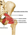
Gluteus Medius
O: Illium between posterior and anterior gluteal lines
I: Greater trochanter
In: Superior Gluteal N.
Fx:
- Hip Abduction
- Hip IR

Gluteus Minimus
O: Illium between posterior and anterior gluteal lines
I: Anteiror boarder of greater trochanter
In: Superior Gluteal N.
Fx:
- Hip Abduction
- Hip IR

Tensor Fascia Lata
O: Anterior iliac crest
I: IT Band, to Gerdy’s Tubercle
In: Superior Gluteal N.
Fx:
- Abduct Hip
- Hip IR
- Assists in Knee Extension

Vastus Lateralis
O: Intertrochanteric Line
I: Quadriceps tendon to patella
In: Femoral N.
Fx: Knee Extension

Vasus Medialis
O: Intertrochanteric Line
I: Quadriceps tendon to patella
In: Femoral N.
Fx: Knee Extension

Vastus Intermedius
O: Superior 2/3 of anterior and lateral surfaces of femur
I: Quadriceps tendon to patella
In: Femoral N.
Fx: Knee Extension

Articularis Genu
O: Anterior surface of distal femur, deep to vastus intermedius
I: Synovial membrane of knee joint
In: Femoral N.
Fx: Pulls suprapatella bursa superiorly during extension to avoid impingement

Biceps Femoris
O:
- Long Head: Ischial Tuberosity
- Short Head: Lateral lip of linea aspera, lateral supracondylar ridge of femur
I: Fibular Head & lateral tibial condyle
In:
- Long: Tibial
- Short: Common Peroneal N.
Fx:
- Knee Flexion
- Hip Extension (long head)
- Tibia ER

Semitendinosus
O: Ischial Tuberosity
I: Anteriomedial Aspect of Tibia (Pes)
In: Tibial Fibres of Sciatic N.
Fx:
- Extend Thigh
- Flex Knee
- Tibia IR

Semimembranosus
O: ischial tuberosity
I:
- Posteiror Medial Tibial Condyle (Deep to MCL)
- Oblique Popliteal Ligament
- Posterior capsule & posterior horn of medial meniscus
- Posterior Oblique Ligament
- Expansion to Aponeurosis of Popliteal Muscle
In: Tibial Fibres of Sciatic N.
Fx:
- Extend Thigh
- Flex Knee
- IR Tibia

Tibialis Anterior
O: Lateral Tibia, IOM
I:
- Medial Cuneiform
- Base 1st Metatarsal
In: Deep Peroneal N.
Fx:
- Ankle Dorsiflexion
- Foot Inversion

Extensor Hallucis Longus (EHL)
O: Medial Fibula, IOM
I: Base of Distal Phalanx, 1st toe
In: Deep Peroneal N.
Fx:
- Ankle Dorsiflexion
- Great Toe Extension

Extensor Digitorum Longus (EDL)
O: Lateral Tibial Condyle, Proximal Fibula
I: Base of Distal Phalanx D2-D5
In: Deep Peroneal
Fx:
- Toe Extension
- Ankle Dorsiflexion

Peroneus Tertius
O: Distal Fibula, IOM
I: Base of 5th Metatarsal
In: Deep Peroneal N.
Fx:
- Ankle Eversion
- Ankle Plantarflexion

Peroneus Longus
O: Proximal Lateral Fibula
I:
- Medial Cuneiform - plantar surface
- Base of 1st Metatarsal - plantar surface
In: Superficial Peroneal N.
Fx:
- Ankle Eversion
- Ankle Plantarflexion
Peroneus Brevis
O: Distal Lateral Fibula
I: Base of 5th Metatarsal
In: Superficial Peroneal N.
Fx: Foot Eversion

Gastrocnemius
O: Posterior Medial and Lateral Condyle of Femur
I: Calcaneus via Achilles
In: Tibial N.
Fx: Ankle Plantarflexion

Soleus
O: Posterior Fibular Head, Soleal Line
I: Calcaneus via Achilles
In: Tibial N.
Fx: Ankle Plantarflexion

Plantaris
O: Lateral Femoral Supracondylar Line
I: Calcaneus
In: Tibial N.
Fx: Ankle Plantarflexion
*runs medial to achilles tendon. Deep to gastrocs, superfiical to soleus

Popliteus
O: Posterior Surface of Tibia (above soleal line)
I: Lateral Condyle, Anterior and Inferior to LCL origin
In: Tibial N.
Fx:
- Unlocks Knee from Extension, Initiates Flexion
- Tibia IR

Flexor Hallicus Longus (FHL)
O: Posterior Fibula
I: Base of Distal Phalanx of Great Toe
In: Tibial N.
Fx: Great Toe Platar Flexion

Flexor Digitorum Longus (FDL)
O: Posterior Tibia
I: Base of Distal Phalanx D2-D5
In: Tibial N.
Fx:
- Toe Plantar Flexion
- Ankle Plantar Flexion

Tibialis Posterior
O: Posterior Tibia, IOM
I: Navicular Tuberosity, Medial Cuneiform
In: Tibial N.
Fx:
- Ankle Inversion
- Ankel Plantar Flexion

Extensor Digitorum Brevis
O: Dorsal Surface Calcaneus
I: Proximal Dorsal Aspect of Middle Phalanx D2-D4
In: Deep Peroneal N.
Fx: Extend Digits 2-4

Extensor Hallucis Brevis
O: Dorsal Calcaneus
I: Proximal Phalanx of D1
In: Deep Peroneal N.
Fx: Extend Hallux

Adductor Hallucis
O: Metatarsals 2-4
I: Proximal Phalanx 1st Toe
In: Lateral Plantar N.
Fx: Adduct 1st Toe

Flexor Hallucis Brevis
O: Cuboid
I: Medial Cuneiform to Proximal Phalanx of 1st Toe
In: Medial Plantar N.
Fx: Great Toe Flexion

Flexor Digitorum Brevis
O: Calcaneus
I: Distal Phalanx D2-D5
In: Medial Plantar N.
Fx: Lesser Toe Flexion

Abductor Halucis
O: Calcaneal Tuberosity
I: Proximal Phalanx 1st Toe
In: Medial Plantar N.
Fx: Great Toe Abduction

Quadratus Plantae
O: Calcaneus
I: FDL Tendon
In: Lateral Plantar N.
Fx: Assist FDL in Flexion of DIP Joint

Abductor Digiti Minimi of the Foot
O: Calcaneal Tuberosity
I: Base of 5th Toe
In: Lateral Plantar N.
Fx: Abduct 5th Toe

Dorsal & Plantar Interossei of the Foot
Dorsal (4) - Abduct (DAB, Bipennate)
Plantar (3) - Adduct (PAD, Unipennate)
O: Metatarsal
I: Proximal Phalanx
In: Lateral Plantar N.
Both in 4th plantar layer with tendon of peroneus longus and tib post.

Lumbricals of the Foot
O: Medial FDL Tendon
I: Medial Proximal Phalanx & EDL Tendon D2-D5
In:
- Lumbrical to 2nd Toe - Medial Plantar N.
- Lumbricals to 3rd-5th Toes - Lateral Plantar N.
Fx: Flex MCP Joints, Extend IP Joints

Levatores Cosatorum
O: Vertebrae - TP C7-T11
I: Ribs inferior to origin
In: Dorsal Rami of thoracic spinal nerves.
Fx: Raise ribs in inspiration

Serratus Posterior Superior
O: Vertebrae - spines of cervical and upper thoracic vertebrae
I: Ribs
In: Intercostal Nerves.
Fx: Raise ribs in inspiration

Serratus Posterior Inferior
O: Verterbae - spines of lower thoraric and upper lumbar
I: Ribs
In: Intercostal Nerves.
Fx: Lower Ribs in Expiration

Spenius
O: T1-T4 spinous process
I:
- Splenius capitus to skull
- Splenic Cervicis to vertebrae
In: Dorsal rami of spinal nerves.
Fx: Extend neck and head (rotate in unilateral action)

Errector Spinae (Illiocostalis, Longissimus, Spinalis)
Illiocostalis
O: Illium and ribs
I: Ribs and transverse procesess
Longissiumus
O: Transverse processes
I: Transverse processes
Spinalis
O: spinous processes
I: spinous processes
In: Dorsal rami of spinal nerves
Fx: Extend trunk and vertebral column

Transversospinalis (Semispinalis, Multifidus, Rotatores)
O: All originate from transverse proceses
I: All insert on spinous process superior to the origin
- Semispinalis extend across 5-6 vertebrae
- Multifidus - extend across 3-4 vertebrae
- Rotatores - extend across 1-2 vertebrae
In: Dorsal rami of spinal nerves
Fx: Extend trunk in bilateral action, rotates vertebral column in unilateral action

