Must know Flashcards

1
Q

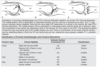
Nonop parameters for metacarpal shaft fractures

A

no rotational deformity
acceptable shaft shortening 2-5m

index/long finger >10 angulation
ring finger <20 angulation
little finger <30 angulation

2
Q

nonop parameters for metacarpal neck fractures

A
  1. Index and middle = <10-15°
  2. Ring = <40°
  3. Small = <60°
  4. No rotation

acceptable shaft shortening 2-5m

3
Q

how should malrotation of a MC fracture be assessed?

A
  • with the fingers in flexion all should point towards the scaphoid tubercle without overlapping adjacent finger (compare to contralateral side)
  • for patients who are unable to perform active flexion, the digital cascade can be observed through the tenodesis effect by flexing and extending the wrist
    • each degree of rotation at the MC results in 5° of rotation at the fingertip, leading to 1.5cm of digital overlap in the closed fist
4
Q

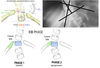
what is the reduction maneuver described for MC neck fractures

A
  • Jahss Maneuver
    • MCP and PIP joints are fully flexed and dorsal force is applied along the long axis of the proximal phalanx and volarly along the MC shaft to reduce the MC head from a flexed position
5
Q

What is acceptable alignment for metacarpal head fractures?

A

No articular displacement acceptable

6
Q

VACTERL

A

The following features are observed with VACTERL association:
V - Vertebral anomalies
A - Anorectal malformations
C - Cardiovascular anomalies
T - Tracheoesophageal fistula
E - Esophageal atresia
R - Renal (Kidney) and/or radial anomalies
L - Limb defects

7
Q

physeal growth plate zones and associated conditions

A

Reserve zone (B)
Gaucher’s
Diastrophic dysplasia
Kneist

Proliferative zone (C)
Achondroplasia
Gigantism
MHE

Hypertrophic zone (D)
Zone of chondrocyte maturation, chondrocyte hypertrophy, and chondrocyte calcification
3 phases: maturation, degenerative, provisional calcification
SCFE (not renal)
Rickets (provisional calcification zone)
Enchondromas
Mucopolysacharide disease
Schmids
Fractures most commonly occur through zone of provisional calcification

primary spongiosa (E)
(metaphysis)
Metaphyseal “corner fracture” in child abuse
Scurvy

secondary spongiosa
(metaphysis)
Metaphyseal “corner fracture” in child abuse
Scurvy

8
Q

most active physes in upper/extremity and lower extremity and mm/y

A

U/E
1. proximal humerus 7mm/y
2. distal radius 5.25mm/y

L/E
1. distal femur 9 mm/y
2. proximal tibia 6mm/y
3. distal tibia 5 mm/y

9
Q

most common causative bacteria in PJI infections of the shoulder

A
  • cutibacterium acnes (38.9%)
    gram-positive,facultative, aerotolerant, anaerobic rod that ferments lactose to propionic acid
    – concentrated in the axilla within the dermal sebaceous glands
    – forms biofilm within 18-90h (found on implant surface and on synovial tissue)&raquo_space; planktonic
    – Mean duration of culture incubation between 7-21 days
  • staph aureus 14.8%
  • staph epidermidis (14.5%)
  • coagulase-negative staph (14%)
10
Q

RF of PJI of shoulder

A
  • male
  • higher BMI
  • younger age
  • immunosuppressed conditions and meds
  • post-truma
  • rTSA
  • previous surgery
11
Q

What is the cause of swan neck deformity & treatment

A

laxity/attenuation of volar plate
characterized by hyperextension of the PIP joint and flexion of the DIP joint due to an imbalance of muscle forces on the PIP.

  • treatment
    • volar plate advancement and PIP balancing with central slip tenotomy
12
Q

what is the cause of boutoniere deformity

A

central slip rupture

13
Q

Goutallier classification

A

0 Normal
1 Some fatty streaks
2 muscle>fat
3 fat = muscle
4 fat>musclemost tear articular sided, less strong

14
Q

RC repair indications

A
  • tear >50% M-L width of supra
  • acute full-thickness
  • bursal sided >3mm/>25% in depth
  • PASTA >7mm of exposed bony footprint w/ >25% healthy bursal sided tissue
  • young pt with acute traumatic tears
  • older pt with degenerative tears
15
Q

when do you do lat dorsi transfer

A

irreparable posterosuperior tears with intact subscap
* young laborer
* radial n + post branch of axillary n. at risk

16
Q

massive RC retear RF

A

increased fatty infiltration,
decreased acromiohumeral space,
smoking,
size of RC tear, and
increase tension on repair

17
Q

RF associated with lower tendon-bone RC healing following repair

A
  • increase age
  • osteoperosis (ind of age)
  • smoker
  • chronic tear
  • large gap
  • large size
  • high tension repair
  • low initial fixation strength
  • fatty infiltration
  • muscle atrophy
18
Q

what are the indications for superior capsular reconstruction?

A
  • massive irreparable supraspinatus and/or infraspinatus tear
  • minimal to no arthritis
  • functioning deltoid
  • not suitable for rTSA (young, active)
19
Q

what tendon transfers can be considered for irreparable RC tear?

A
  • Lat dorsi for posterosuperior tears
  • pec major for irreparable anterosuperior tears
20
Q

Innervation of RC muscles

A
  • supraspinatus
    • suprascapular n.
  • infraspinatus
    • suprascapular n.
  • teres minor
    • posterior branch of axillary n.
  • Subscapularis
    • upper and lower subscap n.
21
Q

what is the rotator crescent

A

thin, crescent-shaped sheet of rotator cuff comprising the distal portions of the supraspinatus and infraspinatus insertions.

22
Q

rotator cable

A

thick bundle of fibers found at the avascular zone of the coracohumeral ligament running perpendicular to the supraspinatous fibers and spanning the insertions of the supra- and infraspinatus tendons.

23
Q

triangular interval

A

3 syllable n.
3 word artery with ‘i’

superior: lower border of teres major
lateral: shaft of humerus
medial: long head of triceps

n: radial
v: profunda brachii artery

24
Q

What are the boundaries of the quadrilateral space? What nerve and vessel run thru the quadrilateral space?

A

superiorly -teres major

Inferiorly - teres major

Laterally - surgical neck

Medially - long head of triceps

Axillary nerve

Posterior circumflex humeral artery

25
Q

What are the borders and the contents of the Triangular Space?

A

3 word artery with ‘s’

Inferior - teres major

Lateral - long head of triceps

Superior - inferior border of teres minor

  • CONTENTS - circumflex scapular artery
26
Q

What is the primary stabilizer to valgus stress at elbow
which band
what is the origin & insertion

A

anterior bundle of medial collateral ligament
central band
origin: median epicondyle
insertion: sublime tubercle
primary statis stabilizer to valgus stress from 20-120 degrees of elbow flexion

27
Q

superficial flexors of forearm and innervation and origin

A

PT, FCR, PL, FDS, FCU
all inervated by median n. except for FCU, supplied by the ulnar n.
all originate from common flexor origin on medial epicondyle
FDS also originates on anterior radius

28
Q

deep flexors of forearm, innervation

A

FDP, FPL, PQ
all innervated by AIN
FDP has dual innervation (ulnar haf by ulnar n., radial half by AIN)

29
Q

muscle of mobile wad and origin

A

brachioradialis, ECRL, ECRB
* The lateral supracondylar ridge of the humerus serves as an attachment point for the brachioradialis and ECRL, which also has attachments onto the superior aspect of the lateral epicondyle
* ECRB originates on the lateral epicondyle
* AllinnervatedbytheRadialn.

30
Q

superficial extensors of forearm and origin

A

anconeus, EDC, EDM, ECU
all innervated by PIN except for anconeus, supplied by radial n.
ECU not innervated by ulnar n.

31
Q

deep extensors of forearm

A
  • Supinator
  • Abductor Pollicis Longus
  • Extensor Pollicis Brevis
  • Extensor Pollicis Longus
  • Extensor indicis proprius

all innervated by PIN

32
Q

Sites of compression of the ulnar nerve at the cubital tunnel

A

Arcade of struthers
Medial intermuscular septum
Medial epicondyle
Osborne’s ligament
Between heads of FCU (fasica)
Aponeurosis of FDS

33
Q

which ligament in the elbow is the primary stabilizer to varus and ER stress

A

LUCL
origin lateral humeral epicondyle
insertion tubercle of supinator crest of ulnar

34
Q

Varus Posteromedial Rotatory Instability (VPMRI) vs.Valgus Posterolateral Rotatory Instabiliy (VPLRI)
1. radial head
2. coronoid fracture
3. MCL
4. LCL
5. P/E

A
  • VPMRI
  • No radial head fracture
  • > 15% (anteromedial facet)
  • Posterior band of MCL ruptured, anterior band intact (attached to anteromedial facet)
  • LCL complex (includes LUCL) avulsion
  • Valgus stress, moving valgus, milking maneuver
  • VPLRI
  • Radial head fracture
  • < 15% (coronoid tip)
  • Anterior band of MCL ruptured
  • LCL complex (includes LUCL) avulsion
  • Varus stress, chair rise, lateral pivot shift
35
Q

approach for LUCL reconstruction

A

kocher approach

36
Q

how to cast simple elbow dislocation post reduction

A

likely LUCL injury if any lig (most common)
cast in pronation
‘thumb away from injured side’

37
Q

mechanism of injury of LUCL resulting in PLRI

A

FOOSH with axial compression, in supination and valgus

38
Q

primary ligament injured that leads to PLRI

A

LUCL

39
Q

P/E for PLRI

A
  • lateral pivot-shift test
    • patient lies supine with affected arm overhead; forearm is supinated and valgus stress is applied while bringing the elbow from full extension to 40 degrees of flexion
    • with increased flexion, triceps tension reduces the radial head
    • often more reliable on anesthetized patient
  • posterior drawer test
    • patient lies supine with affected arm overhead; forearm is supinated and the examiner’s index finger is placed under the radial head and the thumb over it.
    • application of a posterior force will cause posterior subluxation of the radial head
  • prone push up
  • chair push up
  • table top relocation test
40
Q

Kocher approach
interval
indications
advantages
disadvantages

A

interval between the anconeus and extensor carpi ulnaris

indications: radial head #, excision, prosthesis, LUCL recon
advantages: low risk of PIN injury
disadvantage: distal extension may endager PIN
relatively more posterior and thus risks injuring the lateral collateral ligament complex.

41
Q

kaplan approach
interval
indications
advantages
disadvantages

A

interval between the extensor carpi radialis brevis and extensor digitorum communis

Indications Radial head fractures – fixation, excision and prosthesis.
Advantages Good view of the anterior half of the radial head which is a common site of fracture.
No disruption of the LUCL.
Drawbacks Inadvertent injury to the PIN if the incision is too anterior.
Distal extension can endanger the PIN.

Remember: If there is a supinator crest of ulna fracture or LUCL avulsion as in 5% of terrible triad injuries, this approach should not be used.

42
Q

EDC split approach
interval
advantages

A

EDC tendon is dissected directly longitudinally starting at its origin at the lateral epicondyle

The approach offers a slightly more anterior access than the Kocher’s approach.

43
Q

Hotchkiss approach (over-the-top)
interval
indications
advantages
disadvantages

A

Interval: splits the flexor-pronator mass and elevates the anterior part (pronator teres (PT), flexor carpi radialis (FCR), and palmaris longus (PL)) along with brachialis from the anterior elbow capsule
Indications The Hotchkiss or ‘over-the-top approach’ is the most anterior of the medial approaches and provides good access to the tip of the coronoid process and the anterior elbow joint.
Advantages Good view of the coronoid tip fracture site.
Drawbacks Ulnar nerve exploration and visualisation is required.
Possible injury to medial antebrachial cutaneous nerve.

44
Q

Flexor carpi ulnaris (FCU) splitting (Ring) approach
interval
indications
advantages
disadvantages

A

interval: between the heads of the flexor carpi ulnaris
Indications Coronoid fractures.
Advantages Good view of the fracture site especially the anteromedial facet.
Drawbacks Ulnar nerve exploration is required.
Possible injury to medial antebrachial cutaneous nerve.

45
Q

Taylor and Scham approach
approach
interval
indications
advantages
disadvantages

A

Interval - For access to the base consider elevating the entire flexor-pronator mass from posterior to anterior
Indications This is a good approach for a medial plate fixation for a large basilar fracture of the coronoid.
Advantages Good view of the fracture site.
Drawbacks Ulnar nerve exploration is required.
Possible injury to medial antebrachial cutaneous nerve.

46
Q

Which muscle shares the same origin site as the tendon that undergoes angiofibroplastic hyperplasia during the pathogenesis of tennis elbow?

A

anconeus

Lateral epidondylitis is classically thought to be caused by histopathologic angiofibroblastic hyperplasia at the origin of the extensor carpi radialis brevis. ECRB originates from the common extensor wad, that also includes ECRL, ED, ECU. The anconeus shares the same attachment site at the lateral epicondyle as the ECRB

47
Q

which extensor muscle of the forearm originates on lateral supracondylar ridge

A

ECRL

48
Q

what tendon is most commonly implicated in lateral epicondylitis

A

ECRB

49
Q
  • what is the underlying histopathology in lateral epicondylitis
  • what are the histological findings in lateral epicondylitis
A
  • tendinosis (degenerative condition) rather than tendinitis (inflammation)
    • angiofibroblastic hyperplasia
    • characterized by dense populations of fibroblasts, vacular hyperplasia and disorganized collagen
50
Q

how to avoid iatrogenic injury to LUCL with lateral epicondylitis release

A

do not extend beyond equator of radial head
may lead to PLRI

51
Q

what is valgus extension overload

A
  • describes collection of injuries in the medial, lateral and posterior aspects of the overhead thrower’s elbow
  • occurs as a consequence of the large valgus loads and rapid elbow extension during the throwing motion
52
Q

what are the common presenting symptoms of valgus extension overload in over head thrower’s

A
  1. decreased performance (velocity, control, fatigue)
  2. posteromedial elbow pain with full extension of elbow
  3. locking/catching (loose bodies), loss of terminal elbow extension
  4. ulnar nerve symptoms (neuritis, subluxation)
  5. pain in deceleration of throwing phase
53
Q

complication of surgical procedure for valgus extension overload syndrome

A

valgus instability - over-resection of posteromedial ostephy past its native margin or >3mm may lead to increased stress on the MCL and valgus instability

54
Q

what are the components of the lateral collateral ligament of the elbow

A
  1. lateral ulnar collateral ligament
    • inserts on the supinator crest
  2. radial collateral liagement
    • inserts on the annular ligament
  3. annular ligament
    • inserts on the supinator crest
  4. accessory lateral collateral ligament
    • inserts on the supinator crest
55
Q

what are the components of medial ulnar collateral ligaments of the elbow and which is the most important for valgus stability

A
  • anterior bundle
    • strongest and most significant stabilizer to valgus stress
    • insert - sublime tubercle
    • provides stability between 30-120° flexion
    • subdivides into anterior and posterior bands
      • anterior band - 1° restraint to valgus stress, exhibiting nearly isometric strain during elbow ROM
  • posterior bundle
    • greatest change in tension from flexion to extension
    • tighter in flexion
  • transverse bundle
56
Q

what is the most sensitive test for diagnosis of medial elbow instability

A

moving valgus test (100% sensitive, 75% specific)

57
Q

What are preventative strategies to avoid UCL injuries in pitchers?

A
  1. Limit pitching to 100 innings in a calendar year
  2. Do not pitch for multiple teams
  3. Do not pitch all year (3 month rest period advised)
  4. No pitching on consecutive days
  5. No pitching in a game or practice after being removed from a game
  6. No breaking pitches (curveballs/sliders) until puberty
  7. Proper pitching mechanics and year round conditioning should be stressed
  8. Avoid pitching while fatigued
58
Q

where do partial distal biceps tendon tears primarily occur

A

on radial side of tubersosity footprint

59
Q

RF for biceps tendon tear

A

high BMI, smoking, anabolic steroids

60
Q

contents of antecubital fossa

A

medial) median nerve, brachial a., biceps tendon, radial n. (lateral)

61
Q

when is LABC most at risk during surgery for biceps tendon repair

A

during surgical disseciton btw biceps and brachialis

62
Q

distal biceps anatomy and insertion

A
  1. The tendon externally rotates 90°
    • Brings the medial short head fibres anterior and the lateral long head fibres posterior
  2. The tendon inserts with:
    • Short head fibres distal on the radial tuberosity
      • Stronger flexor
    • Long head fibres proximal
      • Stronger supinator
63
Q

What are the reported deficits with nonoperative management of complete distal biceps tears?

A

Supination

  • 79% endurance [50% loss]
  • 21-55% strength [40% loss]

Flexion

  • 10-40% strength [30% loss]
  • 30% endurance
64
Q

intervals for single incision technique for biceps repair

A

proxial - BR and brachialis
distal - BR and PT

65
Q

intervals for single incision technique for biceps repair, how do you protect PIN

A

Forearm held in supination to protect the PIN and bring the radial tuberosity into view

66
Q

If L4/L5 paracentral (posterolateral) disc herniation, what nerve is affected

A

affects traversing/descending/lower nerve root at L4/5 - affects L5 nerve root
most common PLL is weakest here

67
Q

If L4/L5 foraminal disc herniation, what nerve is affected

A

less common (5-10%)
affects exiting/upper nerve root
at L4/5 affects L4 nerve root

68
Q
  • What are the sites of entrapment of the median nerve?
A

Pronator syndrome (SLAPS)
* **Struther’s ligament **Resisted Flexion at 120
* **Lacertus fibrosis **resisted flexion with supinated forearm
* **Arch of FDS **(sublimus arch) resisted FDS middle finger
* Pronator Teres (2 heads) resisted supination with elbow flexed

69
Q
  • What is the motor innervation of the median nerve?
A
  1. Motor
    Order:
    - Forearm
    - Pronator teres
    - FCR
    - Palmaris longus
    - FDS
         - Radial FDP [AIN]
             - Index and middle finger
         - FPL [AIN]
         - Pronator quadratus [AIN]
     - Hand
         - APB
         - FDB (superficial head)
         - OP
         - Lumbricals 1 & 2
70
Q

what is the sensory innervation of median nerve

A
  1. Sensory
    1. volar wrist capsule, radial palm and palmar aspect of thumb, index, long and radial half of ring
      1. terminal AIN
      2. palmar cutaneous branch of median nerve
      3. terminal digital cutaneus branch
71
Q

median nerve course

A
  • Lies medial to brachial a. at the elbow
  • Passes between the two heads of the pronator
    teres and then runs between FDS and FDP
72
Q

Carpal tunnel borders and contents

A
  • Carpal tunnel borders
    • scaphoid tubercle and trapezium radially
    • hook of hamate and pisiform ulnarly
    • transverse carpal ligament palmarly (roof)
    • proximal carpal row dorsally (floor)
  • Carpal tunnel contents
    • four flexor digitorum superficialis (FDS) tendons
    • four flexor digitorum profundus (FDP) tendons
    • flexor pollicis longus (FPL)
      • most radial structure
    • median nerve
73
Q

what is Parsonage-Turner Syndrome

A
  • bilateral AIN signs caused by viral brachial neuritis
  • be suspicious if motor loss is preceded byintense shoulder pain and viral prodrome
74
Q

AIN motor innervation

A
  • FDP (index and middle finger)
  • FPL
  • pronator quadratus
75
Q

pronator vs AIN syndrome

A

AIN Syndrome
* Forearm pain
* Weakness of grip and pinch - FPL, FDP 1&2, PQ
* * unable to make OK sign
* No sensory disturbance

Pronator Syndrome
* Pain in proximal, volar forearm
* Paresthesias in thumb, index, long fingers
* AIN Syndrome PLUS weakness of all digits & wrist flexion
* Exacerbated by resisted pronation
* Tinel’s at anterior elbow

76
Q

AIN sites of compression

A
  • potential sites of entrapment of AIN
    • tendinous edge of deep head of** pronator teres**
      • most common cause
        ** - fibrous arch of the FDS**
        ** - aberrant vessels**
        ** - accessory head of FPL(Gantzer’s muscle)**
    • accessory muscle from FDS to FDP
    • abberant muscles (FCRB, palmaris profundus)
77
Q

how is pronator syndrome different to CTS

A
  • should havecharacteristics differentiating from carpal tunnel syndrome (CTS)
    • aching painover proximal volar forearm
    • sensory disturbances over thedistribution of palmar cutaneous branch of the median nerve(palm of hand)
      • arises 4 to 5 cm proximal to carpal tunnel
    • lack of night symptoms
78
Q

how is cubital tunnel different than ulnar tunnel

A
  • less clawing
  • sensory deficit to dorsum of the hand
  • motor deficit to ulnar-innervated extrinsic muscles
  • Tinel sign at the elbow
  • positive elbow flexion test
79
Q

boundaries of guyon’s canal
what are the contents

A

floor:Transverse carpal ligament, hypothenar muscles
roof: Volar carpal ligament
ulnar border: Pisiform and pisohamate ligament, abductor digiti minimi muscle
radial border: Hook of hamate

ulnar nerve, a/v

80
Q

zones of guyon’s canal

A
  • zone I - proximal to bifurcation (motor and sensory)
  • zone II - deep motor branch (motor only)
  • zone III - superifical sensory branch (sensory only)
81
Q

ulnar nerve motor and sensory innervation

A
  1. Motor
    • FCU
    • Ulnar half of FDP
    • All intrinsics except lateral 2 lumbricals and thenar muscles
      • Except deep head of FPB
  2. Sensory
    • Elbow joint
    • Ulnar palm and dorsum of hand including small finger and ulnar half of ring finger
    • Branches:
      • Articular branch of the elbow
      • Dorsal cutaneous branch
      • Palmar cutaneous branch
        • Only present in 58% of people
      • Terminal superficial branch
82
Q

ulnar nerve sites of compression

A
  1. Medial intermuscular septum
  2. Arcade of Struthers
  3. Triceps fascia
  4. Osborne ligament** (most common site)**
    • Roof of cubital tunnel
  5. Medial epicondyle
  6. FCU
    • Deep fascia
    • Between two heads
  7. FDS/FDP fascia
  8. Anconeus epitrochlearis
    • Anomalous muscle which arises from medial border of olecranon & adjacent triceps & inserts into the medial epicondyle
  9. Canal of Guyon
83
Q

what is ulnar tunnel syndrome characterized by

A
  • numbness/paresthesia of the small and ulnar half of ring finger (ulnar palm and dorsum are spared)
  • intrinsic muscle weakness (thenar spared)
84
Q

site of compression of radial n.

A

FREAS (radial nerve)
* Fascia superficial to radiocapitallar joint
* Radial Recurrent Vessels (Leash of Henry)
* ECRB tendinous proximal margin at origin
* Arcade of Frohse (prox aponeurotic/tendinous edge of sup, most common site of PIN entrapment)
* Supinator (distal edge of)

85
Q

last muscle innervated by radial n.

A

EIP

86
Q

radial tunnel borders

A
  • Borders:
    • Proximal border – begins at radiocapitellar joint
    • Distal border – arcade of Froshe
    • Roof – BR
    • Medial border – brachialis and biceps tendon
    • Lateral border – ECRB, ECRL, BR
87
Q

characteristic features of radial tunnel syndrome

A
  • Pain at lateral forearm distal to lateral epicondyle
  • Lack motor and sensory changes
88
Q

motor and sensory innervation of PIN

A

motor
common extensors
* ECRB (often from radial nerve proper, but can be from PIN)
* Extensor digitorum communis (EDC)
* Extensor digiti minimi (EDM)
* Extensor carpi ulnaris (ECU)
deep extensors
* Supinator
* Abductor pollicis longus (APL)
* Extensor pollicus brevis (EPB)
* Extensor pollicus longus (EPL)
* Extensor indicis proprius (EIP)
sensory
* sensory fibers to dorsal wrist capsule - provided by terminal branch which is located on the floor of the 4th extensor compartment
* no cutaneous innervation

89
Q

motor innervation of radial n. proper

A

triceps brachii, anconeus, brachioradialis, extensor carpi radialis longus, brachialis

90
Q

provocative test for radial tunnel syndrome

A
  • Provocative maneouvers
    • Pressure over supinator muscle in supinated position
    • Pain with resisted wrist or long finger extension
91
Q

potential sites of PIN compression

A
  • fibrous tissue anterior to the radiocapitellar joint
  • between the brachialis and brachioradialis
  • “leash of Henry”
  • are recurrent radial vessels that fan out across the PIN at the level of the radial neck
  • extensor carpi radialis brevis edge
  • medio-proximal edge of the extensor carpi radialis brevis
  • “arcade of Fröhse”
  • which is the proximal edge of the superficial portion of the supinator
  • supinator muscle edge
  • distal edge of the supinator muscle
92
Q

characteristics and provocative maneovers of PIN compression syndrome

A
  • Weakness of PIN innervated muscles
    • Sparing of BR, ECRL
  • Pain in dorsal radial forearm
  • no sensory changes
  • Wrist extension demonstrates radial deviation
    • Intact ECRL
  • resisted supination
    • will increase pain symptoms
93
Q

what is wartenburg syndrome

A
  1. Compression of the superficial radial nerve ~9cm proximal to radial styloid where the nerve passes between BR and ECRL
  2. Characterized by:
    - Dorsal radial forearm pain radiating to dorsoradial hand
    - more common inwomen
    - SRN compressed by scissoring action ofbrachioradialisandECRL tendonsduringforearm pronation
    - provocative test: wrist flexion, ulnar deviation and pronation for one minute
    • De Quervain’s tenosynovitis: pain is not aggravated by wrist pronation,
94
Q

which ligament in the hand is not involved in dupuytrens disease

A

cleland’s ligament (c for feiling
- dorsal to digital nerve

95
Q

which ligaments are critical to prevent bowstring

A

A2 and A4

96
Q

What are the extensor compartments of the wrist and associated pathology?

A
  1. EPB + APL = De Quervain’s tenosynovitis
  2. ECRB + ECRL = intersection syndrome
  3. EPL = drummer’s wrist, traumatic rupture with DR #
  4. EDC + extensor indicis = extensor tenosynovitis
  5. EDM = Vaughn-Jackson syndrome
  6. ECU = snapping ECU
97
Q

what is the disi deformity and findings on xray

A
  • DISI (dorsal intercalated segmental instability)
    • Division of the dorsal component of the SLIL results in gradual collapse of the scaphoid into flexion, while the lunate is pulled into extension to form the dorsal intercalated segment instability pattern
    • Diagnosis of DISI deformity can be made with lateral wrist radiographs showing
      • a scapholunate angle > 70 degrees. (N 30-60)
      • DISI defined by radiolunate angle >15°
98
Q

signs on xrays of SL ligament injury

A
  1. Widened SL distance
    1. gap >3mm with clenched fist view (terry thomas sign)
  2. Cortical ring sign
    1. caused by schaphoid malalignment
  3. Shortening of the scaphoid
  4. Scapholunate angle >70° (normal = 30-60)
  5. Lunate extension
  6. DISI defined by radiolunate angle >15°
99
Q

which SL ligament component is the strongest

A

dorsal

100
Q

most common pattern of wrist arthritis

A

SLAC 55%

101
Q

what is SLAC wrist and radiographic classification

A
  • scapholunate advanced collapse
    • describes a pattern of wrist arthritis that occurs following a scapholunate ligament injury

Watson Classification
Stage I - radial styloid
Stage II - radioscaphoid joint
Stage III - capitolunate joint
[Stage IV - pancarpal involvment (controversial)]

102
Q

Treatment of SLAC

A
  • Stage I - scaphoid and radial styloid arthritis
    • radial styloidectomy
    • PIN and AIN denervation
  • Stage II - scaphoid/scaphoid facet of radius arthritis
    • proximal row carpectomy
      • contraindicated with caputolunate arthritis because capitate articulates with lunate fossa of the distal radius
      • contraindicated if there is an incompetent radioscaphocapitate ligament
      • advantages - greater postop ROM (flexion, extension, total flexion/extension arc), lower complication rate
      • others - earlier ROM no hardware
    • scaphoid excision and 4 corner fusion
    • advantages - greater radial deviation ROM, greater grip strength
  • Stage III - pan carpal arthritis
    • scaphoid excision and 4 corner fusion
    • wrist fusion
      • wrist fusion gives best pain relief and good grip strength at the cost of wrist motion
103
Q

what is the most common complication following PRC

A

synovitis and significant edema

104
Q

major blood supply of scaphoid

A
  • major blood supply isdorsal carpal branch (branch of the radial artery)
    • enters scaphoid in a nonarticular ridge on the dorsal surface and supplies proximal80% of scaphoidvia retrograde blood flow
  • supplies proximal80% of scaphoidvia retrograde blood flow
  • minor blood supply fromsuperficial palmar arch (branch of volar radial artery)
    • entersdistal tubercle andsupplies distal 20% of scaphoid
105
Q

pathomechanics of SNAC wrist development?

A
  1. In the normal wrist:
    • Scaphoid links the proximal and distal carpal rows
      • Proximal row moves with the scaphoid
    • Scaphoid has a tendency to assume a flexed posture
    • Capitate longitudinal load on the lunate is eccentric causing the lunate and triquetrum to extend
    • These forces are balanced as long as the link between the scaphoid and lunate are intact
  2. With scaphoid nonunion
    • The distal scaphoid flexes
    • The proximal scaphoid extends with the lunate and triquetrum
106
Q

radiographic stages of SNAC wrist

A
  • Stage I - radial styloid
  • Stage II - proximal scaphocapitate joint
  • Stage III - capitolunate joint
    • Initially, degeneration occurs between the radial styloid and radial side of the distal scaphoid fragment
    • Degeneration does not progress proximally in the radioscaphoid joint because the proximal scaphoid relationship with the lunate is maintained
    • Degeneration progresses to the midcarpal joint starting with the proximal scaphocapitate joint then the capitolunate joint
107
Q

which MC is the plate for wrist arthrodesis placed

A

3rd metacarpal shaft

108
Q

optimal position of wrist fusion

A

10-15° of extension, slight ulnar deviation

109
Q

What are the ulnar-sided procedures that can be considered in context of distal radius malunion?

A
  1. Hemiresection-interposition
  2. Ulnar shortening osteotomy
  3. ‘Wafer’ resection
  4. Sauve-Kapandji
    • DRUJ fusion with proximal pseudoarthrosis
  5. Darrach
    • Complete distal ulna resection
  6. Arthroplasty
110
Q

describe humpback deformity of scaphoid

A

humpback deformity (distal pole flexes over the volar radioscaphocapitate ligament)

results from volar angulation of the proximal and distal poles of the scaphoid in the setting of scaphoid fracturethrough the waist

The dorsal component forms a ‘humpback’ which can be palpated

111
Q

What is the treatment algorithm for scaphoid nonunion based on specific fracture characteristics?

    1. Delayed union (<6 months)
    2. Established nonunion without humpback
    3. Nonunion with humpback deformity; no AVN
    4. AVN without humpback deformity
    5. AVN with humpback deformity
A
  1. Delayed union (<6 months)
    - ORIF with headless compression screw
    1. Established nonunion without humpback
      • ORIF with headless compression screw + bone graft (cancellous ICBG or distal radius)
    2. Nonunion with humpback deformity; no AVN
      • ORIF via volar approach + corticocancellous bone graft
    3. AVN without humpback deformity
      • Vascularized bone graft via volar or dorsal approach
    4. AVN with humpback deformity
      • Vascularized medial femoral condyle bone graft via volar approach
112
Q

Classification of Kienbock disease

A
  • Lichtman Classification
    • Stage I
      • Normal xray
      • MRI decreased signal T1
      • Bone scan positive
    • Stage II
      • Lunate sclerosis
    • Stage IIIa
      • Lunate collapse (no scaphoid rotation)
      • Carpal height maintained
    • Stage IIIb
      • Lunate collapse
      • Carpal collapse
      • Scaphoid rotation (hyperflexed, RS angle >60°)
        • Cortical ring sign
        • Capitate migrates proximal
        • Decreased carpal height
    • Stage IV
      • Pancarpal arthritis (Kienbock’s disease advanced collapse)
113
Q

treatment options for Kienbock’s disease based on stage of disease?

A
  • Stage I (normal xray, mri decreased signal t1)
    • Nonoperative (3 months immobilization)
  • Stage II (lunate sclerosis)
    • Radial shortening osteotomy
      • If ulnar negative or neutral
    • Capitate shortening
      • If ulnar positive
    • (vascularized bone graft)
  • Stage IIIa (lunate collapse)
    • Same as Stage II
    • Vascularized bone graft (dorsal pedicle)
      • 4,5 ECA graft
      • Vascularized pisiform
      • Free vascularized medial femoral condyle
  • Stage IIIb (lunate/capitate collapse and scaphoid rotation)
    • Scaphocapitate fusion
    • STT fusion
    • PRC
  • Stage IV (pancarpal arthritis)
    • PRC
    • Total wrist arthrodesis
114
Q

What vascularized bone graft would you use for kienbock disease

A

dorsal 4 + 5 extensor compartment artery (ECA) pedicled VBG

115
Q

What are surgical management options for DRUJ arthritis

A
  • what are surgical management options for DRUJ arthritis
    • darrach procedure
      • indications
        • preferred for low demand and nonreconstructable joint
      • technique
        • subperiosteal distal ulna exposure
        • distal ulna resection just proximal to sigmoid notch
        • preserve soft tissue
          • TFCC, ECU sheath, periosteum
    • hemiresection
      • indications -required intact TFCC
      • technique
        • classic -resection of articular distal ulna with remainder left insitu including TFCC attachment
        • hemi-resection interposition technique (HIT)
          • resection as classic
          • soft tissue interposition into void to prevent radioulnar convergence (capsular flap or free tendon)
    • sauve-kapandji procedure
      • indications - preferred for young, active patient with nonreconstructable joint
      • technique
        • dorsal or ulnar approach preserving soft tissue
        • identify and protect the dorsal cutaneous branch of the ulnar nerve
        • ulnar neck resection just proximal to sigmoid (~10-15mm)
        • sigmoid notch and ulnar head prepared for fusion (Cancellous bone)
        • DRUJ fusion with 2 k-wires or 3.5mm screw (neutral ulnar variance)
        • pronator quadratus interposed in osteotomy site (prevents re-ossificaiton)
        • FCU slip can be tenodesed through drill hole in ulnar stump to prevent instability
    • partial ulnar head arthroplasty
      • indication - isolated DRUJ arthritis without instability
        • failed HIT
    • total ulnar head arthroplasty
      • indications - painful instability after failed resection, isolated instability
      • requires stability from native soft tissues
    • total DRUJ arthroplasty
      • indications - incompetent native soft tissues, salvage option after failed distal ulnar resection
116
Q

which tendon could inhibit reduction of DRUJ

A

ECU

117
Q

stabilizing structures of DRUJ (8)

A
  1. bone contour (sigmoid notch of radius and ulnar head)
  2. TFCC
  3. ulnocarpal ligament complex
  4. ECU
  5. ECU tendon sheath
  6. pronator quadratus
  7. interosseous membrane
  8. DRUJ joint capsule
118
Q

management options for DRUJ instability

A
  • nonop
    • acute dislocation
      • closed reduction and splinting in stable position for 6 weeks
        • dorsal radioulnar ligament injury - splint midsupination
        • volar radioulnar dislocaiton - splint midpronation
  • operative
    • acute DRUJ instability indications
      • irreducible
        • open reduction +/- DRUJ pinning +/- TFCC repair +/- ulnar styloid fracture fixation
      • associated fractures
        • ORIF of associated fractures often resolves the instability
        • if remains unstable pin in reduced position
      • TFCC tear
        • open or arthroscopic repair
          • open - dorsal interval between 5 & 5 compartment, TFCC repaired to distal ulnar with anchor or suture tunnels
        • reconstruction if repair fails
    • chronic DRUJ instability
      • in absence of arthritis
        • distal radius malunion
          • indications for correction = >20° of dorsal angulation (controversial)
          • correct distal radius malunion then assess DRUJ stability
            • if still unstable reconstruct the DRUJ
        • reconstruction
          • indications - TFCC or radioulnar ligament repair failure, unrepairable
          • Adams procedure +/- notchplasty (if flat lesser sigmoid)
            • dorsal approach between 5-6 compartments and PL graft radius and ulna
        • bain procedure
          • indication
            • chronic DRUJ instability with a TFCC foveal tear and stable radial attachment
            • positive arthroscopic hook and trampoline test
          • technique
            • dorsal approach via 5th extensor compartment, PL graft in TFCC and ulna
      • in presence of arthritis
        • darrach with ulnar stump stabilization
        • sauve-kapandji
119
Q

xrays to assess for ulnocarpal abutment syndrome

A
  • recommended views
    • AP radiograph with wrist in neutral supination/pronation and zero rotation
      • required to evaluate ulnar variance
    • pronated grip view
      • increases radiographic impaction
  • findings
    • ulna positive variance
    • sclerosis of lunateand ulnar head
120
Q

surgical management of ulnocarpal abutment syndrome

A
  • Ulnar shortening osteotomy
    • Technique
      • Subcutaneous approach to the ulna
      • Osteotomy at junction of distal and middle 1/3
      • Compression plate
        • Volar surface preferred
      • Goal of 0 to -1mm ulnar variance
    • Advantages
      • Addresses ulnar styloid carpal impaction concomitantly
      • Decreases dorsal subluxation of distal ulna
      • Larger shortening can be achieved compared to wafer
      • Stabilizes ulnar ligament complex
        • Preferred if associated LT ligament injury
    • Disadvantages
      • Nonunion
      • Hardware irritation
  • Wafer procedure
    • Technique
      • Open or arthroscopic
      • Resection of thin wafer of dome of ulnar head
    • Advantage
      • Less revision compared to shortening osteotomy (hardware removal)
      • No nonunion
    • Disadvantage
      • Limit resection to 2-3mm
      • Does not address associated ulnar styloid carpal impaction
      • Does not improve dorsal ulnar subluxation
      • Does not tighten ulnar ligament complex
121
Q

What are the components of TFCC

A
  1. Articular disc
    * Extends between the volar and dorsal radioulnar ligaments (hammock)
  2. Meniscus homologue
  3. Volar and dorsal radioulnar ligaments
    * Superficial and deep (ligamentum subcruentum)
    * Major stabilizers of the DRUJ
  4. Sheath of ECU
  5. Ulnar capsule (ulnar collateral ligament)
    * Arises from the ulnar styloid and extends between the ulnotriquetral ligament and the ECU sheath
  6. Ulnolunate and ulnotriquetral ligaments (volar)
122
Q

P/E Findings for TFCC

A
  1. Prominent ulna
  2. Fovea sign
    • Palpation of the depression volar between ulnar styloid, FCU and pisiform
    • Tenderness suggests:
      • Tear of ulnotriquetral ligament
      • Foveal disruption of TFCC
      • Chondromalacia of ulnar aspect lunate
        • Suggestive of ulnocarpal impaction
  3. Ulnocarpal stress test
    • Ulnar deviation with axial loading in alternating supination and pronation
  4. Positive grind test
    • Clicking, crepitus or pain with passive supination and pronation
  5. Lunotriquetral shuck test
    • Pain and laxity when examiner grasps the pisiform/triquetrum and lunate with opposite hands and translates volar and dorsal
123
Q

common mechanism for TFCC tear

A

extended wrist with forearm pronation

124
Q

Treatment of TFCC tears

A
  1. Nonoperative
    • Most tears are initially treated nonoperative
  2. Operative
    • Contraindications:
      • Severe OA
      • Previous infection
      • Severe osteoporosis of ulnar head
    • Open
      • Indicated when fixing distal radius fracture or surgeon not familiar with arthroscopy
    • Arthroscopic
      • Palmar 1A – debridement
      • Palmer 1B, C, D – repair
        • Transosseous or suture anchor fixation
    • Ulnar positive wrists
      • Perform ulnar shortening osteotomy or wafer procedure at time of TFCC repair
        • Better outcomes
125
Q

What are the components of the UCL and RCL ligaments in thumb

[JAAOS 2011;19:287-296]

A
  1. Proper collateral ligament
    * Taut in flexion
  2. Accessory collateral ligament and volar plate
    * Taut in extension
126
Q

Where is the typical thumb UCL and RCL ligament injured?

[JAAOS 2011;19:287-296]

A
  1. UCL
  • Proximal phalanx avulsion (90%)
  • Midsubstance and metacarpal avulsion less common
  1. RCL
    * Variable, more common proximal at metacarpal
127
Q

What is a Stener lesion?

[JAAOS 2011;19:287-296]

A
  1. Distal edge of the avulsed UCL is displaced proximal to the adductor aponeurosis
    * Blocked from reapproximation to its insertion on the proximal phalanx
  2. Surgical management is recommended
128
Q

How do you grade collateral ligament tears?

[JAAOS 2011;19:287-296]

A

Grade 1

  • Sprain with no joint instability

Grade 2

  • Incomplete tear with asymmetric joint laxity
  • Instability does not meet the criteria for a complete tear
    • Characterized by increased laxity with a firm end point, or an incomplete tear

Grade 3

  • Complete tear with joint instability
    • Laxity of >35° in 0° and 30° of flexion
      • Or 15° greater than that of the contralateral side
129
Q

Which injury, UCL or RCL, is more prone to joint subluxation of the MCP joint?

[JAAOS 2011;19:287-296]

A
  1. RCL
    * Adductor pollicis inserts on the proximal phalanx and ulnar sesamoid creating an ulnar and volar deforming force
  2. Assess with anterior drawer test
    * >3mm displacement volar is more common with RCL injury
130
Q

What is the recommended management of thumb collateral ligament injuries based on grade of injury?

[JAAOS 2011;19:287-296]

A

Grade 1 and 2

  • Nonoperative
    • Cast or splint x 4 weeks
    • Grip and pinch strengthening after 6 weeks

Grade 3

  • Surgical
    • Acute collateral ligament injury = repair
      • Avulsion from proximal or distal insertion
        • Suture anchor repair
      • Midsubstance tear
        • Direct repair
    • Chronic collateral ligament injury (>3 weeks)
      • No arthritis
        • Ligament reconstruction with free graft (palmaris longus)
      • Arthritis = arthrodesis
131
Q

msot common site for arthritis in the hand

A

DIP arthritis > CMC arthritis

132
Q

classification for CMC arthritis

A

Eaton and Littler Classification of Basilar Thumb Arthritis

  • Stage I
    • Normal joint with widening
      • Synovitis, effusion, ligamentous laxity
  • Stage II
    • Mild joint narrowing
    • Mild subchondral sclerosis
    • Subchondral cysts and/or periarticular debris
  • Stage III
    • Severe joint space narrowing
    • Subchondral sclerosis/cysts
    • Larger periarticular debris
  • Stage IV
    • Involves scaphotrapezial joint
133
Q

What is the treatment of thumb basal joint arthritis?

A
  1. Nonoperative
    • Exercise therapy, heat, education, magnetotherapy, adaptive equipment, orthoses, NSAIDs, corticosteroid injections
  2. Operative
    • Joint preserving options described (Stage I/II)
      • 1st metacarpal extension osteotomy
      • Arthroscopy and debridement
      • Imbrication of the dorsoradial capsule
      • Volar beak ligament reconstruction
    • Joint sacrificing options described (Stage III/IV)
      • Trapeziectomy
      • Trapeziectomy and ligament reconstruction and tendon interposition (LRTI)
      • Distraction hematoma arthroplasty
      • Suture suspension arthroplasty
      • Prosthesis
      • CMC arthrodesis
134
Q

What are risk factors for thumb basal joint arthritis?

A
  1. Advanced age
    • 36% >80y vs. 6.6% 40-49y
  2. Female
  3. Ligamentous laxity/ higher Beighton score
  4. Occupations
    • Repetitive finger use or heavy manual labour
  5. Post-traumatic
    • Intra-articular fracture
    • Traumatic ligamentous instability
135
Q

What is the direction of trapeziometacarpal (TM) dislocation and ligament most commonly injured

[J Hand Surg Eur Vol. 2015 Jan;40(1):42-50]

A

Dorsoradial
dorsoradial ligament

136
Q

What is the reduction maneuver for a thumb CMC/TM dislocation?

[J Hand Surg Eur Vol. 2015 Jan;40(1):42-50]

A
  1. Traction
  2. Abduction
  3. Pronation
  4. Dorsal pressure on the base of the metacarpal (anatomical snuffbox)
137
Q

What is the pattern of injury in a Bennett fracture dislocation?

[J Hand Surg Eur Vol. 2015 Jan;40(1):42-50]

A
  1. Anteromedial (volar-ulnar) fragment
  • Smaller (less articular surface area)
  • Nondisplaced, attached to volar oblique ligament
  1. Metacarpal fragment
  • Larger (greater articular surface area)
  • Displaced
    • Proximal, dorsal and radial
138
Q

What are the deforming forces of the metacarpal fragment in a Bennett fracture?

[J Hand Surg Eur Vol. 2015 Jan;40(1):42-50]

A
  1. APL = proximal, dorsal, radial
  2. Adductor pollicis = narrowing of 1st web space
139
Q

What is the reduction maneuver for a Bennett fracture dislocation?

A
  1. Traction
  2. Abduction
  3. Pronation
  4. Pressure to the metacarpal base (dorsal to palmer)

*TrAPP/TAPP

140
Q

What is the open approach for fixation of a Bennett fracture?

A

Wagner approach

  • Junction of the glabrous and nonglaborus skin
  • Elevate thenar muscles and perform capsulotomy
141
Q

What is the management of Bennett fracture

A
  1. Large anteromedial fragment
    • Open reduction and direct screw fixation
  2. Small anteromedial fragment
    • CRPP – transarticular or intermetacarpal
  3. Irreducible or intra-articular gap >2mm [J Hand Surg 2009;34A:945–952.]
    • Open reduction
142
Q

What is the pattern of injury in a Rolando fracture?

A

‘T’ or ‘Y’ pattern

  • Extra-articular fracture separating metadiaphysis from epiphysis and a vertical fracture separating the epiphysis into two fragments
  • Central joint depression may be present
143
Q
  • What is the treatment of a Rolando fracture?
A

Open or arthroscopic reduction

- Followed by provisional K-wire fixation and miniplate fixation or definitive k-wire fixation
144
Q

What degree of displacement is acceptable for nonoperative management of an extra-articular 1st metacarpal base fracture?

A
  1. <30° of angulation
    1. >30 results in unacceptable narrowing of the 1st web space
145
Q

What is the mechanism of injury for dorsal fracture dislocation of the PIP joint?

[JAAOS 2013;21:88-98]

A
  1. Extension (most common)
  2. Axial load of a flexed PIP joint
146
Q

What structures are injured in a dorsal fracture dislocation of the PIP joint?

[JAAOS 2013;21:88-98]

A
  1. Volar plate rupture or avulsion of volar base off middle phalanx
  2. Volar avulsion of variable size (loss of buttress effect)
147
Q

How is the stability of a dorsal PIP fracture dislocation determined?

[JAAOS 2013;21:88-98]

A
  1. Stable
  • Articular surface <30% involvement
  • Clinical stability = stable throughout ROM
  1. Tenuous
  • Articular surface = 30-50% involvement
  • Clinical stability = Requires ≤30° of PIP flexion to maintain reduction
  1. Unstable
  • Articular surface >50% involvment
  • Clinical stability = Requires >30° of PIP flexion to maintain reduction
148
Q

What is the management of dorsal PIP fracture dislocations based on stability?

[JAAOS 2013;21:88-98]

A
  1. Stable
    * Buddy taping
  2. Tenuous
  • Extension block splinting (if no hinging)
    • Prevents extension of the PIPJ into the range where it is unstable while permitting motion within the stable range
  1. Unstable
  • Operative options
    • Extension block pinning or transarticular pinning +/- percutaneous pinning of large fracture fragment
      • Indicated when concern for closed reduction failure or digit will not accommodate extension block splint (swollen, short, small digit)
    • Dynamic distraction and external fixation (Suzuki splint)
    • ORIF
      • Large fragments
    • Volar plate arthroplasty
      • Comminuted and impacted volar fragment
      • <50% articular surface involvement
    • Hemi-hamate resurfacing arthroplasty
      • Comminuted and impacted volar fragment
      • >50% articular surface involvement
149
Q

What is the mechanism of injury for volar PIP fracture dislocation?

A
  1. Hyperflexion
  2. Axial load of an extended PIP joint
150
Q

What structures are injured in a volar PIP fracture dislocation?

A
  1. Central slip rupture or avulsion of dorsal base of middle phalanx
  2. Dorsal avulsion of variable size
151
Q

What is the management of volar PIP fracture dislocation?

A
  1. Stable and small minimally displaced fracture
  • <50% articular surface and <2mm displacement
  • PIP joint splinting
  1. Stable and large or displaced fracture
  • >50% articular surface and >2mm displacement
  • CRPP or ORIF
  1. Unstable with minimally displaced fracture
    * Dynamic distraction and external fixation
152
Q

Most common type of DIP dislocation

A

dorsal dislocation

153
Q

What is the associated injury with dorsal DIP fracture dislocation

A

avulsion of volar lip/FDP

154
Q

What is the associated injury with volar DIP fracture dislocation

A

associated with avulsion of dorsal lip/terminal tendon

155
Q

if injury is a closed dorsal DIPJ dislocation what is likely blocking reduction
if injury is open, what is more likely to be blocking reduction

A
  1. volar plate
  2. FDP
156
Q

in volar DIPJ what is most likely to be blocking reduction

A

terminal tendon interposition

157
Q

What is the usual deformity of proximal phalanx fractures?

A

Apex volar

  • Proximal fragment flexes due to interosseous attachments
  • Distal fragment extends due to extensor central slip
158
Q

What is the usual deformity of middle phalanx fractures?

A
  1. Apex dorsal if fracture proximal to FDS insertion
  2. Apex volar if fracture distal to FDS insertion
159
Q

What is the acceptable alignment of phalanx fractures for conservative care?

A
  1. No rotation
  2. <10-15° angulation
  3. <2mm shortening
160
Q

what is a seymour fracture

A
  • displaced distal phalangeal physeal fractures with an associated nailbed injury.
  • resuls from hyperflexion
  • presents asmallet deformity(i.e. apex dorsal) due to
    • terminal tendon attaches to proximal epiphyseal fragment
    • FDP attaches to distal fragment
161
Q

what is the relationship of digital artery and nerve in the finger

A

the digital nerve is palmar/volar to the artery in the finger

sensation is performed with the pads (nerve is palmar) of your fingers and you test for cap refill at the fingernail (artery is dorsal)

162
Q

what is the definition of mallet finger

A
  • injury of the terminal extensor mechanism resulting in loss of active extension at the level of the DIPJ
  • injury is characterized by extensor tendon disruption, either isolated or in combination with a distal phalanx avulsion fracture
163
Q

what is the consequence of an untreated mallet finger?

A
    • D.I.P. joint osteoarthritis
    • Swan Neck Deformity
    • Hyperextension at the level of the PIP joint as a result of proximal retraction of the central slip
164
Q

what are indications for non-op and operative treatment of mallet finger injuries

A
  • nonop
    • no associated fracture
    • no volar subluxation of the distal phalanx
    • fracture involving <30% of the articular surface
  • operative
    • open
    • fracture involving >30% of the articular surface
    • palmar subluxation of the distal phalanx
165
Q

Describe the pulley system for FDP and FDS tendons?

[JAAOS 2018;26:e26-e35]

A

A1 - MCP volar plate

A2 - proximal phalanx

C1 - between A2-A3

A3 - PIP volar plate

C2 - between A3-A4

A4 - middle phalanx

C3 - between A4-A5

A5 - DIP volar plate

166
Q

What flexor pulleys are most important for digital motion and power?

[JAAOS 2018;26:e26-e35]

A

A2 and A4

167
Q

Describe the pulley system for FPL?

[JAAOS 2018;26:e26-e35]

A

A1 - MCP volar plate

Oblique pulley - proximal half of proximal phalanx

A2 - IP volar plate

168
Q

What pulley is most important for preventing bowstringing of the FPL?

[JAAOS 2018;26:e26-e35]

A

Oblique pulley

169
Q

What are the 5 zones of the flexor tendon system?

[JAAOS 2018;26:e26-e35]

A

Zone 1 - distal to FDS insertion

Zone 2 - FDS insertion to proximal A1 pulley

Zone 3 - proximal A1 pulley to distal transverse carpal ligament

Zone 4 - carpal tunnel

Zone 5 - proximal transverse carpal ligament to musculotendinous junction

170
Q

What are the fundamentals of tendon repair in flexor tendon injuries?

[JAAOS 2018;26:e26-e35]

A
  1. Easy placement of sutures in the tendon
  2. Secure knots
  3. Smooth juncture of the tendon ends
  4. Minimal gapping
  5. Minimal interference with tendon vascularity
  6. Sufficient strength
171
Q

The fundamentals of tendon repair are achieved by adhering to the following principles?

[JAAOS 2018;26:e26-e35]

A
  1. Minimal tendon handling to minimize adhesion
  2. Strength of repair is proportional to the number of core sutures and caliber of suture
  3. Core sutures should be 7-10mm from tendon edge
  4. Dorsal placement is biomechanically advantaged
  5. Epitendinous (peripheral) suture improves:
  • Strength
  • Minimizes gapping
  • Reduces CSA
  • Decreases gliding friction
  1. Locking loops increase tensile repair
  2. Internal knots have decreased strength
172
Q

What are the pathways and phases of (flexor) tendon healing?

[JAAOS 2018;26:e26-e35]

A

Pathways

  • Intrinsic = tenocytes within the tendon
  • Extrinsic = inflammatory cells outside the tendon
    • Implicated in adhesions/scarring

Phases

  • Inflammatory (0-72h)
    • Strength of repair is equivalent to strength of suture repair
  • Proliferative (72h-4 weeks)
    • Type III collagen laid down
    • Strength increases
  • Remodeling phase (>4 weeks - 112 days)
    • Type I collagen laid down
    • Strength increases
      • Does not reach preinjury
173
Q

What are the pathological changes that occur in the tendon and the tendon sheath in trigger finger (stenosing tenosynovitis)?

[JAAOS 2015;23:741-750]

A
  1. Tendon sheath
  • Fibrocartilaginous metaplasia
  • Cartilage degradation
  • Vascular ingrowth
  1. Tendon
  • Chronic degenerative tears
    • Absence of inflammatory cells
174
Q

What pulley is most commonly involved in trigger finger?

[JAAOS 2015;23:741-750]

A

A1 pulley

175
Q

What digits are the most commonly involved in trigger finger?

[JAAOS 2015;23:741-750]

A

Ring finger and thumb

176
Q

What are the risk factors for development of trigger finger?

[JAAOS 2015;23:741-750]

A
  1. Female
  2. Diabetes
  3. RA
  4. Crystalline arthropathy
  5. Thyroid disease
  6. Renal insufficiency
  7. Overuse
177
Q

What condition is commonly associated with trigger finger?

[JAAOS 2015;23:741-750]

A

Carpal tunnel syndrome

  • >60% of patients with trigger digits demonstrate clinical or electrodiagnostic evidence of median nerve compression at the wrist
178
Q

What is the cause of pyogenic flexor tenosynovitis (flexor tendon sheath infection)?

[JAAOS 2012;20:373-382]

A
  1. Puncture wound (most common)
  2. Staph aureus (most common)
179
Q

What are the four cardinal signs of Kanavel in flexor tendon sheath infections?

[JAAOS 2012;20:373-382]

A
  1. Symmetric swelling of entire digit
  2. Tenderness along course of tendon sheath
  3. Semiflexed posture
  4. Pain with passive extension of digit
180
Q

What are the etiologic associations with Dupuytren disease?

[JAAOS 2011;19:746-757]

A
  1. Northern European
  2. Caucasian
  3. Male
  4. Family history
    * Autosomal dominant
  5. Advanced age
  6. Smoking
  7. Alcohol
  8. Local trauma
  9. Local infection
  10. Diabetes (type I > type II)
  11. Epileptic medication
  12. Manual labour
181
Q

What cell is responsible for the contractile nature of Dupuytren disease?

[JAAOS 2011;19:746-757]

A

Myofibroblasts

182
Q

What is the most commonly involved cord in the hand in Dupuytren disease?

[JAAOS 2011;19:746-757]

A

Pretendinous cord

[Normal fascial bands become pathologic cords]

183
Q

What ligaments are typically spared in Dupuytren Disease?

[JAAOS 2011;19:746-757]

A
  1. Transverse ligament of the palmar aponeurosis
  2. Cleland ligament
184
Q

What are the clinically relevant cords in Dupuytren Disease?

A
  1. Pretendinous cord = MCP contracture
  2. Central cord = PIP contracture
  3. Lateral cord = PIP or DIP contracture
  4. Natatory cord = web space contracture
  5. Spiral cord = displaces NV bundle superficial and to midline
    * MCP and PIP contracture
185
Q

What is the most commonly affected digit in Dupuytrens Disease?

[JAAOS 2011;19:746-757]

A

Ring finger

  • Followed by:
    • Small
    • Long
    • Index
    • Thumb
186
Q

What joint is affected first in Dupuytren Disease?

[JAAOS 2011;19:746-757]

A

MCP before PIP

187
Q

How does Dupuytren disease progress?

[JAAOS 2011;19:746-757]

A

Palmer to digital

188
Q

Patients with bilateral Dupuytren disease commonly have Garrod nodes (knuckle pads over PIP) which is associated with increased incidence of which ectopic diseases?

[JAAOS 2011;19:746-757]

A
  1. Peyronie disease (penile fibromatosis)
  2. Ledderhose disease (plantar fibromatosis)
189
Q

What is the operative management of Dupuytren Disease?

[JAAOS 2011;19:746-757]

A
  1. Indications
    * ≥30° MCP joint contracture
    * >15° PIP joint contracture
  2. Techniques
    * Fasciotomy
    * Limited fasciectomy
    * Dermofasciectomy
    * Radical fasciectomy (healthy and diseased fascia)
190
Q

What are the features of rheumatoid hand and wrist?

[JAAOS 2006;14:65-77][Orthobullets] [AAOS comprehensive review 2, 2014]

A
  1. Rheumatoid nodules
  2. Caput ulnae syndrome
  • Dorsal subluxation of the distal ulna
    • Prominent ulnar head
  1. Carpal deformity
    * Classically, palmar translation, ulnar translation, supination, radial deviation
  2. DISI or VISI
  3. Arthritis
    * Usually progression from DRUJ, radiolunate/radioscaphoid, pancarpal arthritis
  4. Extensor tendon
  • Tenosynovitis
  • EDQ rupture
  • EDC rupture
    7. Flexor tendon
  • Tenosynovitis
  • Carpal tunnel syndrome
  • FPL rupture over prominent distal scaphoid and trapezium (Mannerfelt syndrome)
  • FDS and FDP rupture
    8. Ulnar drift
    9. Volar subluxation of proximal phalanx to MCP joint
    10. Swan neck deformity
    11. Boutonniere deformity
191
Q

What is the management of caput ulna syndrome?

[JAAOS 2006;14:65-77] [J Hand Surg Am. 2011;36(4):736–747]

A
  1. Darrach procedure
  • Favoured in low demand, elderly patients
  • Main concern in RA is ulnar translation of carpus (weak ligamentous support)
  1. Sauve-Kapandji
  • Favoured in active, younger patients
  • Main concern in RA is less predictable fusion
192
Q

What are the indications for reimplantation?

[JAAOS 2015;23:373-381]

A
  1. Thumb amputation
  2. Multiple digit amputation
  3. Amputations at or proximal to the palm
  4. Pediatric finger amputation at any level
  5. Single digit amputation in Zone I (relative)
193
Q

What are the relative contraindications for reimplantation?

[JAAOS 2015;23:373-381]

A
  1. Single digit amputation through Zone II
  2. Severe crush, mangled or contaminated amputation
  3. Segmental injuries
  4. Prolonged warm ischemia time
  5. Medically unfit
194
Q

What are two clinical poor prognostic signs for reimplantation?

[JAAOS 2015;23:373-381]

A
  1. Red line sign
  • Red stripe along the mid lateral aspect of the avulsed digit
  • Represents hemorrhage along the vessel
  1. Ribbon sign
  • Tortuous, spiraled blood vessels are seen
  • Indicates significant intimal injury
195
Q

What is the most reliable test to diagnose a Boutonniere deformity?

[JAAOS 2015;23:623-632]

A

Elson test

  • The examiner holds the PIP joint in 90° of flexion and instructs the patient to actively extend the DIP joint
  • With an intact extensor mechanism, holding the PIP joint in 90° of flexion causes slack in the lateral bands as they are held distally by their attachments to the central slip
    • The patient is unable to generate any extension power at the DIP joint
  • With a boutonnière injury, flexion of the PIP joint does not advance the lateral bands distally secondary to disruption of the central slip
    • Tension is generated in the terminal tendon because of the disrupted central slip
    • Therefore, the patient is able to generate an abnormal amount of active extension at the DIP joint with the PIP joint held in flexion
196
Q

What test is used to assess for intrinsic muscle tightness (ie. in Swan neck deformity)?

[JAAOS 2015;23:623-632]

A

Bunnell test

  • A patient with intrinsic muscle tightness will have a decrease in passive and active flexion of the PIP joint with the MP joint held in extension
197
Q

What are the 3 types of polydactyly of the hand?

[JAAOS 2018;26:75-82]

A
  1. Postaxial (ulnar) polydactyly (most common)
  2. Preaxial (radial) polydactyly
    * Involves the thumb
  3. Central polydactyly (rare)
198
Q

What is the classification of postaxial polydactyly?

[JAAOS 2018;26:75-82]

A

Type A

  • Well-formed digit with osseous connection

Type B

  • Incompletely-formed with soft tissue connection
    • Nonfunctional
199
Q

What is the Wassel classification of preaxial polydactyly?

[JAAOS 2018;26:75-82]

A

Type I

  • Bifid distal phalanx

Type II

  • Duplicated distal phalanx (2nd most common)

Type III

  • Bifid proximal phalanx

Type IV

  • Duplicated proximal phalanx (most common)

Type V

  • Bifid metacarpal

Type VI

  • Duplicated metacarpal

Type VII

  • Any triphalangeal thumb
200
Q

what is mannerfelt syndrome and treatment

A
  • Mannerfelt syndrome
    • rupture of FPL due to scaphoid spur
    • treatment
      • FDS4 to FPL tendon transfer + excision of scaphoid spurs (may also lead to rupture index FDP2)
      • tendon graft + spur excision
      • IPJ fusion (for advanced disease)
201
Q

what is Vaughan-Jackson syndrome and treatment

A

Vaughan-Jackson syndrome describes the rupture of the hand digital extensor tendons, which start on the ulnar side of the wrist first and then move radially. This is thought to occur from DRUJ instability, resulting in dorsal prominence of the ulnar head, leading to an attritional rupture of the extensor tendons.

Extensor digiti minimi is the extensor tendon commmonly ruptured. (5th compartment)

202
Q

what is caput ulna syndrome and treatment

A

Destruction of the DRUJ leads to caput ulnae syndrome: dorsal dislocation of the distal ulna, supination of the carpus on the radius, and volar subluxation of the extensor carpi ulnaris tendon.

The “piano keyboard sign” is indicative of DRUJ instability and is elicited when the ulnar head is depressed volarly and rebounds as pressure is released.

A “Z collapse pattern” is observed as the carpus translates ulnarly, the metacarpals deviate radially, followed by ulnar deviation of the fingers.

  • Darrach distal ulna resection
    • must also relocate ECU dorsally with a retinacular flap or perform ECU stabilization of ulna
  • ulnar hemiresection
  • Sauvé-Kapandji(ulnar pseudoarthrosis)
    • has advantage of preserving the TFCC
    • good option for younger patients
203
Q

What cranial nerve palsies may develop in association of occipital condyle fractures?

A

CN IX, X, XI

  • travel in jugular foramen adjacent to occipital condyle
204
Q

What are the two main presentations of occipitocervical instability?

[Orthobullets]

A
  1. Traumatic – often fatal
  2. Acquired – often associated with Down’s syndrome
205
Q

What are the radiographic parameters to be assessed on plain film for craniocervical dissociation?

[JAAOS 2014;22:718-729]

A
  1. Harris lines (Harris rule of 12s)
  • Basion-dens interval
    • Normal = <12mm
    • Distance from basion to tip of dens
  • Basion-axis interval
    • Normal = 4-12mm
    • Distance between line parallel to posterior cortex of C2 and basion
  1. Powers ratio [Orthobullets]
  • Distance from basion to posterior arch C1/distance from opisthion to anterior arch C1
    • Normal = 1
    • >1 = anterior dislocation
    • <1 = posterior dislocation, dens fracture, ring of atlas fracture
  1. Wackenheim line
  • Line parallel along the posterior portion of the clivus to the upper cervical spine
    • Normal = tip of dens is <1-2mm from Wackenheim line
206
Q

What are the radiographic parameters to assess for atlas fractures?

[JAAOS 2014;22:718-729]

A
  1. Atlanto-dens interval (ADI)
  • Distance between anterior dens and posterior aspect of anterior arch of C1
  • Normal = <3mm in adults
  • >3mm indicates transverse ligament disruption and C1-C2 instability
  1. Lateral atlanto-dens interval
  • Distance between the lateral surface of the dens and the medial surface of the lateral mass of C1
  • Normal = <2mm of asymmetry
  1. Combined lateral mass overhang
  • Combined horizontal distance from lateral border of C1 to lateral border of C2 on open mouth radiographs or coronal CT
  • Normal = <7mm
  • >7mm indicates transverse ligament rupture and C1-C2 instability
207
Q

What is the classification system for atlas (C1) fractures?

[Orthobullets]

A

Landells Classification

  • Type I - isolated anterior or posterior arch fracture
  • Type II - Jefferson burst fracture (bilateral anterior and posterior arch fractures)
  • Type III - unilateral lateral mass fracture
208
Q

What is the classification system for transverse ligament injuries?

[Orthobullets]

A

Dickman classification

  • Type I - intrasubstance tear
  • Type II - bony avulsion from tubercle at lateral mass of C1
209
Q

What are the radiographic parameters to assess for atlantoaxial instability?

A
  1. Atlanto-dens interval
    * Normal <3mm in adults (<5mm in children)
  2. Space available for the cord – SAC (posterior atlantodens interval – PADI)
    * Normal >13mm
210
Q

What is the classification system for Odontoid fractures?

[JAAOS 2010;18:383-394]

A
  1. Anderson and D’Alonzo Classification
  • Type I - odontoid tip fracture
    • Oblique fracture due to bony avulsion of the alar ligament
  • Type II - base of the dens fracture
    • Does not involve the C2 superior articular facet
    • High non-union rate due to watershed area
  • Type III – C2 body fracture
    • Does involve the C2 superior articular facet
  1. Grauer modification
  • Type IIA - transverse fracture, <1mm displacement
  • Type IIB- oblique fracture extending from anterosuperior to posteroinferior
  • Type IIC- oblique fracture extending from anteroinferior to posterosuperior
    • May be associated with significant anterior comminution
211
Q

What is the treatment based on odontoid fracture type?

[JAAOS 2010;18:383-394]

A
  1. Type I
  • Stable fractures (at least one alar ligament and the transverse ligament is intact)
    • Cervical collar
  • Unstable fractures (associated craniocervical dissociation)
    • Posterior C0-C2 fusion
  1. Type II
  • Young patient
    • No risk factors for nonunion = halo immobilization
    • Risk factors for nonunion = surgery
  • Elderly patient
    • Surgical candidate = surgical stabilization
      • Posterior C1-C2 fusion
    • Not surgical candidate = cervical orthosis
      • Results in fibrous union in most cases
      • Halo vest is associated with high rate of morbidity and mortality in elderly
        3. Type III
  • Cervical orthosis
212
Q

What are the risk factors for nonunion of odontoid fractures?

[JAAOS 2010;18:383-394]

A
  1. Age >40
  2. Posterior displacement >5mm
  3. Angulation >11°
  4. Comminution
  5. Fracture gap >1mm
  6. Delay in treatment (4 day delay)
  7. Concomitant neurological injury
213
Q

What is the classification for traumatic spondylolisthesis of the axis (Hangman’s Fracture)?

A

Levine and Edwards Classification

  • Type I
    • Minimally displaced pars interarticularis fracture
    • Translation <3mm of C2, no angulation
    • MOI = axial load and hyperextension
  • Type Ia
    • Oblique fracture through one pars interarticularis and anterior to the pars within the body of the contralateral side (unstable)
  • Type II
    • Translation >3mm of C2
    • MOI – axial load and hyperextension followed by flexion
  • Type IIa
    • Angulation (kyphosis) more than translation
    • MOI = flexion-distraction
  • Type III
    • Similar pars fracture as type I plus C2/C3 facet dislocation
    • MOI = flexion distraction followed by hyperextension
    • ***Note: hyperextension causes the pars fracture and flexion causes PLL and disc rupture
214
Q

What is the treatment of traumatic spondylolisthesis of the axis (Hangman’s Fracture)?

[JAAOS 2014;22:718-729]

A

Type I - hard cervical orthosis (12 weeks)

Type Ia - halo immobilization

Type II - halo immobilization (12 weeks)

Type IIa

  • C2-C3 ACDF or posterior fixation [JAAOS 2014;22:718-729]
  • Reduction with gentle axial load + hyperextension, then compression halo immobilization for 6-12 weeks [Orthobullets]

Type III - posterior reduction and stabilization

  • C2-C3 or C1-C3 fusion
215
Q

What is the classification system for subaxial cervical spine trauma?

A
  1. Allen and Ferguson
  • 6 classes based on mechanism of injury and static radiographs (used in research)
  • Flexion-compression, vertical compression, flexion-distraction, extension-compression, extension-distraction, lateral flexion
  1. Subaxial Injury Classification System (SLIC)
  • Three components
    • Morphology
    • Integrity of the discoligamentous complex
    • Neurological status
  • Score dictates treatment
    • <4 = conservative treatment
    • 4 = treatment at discretion of surgeon
    • >4 = surgical treatment
216
Q

What are the main subaxial cervical spine factures and their management?

[Rockwood and Green 8th ed. 2015]

A
  1. Compression fracture
  • Characteristics = anterior vertebral height loss, posterior vertebrae not involved
  • Nonoperative
    • Indicated if stable and PLC intact
      • Facet joints are not subluxed or dislocated
      • No vertebral translation
      • Minimal gapping of interspinous spaces
      • Kyphosis <11°
    • Rigid cervical collar for 3 months followed by flex-ex views
  • Operative
    • Indicated if unstable or neurological deficit
      • Facet joint subluxation or dislocation
      • Vertebral translation
      • Gapping of interspinous spaces
      • Kyphosis >11°
      • MRI findings suggestive of PLC disruption
    • Anterior or posterior stabilization
  1. Burst fracture
  • Characteristics = comminuted vertebral body fracture involves the posterior vertebral body often with retropulsed fragments
  • Nonoperative:
    • Indicated if no PLC disruption, no neurological deficit (rare)
    • Halo vest or rigid cervicothoracic orthosis
  • Operative:
    • Indicated if PLC disruption, neurological deficit
    • Anterior corpectomy
    • Plus posterior instrumentation and fusion if PLC is disrupted
  1. Flexion teardrop fracture
  • Characteristics = oblique fracture line from anterior vertebral body to the inferior endplate (quadrangular fragment)
    • May have posterior translation of the posterior vertebral body
    • PLC disruption suggested by interspinous and facet gapping
  • Nonoperative
    • Minimally displaced, little kyphosis, intact PLC
    • Rigid cervical collar
  • Operative
    • Neurological deficit, PLC disruption
      • Posterior VB translation, kyphosis >11°
    • ACDF
  1. Extension teardrop fracture
  • Characteristics = small avulsion from anterior vertebral body
  • Nonoperative
    • Considered a stable injury
    • Rigid cervical collar
217
Q

What is the progression of injury in cervical facet dislocation based on the Allen Ferguson classification of flexion-distraction injuries?

[Neurosurg Clin N Am 28 (2017):125–137][Orthobullets]

A
  1. Facet subluxation
  2. Unilateral facet dislocation (25% displacement on XR)
  3. Bilateral facet dislocation (50% displacement on XR)
  4. Complete dislocation (100% displacement on XR)
218
Q

What is the significance of a traumatic disc herniation associated with a cervical facet dislocation?

[Neurosurg Clin N Am 28 (2017):125–137]

A
  1. Presence of a disc herniation can lead to neurological injury upon reduction/realignment of the cervical spine
  2. Herniations with disc material posterior to the displaced vertebral body are most concerning
219
Q

In what situations should an MRI be obtained and not obtained prior to a closed reduction in context of cervical facet dislocation?

[Neurosurg Clin N Am 28 (2017):125–137]

A
  1. Obtain MRI
  • Neurologically intact patient
    • No urgency to perform reduction, if disc is present you can convert a neurologically intact patient to one with deficits if reduction performed
  • Obtunded, nonexaminable patient
    • If neurologically intact, reduction in presence of disc may lead to neurological deficit
  • Planning posterior approach and reduction
  • Failed closed reduction or neurological deterioration during closed reduction [Orthobullets]
    2. Omit MRI
  • Awake, alert, cooperative patient with incomplete cord injury or worsening neurological deficit
    • Patient would benefit from immediate reduction rather than delaying for MRI
  • Complete cord injury
220
Q

What is the technique for performing a closed reduction for cervical facet dislocation?

[Wiesel 2016]

A
  1. Requires an awake, alert and cooperative patient
  2. Gardner-Wells tongs are applied
  • Pins 1cm above the pinna of the ear in line with the external auditory meatus below the equator of the skull
  • Placement slightly posterior produces flexion moment (often desirable in cervical facet dislocation)
  • Skin is prepped and lidocaine injected subcutaneously and subperiosteally
  • Pins are tightened until indicator pin protrudes at least 1mm
    3. 10lbs of weight is applied initially then 5-10lbs added incrementally
  • After each weight – lateral radiograph and neurological examination
  • Apply additional weight after 10-15mins
  1. Once reduction is achieved the weight can be reduced to approximately 10-20lbs to maintain reduction
221
Q

What are the indications for anterior approach in the management of cervical facet dislocation?

[Rockwood and Green 8th ed. 2015]

A
  1. Presence of disc herniation
  2. Absence of disc herniation (if surgeon prefers over posterior)
222
Q

what are the unique anatomic and radiographic features of the pediatric cervical spine

A
  1. synchondroses between ossification centers
    1. neurocentral synchondroses - between posterior elements and body
    2. dentocentral synchondroses - between dens and body of C2
    3. normal = smooth with subchondral sclerotic lines
  2. increased elasticity of ligaments, capsule and enplates
  3. wedge-shaped vertebral bodies
    1. normal = ≤ 3mm of anterior wedging
  4. horizontally oriented facet joints
  5. virtually absent uncinate processes
  6. pseudosubluxation of C2 and C3
    1. normal = spinolaminar line (swichuk’s line) between C1-3 should pass within 1mm of C2 spinolaminar junction
    2. abnormal = >1.5mm of displacement
  7. loss of cervical lordosis in neutral position
  8. increased ADI
    1. normal = 3-5mm

Note - pediatric spine adops a more adult configuration by age 8

223
Q

what is sciwora

A
  • sciwora = spinal cord injury without radiographic abnormality
    • refers to ‘objective signs of myelopathy resulting from trauma with no evidence of ligamentous injury or fractures on plain radiographs or CT
    • occurs as a result of greater elasticity of spinal structures in pediatric population
  • more common in younger children but may occur at the age of 16
  • traditionally treated by external immobilization for up to 3 months to allow tissues to heal
    • at 3 months a flexion-extension radiograph is performed to ensure no instability
224
Q

Describe the Denis three-column model of spinal stability

[Rockwood and Green 8th ed. 2015]

A
  1. Anterior column = anterior half/part of vertebral body/disc and ALL
  2. Middle column = posterior half/part of vertebral body/disc and PLL
  3. Posterior column = posterior elements including pedicles, facets, lamina, spinous process and ligaments

***NOTE – based on this system fractures extending into the middle column are largely considered unstable

225
Q

What are the radiographic features of an injury to the middle column?

[Orthobullets]

A
  1. AP view = widened interpedicular distance
  2. Lateral view = loss of height of posterior cortex
226
Q

What is the classification system for thoracolumbar spine injuries?

[JAAOS 2010;18:63-71]

A
  1. Thoracolumbar Injury Classification and Severity Score (TLICS)
  • 3 injury characteristics
    • Injury morphology
    • Neurological status
    • Integrity of PLC
  • Score dictates treatment
    • <4 = nonsurgical
    • 4 = nonsurgical or surgical
    • >4 = surgical
  1. AO Thoracolumbar Classification (Morphology)
  • TYPE A = Compression Injuries
    • A0 = Minor, nonstructural fractures
      • Fractures which do not compromise the structural integrity of the spinal column (eg. transverse process and spinous processes)
    • A1 = Wedge-compression
      • Fracture of a single endplate without involvement of the posterior wall of the vertebral body
    • A2 = Split
      • Fracture of both endplates without involvement of the posterior wall of the vertebral body
    • A3 = Incomplete burst
      • Fracture with any involvement of the posterior wall; only a single endplate fractured
      • Vertical fracture of the lamina is usually present and does not constitute a tension band failure
    • A4 = Complete burst
      • Fracture with any involvement of the posterior wall and both endplates
      • Vertical fracture of the lamina is usually present and does not constitute a tension band failure
  • TYPE B = Distraction Injuries
    • B1 = Transosseous tension band disruption (Chance fracture)
      • Monosegmental pure osseous failure of the posterior tension band
    • B2 = Posterior tension band disruption
      • Bony and/or ligamentary failure of the posterior tension band together with a Type A fracture
      • Type A fracture should be classified separately
    • B3 = Hyperextension
      • Injury through the disc or vertebral body leading to hyperextended position of the spinal column
      • Commonly seen in ankylotic disorders
      • Anterior structures, especially ALL are ruptured but there is a posterior hinge preventing further displacement
  • TYPE C = Translation Injuries
    • C = Displacement/dislocation
      • There are no subtypes because various configurations are possible due to dissociation/dislocation
      • Can be combined with subtypes of A or B
227
Q

What patients are the best candidates for nonoperative management of thoracolumbar fractures?

[AAOS comprehensive review 2, 2014]

A
  1. Neurologically intact
  2. <25° kyphosis
  3. <50% vertebral height loss
  4. <50% canal compromise
  5. Intact PLC
228
Q

What is the ASIA spinal cord injury scale?

A

Asia A: Complete

  • No motor or sensory function preserved in sacral elements

Asia B: Incomplete

  • Sensory but not motor function preserved below neurological level

Asia C: Incomplete

  • Greater than half the muscles below affected level are < antigravity power (<3/5)

Asia D: Incomplete

  • Greater than half the muscles below affected level are > antigravity (>3/5)

Asia E: Normal

229
Q

What are the clinical features of an upper motor neuron vs lower motor neuron lesion?

A
230
Q

what is spinal shock

A
  1. temporary loss of motor (flaccid paralysis), sensation and reflexes as a result of an acute spinal cord injury
  2. state of complete areflexia as demonstrated by loss of bulbocavernosus reflex secondary to an acute spinal cord injury
  3. the significance is that thee extent of the neurologic injury cannot be determined until the spinal shock has resolved
  4. spinal shock is resolved upon return of the bulbovacernosus reflex
    1. usually resolves within 24h from time of injury
    2. bulbocavernosus test = clinical test to assess the integrity of the intact S3-S4 arc, performed by squeezing the glans penis, placing pressure on the clitoris, or tugging on a foley catheter
    3. an intact reflex will result in contraction of the anal sphincter
231
Q

what is neurogenic shock

A
  1. hypotension and bradycardia 2° to loss of sympathetic tone as a result of an acute spinal cord injury
  2. typically occurs with an acute spinal cord injury above the level of T6
232
Q

what is the clinical presentation of brown-sequard syndrome

A
  1. ipsilateral loss of all sensory modalities at the level of the lesion
  2. ipsilateral flaccid paralysis at the lecel of the lesion
  3. ipsilateral spastic paraparesis below the lesion
  4. ipsilateral loss of vibration and position sense below the lesion
  5. contralateral loss of pain and temperature below the lesion
233
Q

what is the clinical presentation of anterior cord syndrome

A
  1. loss of pain, temperature, crude touch sensations below the level of the lesion
  2. loss of motor below the level of the lesion
  3. orthostatic hypotension, bladder and/or bowel incontinence and sexual dysfunction
  4. preservation of fine touch, proprioception and vibration
234
Q

clinical presentation of posterior cord syndrome

A
  1. loss of fine-touch, proprioception and vibration below the level of the lesion
  2. preservation of motor, pain, temperature, crude touch
235
Q

what is the clinical presentation of conus medullaris syndrome

A
  1. lower extremity weakness (mixed UMN and LMN deficits)
  2. main difference between cauda equina syndrome (only LMN deficits)
  3. saddle anesthesia
  4. bowel and bladder dysfunction
  5. impotence
236
Q

At what level does the spinal cord end?

[JAAOS 2008;16:471-479]

A

L1 vertebral body (T12-L2 vertebra)

237
Q

What is the cauda equina?

[JAAOS 2008;16:471-479]

A
  1. Collection of peripheral nerves (L1-S5) in a common dural sac within the lumbar spinal canal
  2. Therefore, lesions involving the cauda equina are lower motor neuron lesions
238
Q

What nerve root is affected in a cervical disc herniation?

[JAAOS 2007;15:486-494]

A
  1. Cervical nerve roots exit above their numbered pedicles
    * Except C8 which exits above T1
  2. Exiting nerve roots affected
    * Eg. C5-C6 disc herniation affects the C6 nerve root
239
Q

What is the most common levels of nerve root involvement for cervical radiculopathies?

[JAAOS 2007;15:486-494]

A

C6 and C7

240
Q

What are the presenting symptoms of a cervical radiculopathy?

[JAAOS 2007;15:486-494]

A
  1. Unilateral neck pain
  2. Upper trapezial and interscapular pain
  3. Radiculopathy patterns/Symptoms
  • C2
    • Posterior occipital headaches, temporal pain
  • C3
    • Occipital headache, retro-orbital or retroauricular pain
  • C4
    • Base of neck, trapezial pain
  • C5
    • Lateral arm pain
    • Motor loss - Deltoid
    • Reflex - Biceps
  • C6
    • Radial forearm pain, pain in the thumb and index fingers
    • Motor loss - Biceps, wrist extension
    • Reflex - Brachioradialis
  • C7
    • Middle finger pain
    • Motor loss - Triceps, wrist flexion
    • Reflex - Triceps
  • C8
    • Pain in the ring and little fingers
    • Motor loss - Finger flexors
  • T1
    • Ulnar forearm pain
    • Motor loss - Hand intrinsics
241
Q

What are risk factors for developing CSM?

[JAAOS 2015;23:648-660]

A
  1. Inherited predisposition
  2. Congenital stenosis
242
Q

What structures/pathology is responsible for narrowing of the spinal canal in CSM?

[JAAOS 2015;23:648-660]

A
  1. Degenerative disc (anterior)
  2. Uncovertebral joint osteophyte (anterior)
  3. Hypertrophied/infolded ligamentum flavum (posterior)
  4. Facet joint degeneration (posterior)
243
Q

What is a simplified treatment algorithm for the management of cervical spondylotic myelopathy?

[Orthobullets]

A
  1. >10° rigid kyphosis
  • 1 or 2 levels of compression = anterior approach
    • ACDF/Corpectomy
  • 3+ levels of compression = combined anterior and posterior
    • Anterior corrects kyphosis and decompresses
    • Posterior decompresses
  1. <10° rigid kyphosis
  • 1 or 2 levels of compression = anterior approach
    • ACDF/Corpectomy
  • 3+ levels of compression = posterior approach
    • Laminectomy + fusion OR laminoplasty
244
Q

What is the most common nerve root palsy following surgery for CSM?

A

C5 palsy (4.6% of patients)

  • Thought to be due to posterior migration of the spinal cord with tethering of the nerve root
245
Q

Mri findings for cervical myelopathy that would demonstrate poor prognosis

A

low signal on T1
clasically also have high signal on T2

246
Q

What is Ossification of the Posterior Longitudinal Ligament (OPLL) by definition?

[JAAOS 2014;22:420-429]

A

Replacement of the PLL with lamellar bone

247
Q

What are risk factors for the development of OPLL?

[JAAOS 2014;22:420-429]

A
  1. East Asians
  2. Male
  3. DISH
  4. Hyperparathyroidism
  5. Hypophosphatemic rickets
  6. Hyperinsulinemia
  7. Obesity
248
Q

What is the kyphosis line (K-line) on a lateral radiograph and what is its significance in OPLL?

[JAAOS 2014;22:420-429]

A
  1. Line from center of the spinal canal at C2 to center of canal at C7
  2. Assesses the affect of the size of OPLL and the cervical lordosis
  3. Negative K-line = OPLL protrudes posterior to the K-line
  4. Positive K-line = OPLL protrudes anterior to the K-line
  5. Significance = negative K-line is a negative predictor of outcome for posterior surgery alone
249
Q

When is anterior surgery preferred over posterior for OPLL?

[JAAOS 2014;22:420-429]

A
  1. OPLL occupies >60% of the canal
  2. Loss of cervical lordosis
250
Q

What are risk factors for dural tear in spine surgery?

[JAAOS 2010;18:537-545]

A
  1. OPLL (greatest risk factor for cervical spine surgery)
  2. Revision surgery
  3. Surgeon inexperience
  4. Age
  • Due to:
    • Narrowing of the spinal canal
    • Thicker ligamentum flavum
    • Osteophyte formation
    • Redundant dura due to shortening of the spine
251
Q

What are the general principles of management of intraop dural tears?

[JAAOS 2010;18:537-545]

A
  1. Ensure proper visualization
  • Dry field
  • Adequate hemostasis
  • Loupes or microscope
  1. Primary repair when possible
  • Augment as necessary
    • Fat grafts, fibrin glue, collagen matrix, hydrogels
  • Dural grafts when primary closure not possible
    • Fascia lata, lumbodorsal fascia
  1. Test the repair for watertight closure
  • Deflated dura should inflate in pulsatile fashion
  • Test repair with Valsalva
    • Duration of 15-20 sec, supine posture, and 40 mmHg intrathoracic pressure [Can J Anesth (2018) 65: 578.]
  1. Tight fascial and wound closure in layers
  2. Bedrest until symptoms of CSF leak resolve
  • Cervical durotomy – position patient upright
  • Lumbar durotomy – position patient supine
  • Symptoms managed with opiods, NSAIDs, antiemetics, caffeine
252
Q

What are signs of persistent CSF leaks postoperative?

[JAAOS 2010;18:537-545]

A
  • Positional headache
  • Nausea
  • Photophobia
  • CSF leak from wound or subfascial drain
253
Q

What test is available to assess for CSF leak?

[JAAOS 2010;18:537-545]

A

B-2 transferrin assay

254
Q

What is the most common level of lumbar disc herniations?

[JAAOS 2017;25:489-498]

A

Up to 95% at L4-L5 and L5-S1

255
Q

What are the locations of lumbar disc herniations?

[Orthobullets]

A
  1. Central
  • Causes back pain
  • Can cause cauda equina
  1. Paracentral (posterolateral)
    * Affects the descending nerve root (L4/5 affects L5)
  2. Foraminal (far lateral)
    * Affects the exiting nerve root (L4/5 affects L4)
256
Q

What are the 3 lumbar disc herniation morphologies?

[AAOS comprehensive review 2, 2014]

A
  1. Protrusion
    * Eccentric bulging through an intact anulus fibrosus
  2. Extrusion
    * Disc material crosses the disrupted anulus fibrosus but is continuous with the disc space
  3. Sequestered
  • Free fragment
  • Disc material is not continuous with the disc space
257
Q

What level is most commonly involved in lumbar spinal stenosis?

[JAAOS 2016;24:843-852]

A

L4/5

258
Q

What inherited condition is associated with lumbar spinal stenosis?

[JAAOS 2016;24:843-852]

A

Achondroplasia

  • Congenitally short pedicles
  • Thick lamina
  • Interpedicular distance that decreases caudally
259
Q

What are the locations of lumbar spinal stenosis?

[JAAOS 2016;24:843-852]

A
  1. Central
    * Disc-osteophyte complex and ligamentum flavum hypetrophy
  2. Lateral recess
    * Facet hypertrophy and osteophytes
  3. Foraminal
  • Loss of disc height
  • Foraminal disc protrusion
  • Osteophyte
  • Scoliosis
  1. Extraforaminal
    * Far lateral disc herniation
260
Q

What is the underlying pathology leading to slippage in degenerative spondylolisthesis?

[Miller’s, 6th ed.]

A

Facet arthrosis

261
Q

What are risk factors for the development of degenerative spondylolisthesis?

[Miller’s, 6th ed.]

A
  1. Sagittally oriented facets (congenital)
  2. Transitional lumbosacral L5 /sacralization of L5
262
Q

What is the most common level affected in degenerative spondylolisthesis?

[Miller’s, 6th ed.]

A

L4/5

263
Q

What is the pathology that leads to neurological symptoms in spondylolisthesis?

[Orthobullets]

A

1.Central and lateral recess stenosis

  • A. Caused by slippage, hypertrophied ligamentum flavum, facet arthrosis
  • B. Affects descending L5 nerve root

2.Foraminal stenosis

  • A. Caused by
    • i. Vertical stenosis – due to loss of disc height and posterolateral osteophytes from vertebral body compressing nerve root against inferior pedicle
    • ii. Anterosuperior stenosis – due to facet arthrosis and posterior vertebral body osteophytes
  • B. Affects exiting L4 nerve root
264
Q

What is the underlying pathology in isthmic spondylolisthesis?

[Miller’s, 6th ed.]

A

Pars interarticularis defect (spondylolysis)

265
Q

What is the most common level affected in isthmic spondylolisthesis?

[Miller’s, 6th ed.]

A

L5 spondylolysis (L5 on S1)

266
Q

What is a high grade vs. low grade slip in spondylolisthesis (Meyerding Classification)?

[Int J Spine Surg 2015; 9-50]

A
  1. Slip <50% = low grade (Grade I-II)
  2. Slip >50% = high grade (Grade III-V)
267
Q

What are the features of degenerative vs. isthmic spondylolisthesis?

[CORR course]

A
  1. Degenerative
  • Level = L4-5
  • Central Stenosis = present
  • L5 nerve compression = at origin
  1. Isthmic
  • Level = L5-S1
  • Central Stenosis = absent
  • L5 nerve compression = foramen
268
Q

How is pelvis and spine balance determined?

[OKU 5 Spine]

A

1.Pelvic balance

  • Balanced = SS > PT
  • Unbalanced = SS < PT

2.Spinopelvic balance

  • Balanced = C7 plumb line falls over or behind the femoral heads
  • Unbalanced = C7 plumb line falls infront of the femoral heads
269
Q

What is the most common organism in bacterial spine infections?

[JAAOS 2016;24:11-18]

A

Staph aureus

270
Q

What is the most common location of vertebral osteomyelitis, discitis, and epidural abscess?

[JAAOS 2016;24:11-18]

A

Lumbar spine

271
Q

What is the most common bony location of extrapulmonary TB involvement?

[JAAOS 2015;23:529-538]

A

Thoracic spine

272
Q

What type of granuloma forms with TB?

[JAAOS 2015;23:529-538]

A

Caseating granuloma

273
Q

What are the 3 major patterns of vertebral involvement in spinal TB?

[JAAOS 2015;23:529-538]

A
  1. Peridiscal (most common)
  • Begins adjacent to a single vertebral end plate (metaphysis)
  • Spreads peripherally to the adjacent intervertebral disc
    • Less severely affected and relatively preserved
  • Tracks deep to the ALL to spread to an adjacent vertebra
  1. Central
  • Abscess formation in the central vertebral body
  • Leads to vertebral collapse and spinal deformity
  1. Anterior
  • Begins anterior to the vertebral body and posterior to the ALL
  • Spreads under ALL, scallops vertebral body and may extend multiple levels
274
Q

What diagnostic testing is indicated for TB of the spine?

[JAAOS 2015;23:529-538]

A
  1. Nonspecific – WBC, ESR, CRP
  2. Tuberculin skin test
    * Positive = induration >5-15mm after 48-72 hours
  3. Interferon gamma release assay
    * Blood test equally sensitive but more specific
  4. PCR
  5. CXR
    * Segmental or lobar infiltrates with ipsilateral hilar or mediastinal lymphadenopathy
  6. Thoracolumbar xray
  • Osteolysis of affected vertebra
  • Kyphotic deformity (late)
  1. MRI
  • T1 - Homogenous low signal with subligamentous spread
  • T2 – Heterogenous high signal with subligamentous spread
  1. CT guided biopsy
  • Diagnostic yield = 42-76%
  • If nondiagnostic consider open biopsy
  • Send biopsy for:
    • Aerobic, anaerobic and tuberculous-specific culture (broth cultures)
    • Acid-fast bacilli smear microscopy
    • PCR
    • Pathology (detects caseating granuloma)
275
Q

What is the usual order of development of instability patterns in RA spine?

[JAAOS 2005;13:463-474]

A

Atlantoaxial instability → atlantoaxial impaction → subaxial instability

276
Q

What is the underlying pathophysiology of each instability pattern in RA spine?

[JAAOS 2005;13:463-474]

A
  1. Atlantoaxial impaction
  • Collapse of the lateral masses due to involvement of the atlanto-occipital and atlantoaxial joints
    • Erosion of O-C1 and C1-2 joints such that odontoid appears to enter foramen magnum
      • Weakening and collapse of the lateral masses
  1. Atlantoaxial instability
  • Due to weakening or rupture of the transverse, alar and apical ligaments
    • Synovitis and pannus weaken the transverse, alar and apical ligaments
  • Repetitive strain leads to stretching or rupture
  • Bony erosion of dens or atraumatic dens fractures possible
  1. Subaxial instability
  • Due to destabilization of the facet joints with weakening of the facet capsule and interspinous ligaments
    • Destruction of facet joints and uncovertebral joints
      • Weakening of interspinous ligaments
  • Lack of osteophyte formation results in relative instability
    • Versus stability in OA due to osteophytes
  • Disc and annulus also degenerative but synovitis does not develop
    • Thus, anterior disease does not directly contribute to instability
277
Q

What are the radiographic features of Rheumatoid spine?

[JAAOS 2005;13:463-474]

A
  1. Atlantoaxial impaction
  • McCrae’s line
    • Line between basion and opisthion
    • Normal = tip of dens should be below line
  • McGregor’s line
    • Line between hard palate and base of the occiput
    • Abnormal = tip of dens 4.5mm above line
  • Ranawat Index
    • Distance from midpoint of C2 pedicle along centre of odontoid to horizontal line through C1
    • Abnormal = <15mm in males, <13mm in females
  • Cervicomedullary angle (MRI)
    • Angle between anterior aspect of cervical cord and anterior medulla
    • <135° indicates impending neurological impairment
  • Clark’s station
    • C2 divided into thirds (1 is superior, 3 is inferior)
    • Abnormal = anterior ring of C1 is in the 2nd or 3rd station
    • Simplest method to use as the relationship does not change in flexion, extension or neutral
  • ***McRae and McGregors inaccurate with odontoid erosion
    2. Atlantoaxial instability
  • ADI (atlantodental interval)
    • Distance between the anterior dens and the posterior anterior arch of C1
    • Abnormal = >3.5mm
      • >5mm suggests instability
      • >10mm suggests complete loss of ligamentous integrity
  • PADI/SAC
    • Posterior atlantodental interval/space available for the cord
    • Distance between the posterior dens and the posterior arch of C1
    • Abnormal = ≤14mm is an indication for surgery
      • Need 14mm for canal (10 cord, 1 dura, 1 CSF)
      • <10mm assoc with poor neuro outcomes
      • ≥14mm assoc with excellent neuro outcomes
    • Better predictor of neuro outcomes than ADI
  • ***ADI and PADI can not evaluate cord compression by soft tissues (i.e. pannus)
    3. Subaxial instability
  • C2/3 and C4/5 most commonly involved
  • Kyphosis
  • “staircase” when multiple levels involved
  • Facet joint erosions and widening
  • Spindling of the spinous processes
  • Subluxation
    • Abnormal = >4mm or 20% listhesis of vertebral body diameter
278
Q

What are the indications for surgery in Rheumatoid Spine?

[JAAOS 2005;13:463-474]

A
  1. Intractable pain
  2. Neurologic deficits
  3. ADI >10 [Orthobullets]
  4. PADI ≤14mm
  5. Dens migration ≥5mm rostral to McGregor’s line
  6. Subaxial subluxation with canal diameter ≤14mm
  7. Cervicomedullary angle <135°

***Increased function and lifespan compared to non-op

279
Q

What is the diagnostic criteria for Diffuse Idiopathic Skeletal Hyperostosis (DISH)?

A
  1. Flowing ossification along the anterolateral aspect of at least 4 contiguous vertebra
  2. Preservation of disc height and relative absence of degenerative changes
  3. Absence of facet joint or SI joint ankylosis
280
Q

What are the differentiating features of DISH compared to Ankylosing Spondylitis?

[Orthobullets]

A
281
Q

What area of the spine is most commonly in DISH?

A

Thoracic spine (right sided due to aorta)

282
Q

What is the most common location for spinal fracture in AS?

[JAAOS 2016;24:241-249]

A

Lower cervical spine followed by thoracolumbar junction

283
Q

What are the spinal radiographic features of ankylosing spondylitis

A
    1. bilatearl symmetric sacroiliitis
      1. initial widening, progressive sclerosis to eventual fusion
    2. marginal syndesmophytes development
      1. shiny corner sign (sclerosis of the annulus fibrosus insertion into the endplate)
      2. romanus lesion (erosion of the annulus fibrosus insertion into the endplate)
      3. squaring of the vertebral body
      4. marginal syndesmophyte (ossification of the outer annulus fibrosus fibers)
    3. bamboo spine
      1. continuous marginal syndesmophytes giving a bamboo stalk appearance indicating ankylosis
    4. ankylosis of the facet joints
    5. trolley track sign on AP view
    6. central line of ossification (supraspinous and interspinous ligaments ) with 2 lateral lines of ossificaiton (facet joints)
    7. osteopenia
    8. dural ectasia
284
Q

what is the radiographic definition of scheuermann kyphosis

A

thoracic kyphosis with anterior wedging of ≥ 5° of at least 3 consecutive vertebral bodies
(rigid thoracic hyperkyphosis >45°, apex usually T6-8)

285
Q

what are the associated radiographic findings with scheuermann kyphosis

A

narrowed disc space, irregular endplates, schmorl nodes

286
Q

What are the indications and contraindications of vertebroplasty and kyphoplasty?

[JAAOS 2014;22:653-664]

A

Indications

  • Painful osteoporotic compression fracture that does not improve with 2 to 3 weeks of nonsurgical care
  • Patient hospitalized as a result of painful osteoporotic fracture
  • Painful pathologic fracture
  • Aggressive hemangioma of the spine
  • Kümmell disease
    • Post traumatic AVN of vertebral body

Absolute Contraindications

  • Asymptomatic fractures
  • History of vertebral body osteomyelitis
  • Allergy to bone fillers or opacification agents
  • Irreversible coagulopathy

Relative Contraindications

  • Presence of radiculopathy
  • Bone retropulsion against neural structures
  • Greater than 70% collapse of vertebral body height
  • Multiple pathologic fractures
  • Lack of surgical backup to manage potential complications
287
Q

What is the definition of Atlantoaxial Rotatory Subluxation (AARS)?

[JAAOS 2015;23:382-392]

A

Rotation of the atlantoaxial complex that is held in a fixed position as a result of muscle spasm or a mechanical block to reduction

288
Q

What is the presentation of AARS?

[JAAOS 2015;23:382-392]

A
  1. New, fixed torticollis
  2. Head held in classic “cock robin” position
    * Head tilted to one side and chin rotated to the side opposite the facet subluxation
  3. Pain in the neck or jaw
  4. No preexisting history of congenital muscular torticollis, significant trauma or congenital abnormality
289
Q

What is the most common site of metastasis in the skeletal system?

[JAAOS 2015;23:38-46]

A

Spine

  • Thoracic > lumbar > cervical
290
Q

What scoring system can help predict prognosis and guide treatment decisions in metastatic spine disease?

[JAAOS 2015;23:38-46]

A

Modified Tokuhashi scoring system
* high score is good, low score is bad

  • 6 components
    • Karnofsky performance status
    • Number of extraspinal metastases foci
    • Number of metastases in the vertebral body
    • Metastases to the major internal organs
    • Primary site of cancer
    • Palsy (neurological status)
  • Score:
    • 0-8 = life expectancy ≤ 6 months
      • Conservative treatment versus palliative
    • 9-11 = life expectancy ≥ 6 months
      • Palliative surgery
    • 12-15 = life expectancy ≥1 year
      • Excisional surgery
291
Q

What classification system can predict spinal instability in spine tumors?

[JAAOS 2015;23:38-46]

A

SINS (Spinal Instability Neoplastic Score)

  • 6 components
    • Location
    • Pain
    • Bone lesion
    • Radiographic spinal alignment
    • Vertebral body collapse
    • Posterolateral involvement of spinal elements
  • Score:
    • 0-6 = stable
    • 7-12 = impending instability
    • 13-18 = unstable
292
Q

What is the NOMS framework?

[The Oncologist 2013;18:744 –751]

A
  1. A decision making framework for making treatment decisions in patients with spinal metastasis
  2. Considers 4 factors:
  • Neurologic
    • Degree of spinal cord compromise
      • Radiographic assessment (degree of epidural spinal cord compression)
  • Oncologic
    • Radiosensitive vs. radioresistant tumors
  • Mechanical (instability)
    • Mechanical pain (movement related)
    • SINS score
  • Systemic
    • Ability of patient to tolerate intervention
      1. Determines treatment options based on above factors including:
  • Conventional external beam radiation
  • Stereotactic radiosurgery
  • Decompression/separation surgery
  • Stabilization surgery
293
Q

What are the radiosensitive and radioresistant tumors?

A

Radiosensitive = “NOMS LBP”

  • Neuroendocrine (carcinoid/pancreatic)
  • Ovarian
  • Myeloma
  • Seminoma
  • Lymphoma
  • Breast
  • Prostate

Radioresistant = “SMRT Cancers”

  • Sarcoma
  • Melanoma
  • Renal
  • Thyroid
  • Colorectal
  • adenocarcinoma lung
294
Q

Osteoid Osteoma of the Spine

Location

Presentation

Imaging

Treatment

A
  1. Location = primarily posterior elements
  2. Presentation
  • Back pain, often night pain, relieved with NSAIDs
  • Avg age is 19
  • Most common cause of painful scoliosis in adolescents
    • Lesion typically on concavity of curve
  1. Imaging
  • Radiographs – area of sclerosis
  • CT – better defines lesion
  • MRI – better for preop planning to assess proximity to neural structures
  1. Treatment
  • Nonoperative – first line (NSAIDs)
  • Radiofrequency thermal ablation – failed nonop
  • En bloc resection – indicated for:
    • Fixed spinal deformity
    • Neurological compression
    • RFA unsafe
295
Q

Osteoblastoma of the Spine

Location

Presentation

Imaging

Treatment

A
  1. Location = posterior elements
  2. Presentation
  • Dull back pain, possible neural compression
  • Differs from osteoid osteoma in that not worse at night, does not respond to NSAIDs, less frequently associated with scoliosis
  1. Imaging
  • Radiographs
    • Compared to osteoid osteoma:
      • Multiple calcifications
      • Aggressive bony destruction
      • Infiltration into surrounding tissues
  • CT – better defines lesion and location
  • MRI – better assess neural and soft tissue involvement
  1. Treatment
    * En bloc resection +/- fusion if instability created
296
Q

anterior and posterior lesions in the spine

A

Anterior lesions:
1. hemangioma
2. EG
3. GCT
4. osteosarc
5. ewing (ant +sacrum)

Posterior lesions:
1. Osteoblastoma
2. Osteoid osteoma
3. Osteochondroma
4. ABC (start posterior)
5. Metastasis

297
Q

What are the risk factors for curve progression in AIS?

[AAOS comprehensive review 2, 2014]

A
  1. Curve magnitude
  • Thoracic curve >50° and lumbar curve >40° progress 1° per year after skeletal maturity
  • Curve >30° at peak growth velocity will likely require surgery
  1. Skeletal maturity
  • Tanner stage
    • Females with Tanner <3 have greatest risk of progression
  • Risser grade
    • Peak growth velocity is Grade 0
  • Age of menarche
    • Peak growth velocity is just before onset of menses
  • Triradiate cartilage
    • Open triradiate have the greatest risk of progression
298
Q

When should MRI be considered in evaluation of scoliosis?

[OKU Spine 5] [AAOS comprehensive review 2, 2014]

A
  1. Age <10
  2. Males
  3. Abnormal curve pattern
    * Left thoracic or right lumbar curve
  4. Rapid curve progression
  5. Abnormal neurological exam
  6. Apical thoracic kyphosis
  7. Persistent neck pain and headache
  8. Preoperative planning to evaluate for dural ectasia in patients with neurofibromatosis, Ehlers-Danlos and Marfans
299
Q

What is the classification system for adolescent idiopathic scoliosis?

[AOfoundation]

A

Lenke classification

  • Requires upright AP and lateral and supine left and right bending
  • 4 steps
    • STEP 1 – divide spine into 3 regions
      • Proximal thoracic – Apex at T3, T4 or T5
      • Main thoracic – Apex between T6 and the T11-T12 disc
      • Thoracolumbar/Lumbar – T/L apex between T12 and L1, and lumbar apex between the L1-L2 disc and L4
    • STEP 2 – determine the major and minor curves
      • Curve with largest Cobb angle is the major curve
      • Other curves are minor
    • STEP 3 – determine if minor curves are structural or nonstructural
      • Curve is structural if:
        • Residual curve >25° in coronal plane on the bending film
        • Kyphosis >20 in sagittal plane (regardless of coronal flexibility)
    • STEP 4 – based on above information determine the curve type
      • For LENKE 4 – Main Thoracic OR TL/L can be the Major Curve
    • STEP 5 – determine lumbar and sagittal modifiers
      • Lumbar modifier – draw the CSVL and compare to lumbar apical vertebra
        • A = line is between pedicles of apical vertebra
        • B = line touches pedicle
        • C = line does not touch vertebra or pedicle
      • Sagittal modifier – assess kyphosis from T5-T12
        • ‘-‘ (hypo) = kyphosis <10
        • ‘N’ (Normal) = kyphosis 10-40
        • ‘+’ (hyper) = kyphosis >40
300
Q

What are the radiographic parameters to be evaluated during surgical planning in AIS?

[Scoliosis Research Society]

A
  1. Cobb angle
    * Superior endplate of the upper end vertebra, to the inferior endplate of the lower end vertebra
  2. End vertebra
  • Define the ends of a curve in a frontal or sagittal projection
    • Cephalad EV – first vertebra from apex whose superior surface is tilted maximally toward the concavity of the curve
    • Caudal EV – first vertebra from apex whose inferior surface is tilted maximally toward the concavity of the curve
  1. Neutral vertebra
    * Vertebra without axial rotation
  2. Stable vertebra
    * Thoracic or lumbar vertebra cephalad to a lumbar scoliosis that is most closely bisected by CSVL assuming the pelvis is level
  3. Apical vertebra
    * Vertebra most deviated laterally from the CSVL
301
Q

When is bracing indicated for AIS?

[JAAOS 2016;24:555-564]

A
  1. Curves 20-40° and Risser 0-1
  2. Curves 30-45° and Risser 2-3 should also be considered for bracing
  3. Curves >25 during growth [SRS Guidelines]
302
Q

What are the bracing options in AIS?

A
  1. Curve apex above T7 = Milwaukee CTLSO [Orthobullets]
  2. Curve apex below T7 = Boston TLSO [Orthobullets]
303
Q

When is surgery indicated for adolescent idiopathic scoliosis?

[OKU Spine 5] [AAOS comprehensive review 2, 2014]

A
  1. Thoracic curve >50°
  2. Lumbar curve >45°
304
Q

What are the etiologies of early onset scoliosis?

A
  1. Congenital/Structural/Thoracogenic
  2. Neuromuscular
  3. Syndromic
  4. Idiopathic
305
Q

What are the indicators of curve progression in early onset idiopathic scoliosis?

[JAAOS 2006;14:101-112]

A
  1. Rib-Vertebra Angle Difference (RVAD)
  • AKA Mehta angle
  • Most reliable indicator
  • RVAD = angle formed between a line perpendicular to the end plate of the apical vertebra and a line from the midpoint of the rib head to the midpoint of the rib neck
    • Concave RVA – Convex RVA = RVAD
  • RVAD >20° = high risk of curve progression
  • RVAD <20° = curve more likely to resolve
    2. Phase 2 relationship
  • Rib head overlaps the apical vertebra
  • Implies that progression is certain
  1. Cobb angle >20o
306
Q

What are the causes of congenital scoliosis?

[OKU Spine 5]

A
  1. Failure of vertebral body formation
  • Hemivertebra (unilateral formation failure)
    • Fully segmented, partially segmented, unsegmented
      • Segmented if normal disc present
    • Incarcerated or nonincarcerated
      • Incarcerated = within the curve
        • Pedicle of hemivertebra is inline with adjacent pedicles
        • Adjacent vertebral bodies conform in their shape
        • Less disruption of spinal curvature
      • Nonincarcerated = outside of the curve
        • Pedicle of hemivertebra is outside the line of adjacent pedicles
        • Typically fully segmented
        • More disruption of spinal curvature (adjacent vertebrae do not accommondate)
  • Wedge vertebra (unilateral partial formation failure)
  1. Failure of vertebral body segmentation
  • Block vertebra
  • Unilateral bars
  • Unilateral bars with contralateral hemivertebra
  1. Multiple congenital rib fusions (thoracogenic)
307
Q

What is the work up of congenital scoliosis?

[Burrow]

A
  1. Echocardiogram
  2. Renal US
  3. MRI spine
  4. C-spine radiographs
308
Q

What is the typical neuromuscular scoliosis pattern?

[OKU Spine 5]

A
  1. Long, C-shaped curve
  2. Pelvic obliquity
  3. Kyphosis
309
Q

What fusion levels are typically used for neuromuscular scoliosis?

A

T2-pelvis

310
Q

What syndromes are commonly associated with scoliosis?

[OKU Spine 5]

A
  1. Down syndrome
  2. Marfan and Ehlers-Danlos
  3. Neurofibromatosis
  4. OI
  5. Klippel-Feil
  6. VACTERL