Musculoskeletal Pathology Flashcards
What is Achondroplasia?
$ What causes it?
Impaired cartilage proliferation in the growth plate;
$ Due to an activating mutation in fibroblast growth factor receptor 3 (FGFR3); autosomal dominant
- Overexpression of FGFR3 inhibits growth.
- Most mutations are sporadic and related to increased paternal age.
$ What are the clinical features of Achondroplasia?
What is the difference between endochondral and intramembranous bone formation?
How does this disease affect mental function, life span, and infertility?
Short extremities with normal-sized head and chest - due to poor endochondral bone formation; intramembranous bone formation is not affected.
Impaired proliferation of cartilage at the growth plate
Gene mutations increase with paternal age
Endochondral bone formation is characterized by formation of a cartilage matrix, which is then replaced by bone; it is the mechanism by which long bones grow.
Intramembranous bone formation is characterized by formation of bone_ without a preexisting cartilage_ matrix; it is the mechanism by which flat bones (e.g., skull and rib cage) develop.
Mental function, life span, and fertility are not affected.
What is Osteogenesis imperfecta?
Cause?
Congenital defect of bone resorption resulting in structurally weak bone
Most commonly due to an autosomal dominant defect in collagen type I synthesis
$ Clinical features of Osteogenesis imperfecta?
Treatment?
1.. Multiple fractures of bone (can mimic child abuse, but bruising is absent)
$ 2. Blue sclera- Thinning of scleral collagen reveals underlying choroidal veins.
3. Hearing loss- Bones of them iddle ear easily fracture.
Bisphosphonates to increase bone mineralization
What is Osteopetrosis?
The cause is due to poor functioning of this cell?
$ Explain the mechanism of how the normal process involving bone goes wrong.
Inherited defect of bone resorption resulting in abnormally thick, heavy bone that fractures easily
Due to **poor osteoclast function. **deficiency of osteoclasts; “too much bone”
Multiple genetic variants exist; carbonic anhydrase II mutation leads to **loss of the acidic microenvironment required for bone resorption. **Overgrowth and sclerosis of cortical bone (“too much bone”
$ What are the clinical findings in osteopetrosis (“marble bone disease”)
- Bone fractures
- Anemia, thrombocytopenia, and leukopenia with extramedullary hematopoiesis- due to bony replacement of the marrow (myelophthisic process,
- Vision and hearing impairment-due to impingement on cranial nerves
- Hydrocephalus-due to narrowing of the foramen magnum
- $ Renal tubular acidosis- seen with carbonic anhydrase II mutation
i. Lack of carbonic anhydrase results in decreased tubular reabsorption of HC03 -,leading to metabolic acidosis.
$ What is the treatment for Osteopetrosis? Why does this treatment work?
Treatment is bone marrow transplant; osteoclasts are derived from monocytes
- Pathogenesis in this disease is a Deficiency of osteoclasts*
- Normal balance of osteoblasts making bone and osteoclasts breaking down bone is disrupted favoring increased bone formation.*
- Osteopetrosis: deficiency of osteoclasts; “too much bone”*
What is osteomalacia?
What causes this condition?
Defective mineralization of osteoid
Osteoblasts normally produce osteoid, which is then mineralized with calcium and phosphate to form bone.
Due to low levels of vitamin D, which results in low serum calcium and phosphate
Rickets is due to low vitamin D in children, resulting in abnormal bone mineralization.
Where is Vitamin D normally derived from?
How is it activated?
Vitamin Dis normally derived from the skin upon exposure to sunlight (85%) and from the diet (15%).
Activation requires 25-hydroxylation by the liver followed by 1-alpha-hydroxylation by the proximal tubule cells of the kidney.
Name 3 ways Active vitamin D raises serum calcium and phosphate.
Active vitamin D raises serum calcium and phosphate by acting on
i. Intestine- increases absorption of calcium and phosphate
ii. Kidney-increases reabsorption of calcium and phosphate
iii. Bone-increases resorption of calcium and phosphaLe
What conditions cause Vitamin D deficiency?
Vitamin D deficiency is seen with decreased sun exposure (e.g., northern latitudes), poor diet, malabsorption, liver failure, and renal failure.
$ What age does Rickets usually present? How does this condition commonly present?
Most commonly arises in children < 1 year of age; presents with:
i. Pigeon-breast deformity-inward bending of the ribs with anterior
protrusion of the sternum
ii. Frontal bossing (enlarged forehead)-due to osteoid deposition on the skull
iii.$ Rachitic rosary - due to osteoid deposition at the costochondral junction
iv. Bowing of the legs may be seen in ambulating children.

$ Lab findings in Osteomalacia in adults?
Laboratory findings include
- ↓ serum calcium
- ↓ serum phosphate
- ↑ PTH
- ↑ alkaline phosphatase (any time osteoblasts are activated)
Inadequate mineralization results in weak bone with an increased risk for fracture.
Osteoporosis
- Reduction in trabecular bone mass
- Results in porous bone with an increased risk for fracture
What is risk of osteoporosis based on?
Risk of osteoporosis is based on peak bone mass (attained in early adulthood) and rate of bone loss that follows thereafter.
- Peak bone mass is achieved by 30 years of age and is based on (1) genetics (e.g., vitamin D receptor variants), (2) diet, and (3) exercise.
- Thereafter, slightly less than 1% of bone mass is lost each year; bone mass is lost more quickly with lack of weight-bearing exercise (e.g., space travel), poor diet,
or decreased estrogen (e.g., menopause).
Most common forms of osteoporsis?
Most common forms of osteoporosis are senile and postmenopausal.
Clincal findings in osteoporosis?
How is bone density measured?
- Bone pain and fractures in weight-bearing areas such as the vertebrae (leads to loss of height and kyphosis), hip, and distal radius
- Bone density is measured using a DEXA scan.
Most common metabolic abnormality of bone
Osteoporosis
Classification of Osteoporosis?
- (1) Primary**
(a) Most common type (80% women, 60% men)
(b) Idiopathic type in children and young adults
(c) Postmenopausal type (most common)
(d) Senile type in men and women
- (1) Primary**
** (2) Secondary**
** Secondary causes: ↑ cortisol, heparin, hypogonadism, malnutrition, space travel**
    (a) Underlying disease (e.g., hypercortisolism)
     (b) Drugs (e.g., heparin)
     (c) Hypogonadism (e.g., hypopituitarism)
     (d) Malnutrition (e.g., anorexia nervosa)
     (e) Space travel
         Lack of gravity reduces bone stress.
$ What do serum labs show in osteoporosis? What other disease do labs allow you, the physician, to differentiate between?
Serum calcium, phosphate, PTH, and alkaline phosphatase are normal; labs help to exclude osteomalacia (which has a similar clinical presentation).
Pathogenesis of Postmenopausal osetoporosis?
Pathogenesis of Senile osteoporosis?
Due to estrogen deficiency
(1) Increased resorption of bone by osteoclasts
    * *↓ Estrogen: ↑ osteoclastic activity, ↓ osteoblastic activity**
(2) Decreased formation of bone by osteoblasts
Senile osteoporosis
** Decreased ability of osteoblasts to divide and produce osteoid**
Treatment for osteoporosis?
- Exercise. vitamin D. and calcium-limit bone loss
- Bisphosphonates-induce apoptosis of osteoclasts
- Estrogen replacement therapy is debated (currently not recommended).
- Glucocorticoids are contraindicated (worsen osteoporosis).
What is Paget’s disease of the bone?
Describe the pathogenesis?
Osteitis deformans
Imbalance between osteoclast and osteoblast function
Usually seen in late adulthood (average age> 60 years)
Etiology is unknown; possibly viral
Three distinct stages are
(1) osteoclastic,
(2) mixed osteoblastic-osteoclastic, and
(3) osteoblastic.
I. End result is thick, sclerotic bone that fractures easily.
- Paget’s disease: osteoclastic phase followed by an osteoblastic phase*
- Early phase of osteoclastic resorption of bone
Causes shaggy-appearing lytic lesions
Late phase of increased osteoblastic bone formation
(1) Markedly increased serum alkaline phosphatase* - Paget’s disease: ↑ alkaline phosphatase in osteoblastic phase*
- (2) Production of thick, weak bone (mosaic bone*
$ What are the clinical findings in Paget’s disease?
- *1. Bone pain - due to microfractures of weak, thick, vascular bone (most common complaint)**
2. Increasing hat size - Skull is commonly affected.
3. Hearing loss - impingement on cranial nerve
4. Lion-like facies - involvement of craniofacial bones - *5. $Isolated elevated alkaline phosphatase- most common cause of isolated elevated alkaline phosphatase in patients> 40 years old**
Treatment for Paget’s disease
- Calcitonin- inhibits osteoclast function
- Bisphosphonates-induces apoptosis of osteoclasts
$ Complications of Paget’s disease?
High-output cardiac failure- due to formation of AV shunts in bone
Osteosarcoma
What is osteomyelitis? Whom does it normally occur in?
Infection of marrow and bone
1. Usually occurs in children
$ What part of the bone is osteomyelitis seen in children vs. adults?
Most commonly bacterial; arises via hematogenous spread
- *1. Transient bacteremia (children) seeds metaphysis.
2. Open-wound bacteremia (adults) seeds epiphysis.**
Causes of Osteomyelitis?
- Staphylococcus aureus-most common cause (90% of cases)
- N gonorrhoeae-sexually active young adults
- **Salmonella paratyhpi **-sickle cell disease
- Pseudomonas- Most often due to puncture of foot through rubber footwear, diabetics or IV drug abusers
- Pasteurella-associated with cat or dog bite/scratches
- Mycobacterium tuberculosis-usually involves vertebrae (Port disease)
Neutrophils enzymatically destroy bone
Findings in osteomyelitis?
How is diagnosis made?
Bone pain with systemic signs of infection (e.g., fever and leukocytosis)
** Lytic focus** (abscess) surrounded by sclerosis of bone on x-ray; lytic focus is called sequestrum, and sclerosis is called involucrum.
Diagnosis is made by blood culture.
Avascular (Aseptic) necrosis
Cause?
Complications?
A. Ischemic necrosis of bone and bone marrow
B. Causes include trauma or fracture (most common), steroids, siclde cell anemia, and caisson disease.
C. Osteoarthritis and fracture are major complications.
3 major causes of aseptic necrosis of bone?
Causes
(1) Corticosteroids (35%)
 (2) Alcohol (22%)
 (3) Other causes (43%)
     (a) Idiopathic
     (b) Fractures
Aseptic necrosis: femoral head most common site
What is Osteochondrosis?
Aseptic necrosis of ossification centers in children
Legg-Calvé-Perthes disease
** Legg-Calvé-Perthes disease: aseptic necrosis of femoral head ossification center**
    (1) Aseptic necrosis involving the femoral head ossification center
     (2) Occurs most often in boys 3 to 10 years of age
     (3) Presents with pain in the knee or a limp
     (4) Secondary osteoarthritis is common.
Fibrous dysplasia
Fibrous dysplasia is a lesion in which portions of the bone are replaced by fibrous connective tissue and poorly formed trabecular bone.
Fibrous dysplasia may occur in single or multiple bones (monostotic and polyostotic fibrous dysplasia, respectively). The polyostotic form of fibrous dysplasia is known as McCune-Albright syndrome (or Albright syndrome, MIM #174800) and is associated with endocrine abnormalities and café-au-lait spots
Defect in osteoblastic differentiation and maturation, Skeletal developmental anomaly
Defect in bone-forming mesenchyme with replacement of medullary bone by fibrous tissue
Defect in osteoblastic differentiation and maturation
Fibrous dysplasia: medullary bone replaced by fibrous tissue with cyst formation
Cysts may develop in the fibrous tissue matrix that manifests as a defect in osteoblastic differentiation and maturation.
Occurs between 10 and 30 years of age
Clinical findings
Pain overlying the bone
Swelling of bone
Complications: pathologic fracture, osteogenic sarcoma, fibrosarcoma

Most common site of fibrous dysplasia?
ribs most common site
Albright’s syndrome.
Polyostotic bone involvement is associated with Albright’s syndrome.
** Albright’s syndrome: polyostotic bone involvement; café au lait spots; precocious puberty**
Café au lait spots on skin
 Precocious sexual development
Clinical findings
Pain overlying the bone
 Swelling of bone
What is an osteoma?
$ Where do they commonly arise?
$ Association?
Benign tumor of bone
$ Most commonly arises on the surface of facial bones
$ Associated with Gardner syndrome
What is an osteoid osteoma?
Benign tumor of osteoblasts (that produce osteoid) surrounded by a rim of reactive bone
Occurs in young adults < 25 years of age (more common in males)
Osteoid osteoma is a benign bone-forming tumor that is characterized by a small radiolucent nidus (usually <1 to 1.5 cm in diameter). The nidus produces high levels of prostaglandins. In addition to prostaglandins, there is some evidence that osteoid osteomas may secrete osteocalcin
Where do osteoid osteomas most oftenly arise?
$ How do they present?
Arises in cortex of long bones (e.g., femur)
Presents as bone pain that resolves with aspirin
*Patients with osteoid osteoma typically complain of progressively increasing pain that is worse at night and unrelated to activity [3]. The pain is relieved by aspirin or other nonsteroidal antiinflammatory medications (ie, prostaglandin inhibitors), usually within 20 to 25 minutes [12,14,15]. Lack of relief by nonsteroidal antiinflammatory agents should prompt consideration of other diagnoses *
Osteoid osteoma typically presents during the second decade [11,12]. The lower extremity is most frequently affected; the proximal femur is the most common site. Other common locations are the tibia, the remainder of the femur, and the spine. Boys are affected two to three times as often as girls
What would imaging of an osteoid osteoma show?
Imaging reveals a bony mass (< 2 cm) with a radiolucent core (osteoid).
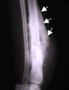
A) Full (look at the femur) and B) coned views of the midshaft of the femur demonstrate a dense sclerotic zone of cortical thickening laterally, which contains a small oval lucent nidus (arrow).

A 15 year old boy presents to your clinic with a limp. He complains of pain in his hip. No relief is obtained with NSAIDs. Diagnosis?
$ How do you distinguish between osteoid osteoma and osteoblastoma?

Osteoblastoma is similar to osteoid osteoma but is larger (> 2 cm), arises in vertebrae, and presents as bone pain that does not respond to aspirin.
rare benign bone-forming tumor of unknown etiology.
The radiographic findings of osteoblastoma are variable. Above, an expansile, eccentric mass in the proximal humerus causes thinning of the cortex (arrows)
Most common benign tumor of bone?
An osteochondroma (osteocartilaginous exostosis) is a bony spur arising on the external surface of a bone. A cartilaginous cap overlies the bony spur and is the source of growth
Tumor of bone with an overlying cartilage cap; most common benign tumor of bone
Arises from a lateral projection of the growth plate (metaphysis); bone is continuous with the marrow space.
Overlying cartilage can transform (rarely) to chondrosarcoma.
What is an osteosarcoma?
$ Peak incidence?
$ Risk factors?
$ Which part of bone?
**Malignant proliferation of osteoblasts - **primary malignant tumors of bone that are characterized by the production of osteoid or immature bone by the malignant cells. Uncommon.
Peak incidence is seen in** $ teenagers**; less commonly seen in the elderly Osteosarcoma is the most common primary bone tumor affecting children and young adults; the peak age is between 13 and 16.
- Risk factors include** $ familial retinoblastoma, Paget disease (especially in patients >40 years old), and radiation exposure - **Osteosarcoma is the most frequent second primary cancer occurring during the first 20 years following radiation therapy for a solid cancer in childhood **
- Arises in the** $ metaphysis of long bones, usually the distal femur or proximal tibia **(region of the knee)
Draw the major components of a long bone.

How does osteosarcoma present?
Presents as a pathologic fracture or bone pain with swelling.
- localized pain, typically of several months’ duration.
- Pain frequently begins after an injury, and may wax and wane over time. Systemic symptoms such as fever, weight loss, and malaise are generally absent.
- The most important finding on physical examination is a soft tissue mass, which is frequently large and tender to palpation.
Imaging of Osteosarcoma?
$ Biopsy?
Imaging reveals a destructive mass with a ‘sunburst’ appearance and lifting of the periosteum
Biopsy reveals $ pleomorphic cells that produce osteoid
malignant osteoid (arrows) with pleomorphic malignant osteoblasts.

Diagnosis?
OSTEOSARCOMA
Imaging reveals a destructive mass with a ‘sunburst’ appearance and lifting of the periosteum (Codman triangle
Giant cell tumor is composed of which type of cells. Whom does it commonly occur in?
$ Where does it commonly arise?
- Tumor comprised of multinucleated giant cells
- Occurs in young adults
- $ Arises in the EPIPHYSIS of long bones, usually the distal femur or proximal tibia (region of the knee)
Epi of Osteosarcoma
- 2nd most common 1⁰ MALIGNANT tumor of bone (1st, multiple myeloma)
- Peak incidence in men 10-20 y/o
- Poor prognosis
$ Diagnosis?

$ ‘Soap-bubble’ appearance on x-ray
Locally aggressive tumor; may recur
Giant cell tumor
$ Ewing Sarcoma
- Malignant proliferation of poorly-differentiated cells derived from NEUROECTODERM
- Arises in the diaphysis of long bones; usually in male children(< 15 years of age)
How does Ewing Sarcoma appear on an x-ray?
$ What does biospy reveal in Ewing Sarcoma?
What can it be confused with?
Genetics?
Often presents with?
**$ Biopsy reveals small, round blue cells that resemble lymphocytes **
1. Can be confused with lymphoma or chronic osteomyelitis
2. (11;22) translocation is characteristic.
Often presents with metastasis; responsive to chemotherapy
What is a chondroma?
$ Where does it usually arise?
Benign tumor of cartilage
$ Usually arises in the medulla of small bones of the hands and feet
$ What is a chondrosarcoma?
- Malignant cartilage-forming tumor
- Arises in medulla of the pelvis or central skeleton
Most common type of bone tumor?
$Typical type of lesions? $ Exception?
A. More common than primary tumors
B. Usually result in osteolytic (punched-out) lesions
1. Prostatic carcinoma classically produces osteoblastic lesions.
Osteoarthritis
- Progressive degeneration of articular cartilage;
- most common type of arthritis
- Major risk factor is age (common after 60 years); additional risk factors include obesity and trauma.
$ Which joints are affected in OA?
- Affects a limited number of joints (oligoarticular)
- hips
- Lower lumbar spine
- knees
- distal interphalangeal joints (DIP) and
- **proximal interphalangeal joints (PIP) **of fingers are common sites.
$ Classic presentation of OA?
Classic presentation is joint stiffness in the morning that worsens during the day.
$ Pathologic features of OA?
- Disruption of the cartilage that lines the articular surface fragments of cartilage floating in the joint space are called ‘ joint mice.’
- Eburnation of the subchondral bone
- Osteophyte formation (reactive bony outgrowths); classically arises in the DIP (Heberden nodes) and PIP (Bouchard nodes) joints of the fingers
Rheumatoid arthritis?
Affects?
$ Association?
Chronic, systemic autoimmune disease
- Classically arises in women of late childbearing age
* *2.$ Associated with HLA-DR4**
$ What is the hallmark of Rheumatoid arthritis?
Characterized by involvement of joints
- *1. Hallmark is synovitis leading to formation of a pannus (inflamed granulation tissue).**
- *2. Leads to destruction of cartilage and ankylosis (fusion) of the joint**
Clinical features of RA?
-
Arthritis with morning stiffness that improves with activity.
i. Symmetric involvement of PIP joints of the fingers (swan-neck deformity),
wrists (ulnar deviation), elbows, ankles, and knees is characteristic ; DIP is usually spared (unlike osteoarthritis).
ii. Joint-space narrowing, loss of cartilage, and osteopenia are seen on x-ray. - Fever, malaise, weight loss, and myalgias
- Rheumatoid nodules-central zone of necrosis surrounded by epithelioid histiocytes; arise in skin and visceral organs
- Vasculitis - Multiple organs may be involved.
- Baker cyst - swelling of bursa behind the knee
- Pleural effusions, lymphadenopathy, and interstitial lung fibrosis
$ Lab findings in Rheumatoid arthritis?
1. $ IgM autoantibody against Fc portion of lgG (rheumatoid factor); marker of tissue damage and disease activity
2. Neutrophils and high protein in synovial fluid
$ Complications of RA?
Complications include anemia of chronic disease and $ secondary amyloidosis. (SAA -> AA -> Deposits)
What are the seronegative spondyloarthropathies characterized by?
Group of joint disorders characterized by
- . Lack of rheumatoid factor
- Axial skeleton involvement
- HLA-B27 association
Ankylosing spondyloarthritis
Ankylosing spondyloarthritis involves the sacroiliac joints and spine.
- Arises in young adults, most often male
- Presents with low back pain; involvement of vertebral bodies eventually arises,
leading to fusion of the vertebrae (‘bamboo spine’). - Extra-articular manifestations include uveitis and aortitis (leading to aortic
regurgitation).
Reiter syndrome
Reiter syndrome is characterized by the triad of arthritis, urethritis, and conjunctivitis.
1. Arises in young adults (usually males) weeks after a GI or Chlamydia trachomatis infection
Psoriatic arthritis
$ Most commonly affected joints?
Involves axial and peripheral joints;** $ DIP joints of the hands and feet are most commonly affected, leading to “sausage” fingers or toes.**
What is the most common overall cause of infectious arthritis? Second most common cause?
N. gonorrhoeae
- young adults
S. aureus
- older children and adults
Infectious arthritis
Arthritis due to an infectious agent, usually bacterial
Causes include
1. N gonorrhoeae- young adults; most common cause
2. S aureus-older children and adults; 2nd most common cause
Classically involves a single joint, usually the knee
Presents as a warm joint with limited range of motion; fever, increased white count,
and elevated ESR are often present.
Gout
Deposition of monosodium urate (MSU) crystals in tissues, especially the joints
Due to hyperuricemia; related to overproduction or decreased excretion of uric acid
l. Uric acid is derived from purine metabolism and is excreted by the kidney.
Most common form of gout
Primary gout is the most common form; etiology of hyperuricemia is unknown.
What is secondary gout seen with?
- Leukemia and myeloproliferative disorders-Increased cell turnover leads to hyperuricemia.
- Lesch-Nyhan syndrome-X-linked deficiency of hypoxanthine-guanine phosphoribosyltransferasc (HGPRT); presents with mental retardation and selfmutilation
- Renal insufficiency- decreased renal excretion of uric acid
$ Presentation of gout
$ Mechanism of injury in gout?
Presents as exquisitely painful arthritis of the great toe (podagra)
- *1. $MSU crystals deposit in the joint, triggering an acute inflammatory reaction. $ Crystals activate neutrophils**
2. Alcohol or consumption of meat may precipitate arthritis.
What can chronic gout lead to?
Development of tophi-white, chalky aggregates of uric acid crystals with fibrosis and giant cell reaction in the soft tissue and joints
2. Renal failure- Urate crystals may deposit in kidney tubules (urate nephropathy).
$Interpret these findings under polarized light.

Pseudogout resembles gout clinically, but is due to deposition of calcium pyrophosphate dihydrate (CPPD);
synovial fluid shows rhomboid-shaped crystals
with weakly positive birefringence under polarized light.
$ Lab findings in Gout?
Development of tophi-white, chalky aggregates of uric acid crystals with fibrosis and giant cell reaction in the soft tissue and joints
What is dermatomyositis?
Inflammatory disorder of skin and skeletal muscle
Unknown etiology
Associated with gastric carcinoma
$ What are the clinical features of dermatomyositis?
- Bilateral proximal muscle weakness; Distal involvement can occur late in disease
- Rash in upper eyelid (helitrope rash)
- Malar rash
- Red papules on elbows, knuckles, knees (Grotton lesion)
$ What are the lab findings of dermatomyositis?
Increased creatine kinase
- *Positive ANA** and anti-Jo-1 antibody
- *Perimysial inflammation with perifascicular atrophy on biopsy**
What is the treatment for dermatomyositis?
Corticosteroids
What is polymyositis?
- Inflammatory disorder of skeletal muscle
- Resembles dermatomyositis clinically but skin not involved
- Endomysial inflammation with necrotic muscle fibers on biopsy (CD8)
What is Becker Muscular dystrophy?
Mutated dystrophin
Milder disease
X-Linked Muscular Dystrophy?
$ What causes it?
Degenerative disorder characleri:t:ed by muscle wasting and replacement of skeletal muscle by adipose tissue.
- *Due to mutations of dystrophin
l. Dystrophin is important for anchoring the muscle cytoskeleton to the extracellular matrix.
2. Mutations are often spontaneous; large gene size predisposes to high rate of mutation.**
$ What causes Duchenne Muscular dystrophy?
- *Duchenne muscular dystrophy is due to deletion of dystrophin.**
l. Presents as proximal muscle weakness at 1 year of age; progresses to involve distal muscles
i. Calf pseudohypertrophy is a characteristic finding.
ii. Serum creatinine kinase is elevated.
What is the cause of death in Duchenne muscular dystrophy?
Death results from cardiac or respiratory failure; myocardium is commonly involved.
Myasthenia gravis
A. Autoantibodies against the postsynaptic acetylcholine receptor at the neuromuscular junction
B. More commonly seen in women
C. Clinical features
- Muscle weakness that worsens with use and improves with rest; classically involves the eyes, leading to ptosis and diplopia
- Symptoms improve with anticholinesterase agents.
- Associated with thymic hyperplasia or thymoma; thymectomy improves symptoms.
Lambert-Eaton Syndrome
$ When does it arise?
$ Clinical findings?
Antibodies against presynaptic calcium channels of the neuromuscular junction
Arises as a paraneoplastic syndrome, most commonly due to $ small cell carcinoma of the lung
C. Leads to impaired acetylcholine release
1. Firing of presynaptic calcium channels is required for acetylcholine release.
Clinical features
- Proximal muscle weakness that improves with use; eyes are usually spared.
- Anticholinesterase agents do not improve symptoms.
- Resolves with resection of the cancer
Most common benign soft tissue tumor?
Lipoma
A. Benign tumor of adipose tissue
B. Most common benign soft tissue tumor in adults
Most common malignant soft tissue tumor in adults
LIPOSARCOMA
A. Malignant tumor of adipose tissue
B. Most common malignant soft tissue tumor in adults
C. Lipoblast is the characteristic cell.
Rhabdomyoma
$ Association
A. Benign tumor of skeletal muscle
B. Cardiac rhabdomyoma is $associated with tuberous sclerosis.
Most common malignant soft tissue tumor in children
$ Most common location
RHABDOMYOSARCOMA
A. Malignant tumor of skeletal muscle
B. Most common malignant soft tissue tumor in children
C. Rhabdomyoblast is the characteristic cell; desmin positive
D. Most common site is the head and neck; vagina is the classic site in young girls.
