MSK Upper and Lower Extremity Injuries Flashcards
Where is the most common site for clavicle fx?
Middle 1/3
MOI of a clavicle fx?
- Fall onto shoulder – 87%
- Direct blow from an object
- Fall on outstretched hand
List the clinical manifestations of a clavicle fx:
- Pain
- Deformity
- Skin ‘tenting’
- (+) ecchymosis (bruising)
- (+) crepitus, palpable tenderness
How would we tx a non-displaced clavicle fx?
- Sling
- Limited shoulder ROM
- Frequent ROM at elbow to prevent stiffness
- Ice
- Analgesics
- NSAIDs (controversial)
- Narcotics x 3-5 days
- PT
WHat procedure would we do to fix a displaced clavicle fx?
•ORIF – plate/screws
What injury would result from a direct blow/force to lateral shoulder with arm adducted?
AC Joint separation
Describe how we would conduct a Cross-Arm test?
What injury is this test used to help dx?
- Pt flexes shoulder to 90 degrees, then actively adducts it
- (+) test if pain produced at AC joint
AC Joint separation

Define the Grading criteria for AC joint separation:
- Grade I – ‘sprain’ of AC ligament (conservative tx)
- Grade II – tearing AC ligament (conservative tx)
- Grade III – tearing both AC and CC (coracoclavicular) ligaments (surgical tx)
When would we surgically treat an AC joint separation?
> Grade III injuries based on age and activity level
Pt arrives at clinic after a fall from standing. On immediate assessment you see:
Most likely Dx?

Proximal Humerus Fx
What images would we order when looking for a proximal humerus fx?
- X-rays – 2v-3v
- CT (non-contrast)
- Evaluate joint/articulation
- Rule in/out surgery
Pt presents to clinic in severe pain: she is holding involved arm adducted to side, you notice swelling and ecchymosis. She reports loss of ROM and has almost no shoulder movement at all.
Dx?
Proximal Humerus Fracture
Identify the fx:

Proximal Humerus Fracture
Is a Proximal Humerus Fracture an indication for narcotics?
yes
conservative tx for Proximal Humerus Fracture
~ 80% are impacted/Nondisplaced
Sling, cuff and collar
Ice
Analgesics
(+) indication for narcotics
Suggest ‘recliner chair’ for rest/sleep
ROM elbow/wrist/hand
PT/OT at 2-week mark
What is the most common cause of shoulder pain visits in primary care
shoulder impingement
Most common cause of shoulder pain?
rotator cuff disease (supraspinatus)
Pt presents w/ diffuse tenderness anterolateral shoulder and decreased Active ROM. The rotator cuff is in tact but shows some weakness.
What test could you perform to add to your suspected dx?
Impingment test
Which variation in acromion shape is most likely to lead to an impingment?
Type III
Images to look at shoulder impingement?
3vshoulder X-ray
- AP (Grashey), Axillary, Outlet or Y scapular
- “Type” of acromion
MRI- not indicated
Pt presents with pain with overhead activities, Sleeping on shoulder and has difficulty putting on jacket.
Suspected dx?
Shoulder impingement
surgical tx for shoulder impingement:
Arthroscopic ‘acromioplasty’
Removal of the ‘hook’/spur under the acromion
Type III → Type I
Bursectomy
Debridement of rotator cuff
Name the 3 grades of rotator cuff tears:
- Intra-substance
- Partial thickness
- Full thickness **
What image/test would we order when you suspect a complete tear od the rotator cuff?
•MSK Ultrasound
NSAIDs appropriate for rotstor cuff tears:
Meloxicam 15mg 1 po qd x 3 weeks
Aleve 440mg po bid with meals x 3 weeks
Advil 400-600mg po tid with meals x 3 weeks
Name the muscles of the rotstor cuff?
which is the most likely to tear?
ubscapularis, teres minor, supraspinatus, and infraspinatus
supraspinatus
Pt comes into clinic and yells “I think I broke my shoulder” you ask the pt how they fell and they reply that they have not fallen, they “woke up like this”.
the patient is in obvious pain and holding the affected shoulder.
probable dx?
Calcific Tendonitis
Calcific Tendonitis is the deposition of _____ ________ within substance of tendon
calcium hydroxyapatite
Tx for calcific tendonitis
- Analgesic/anti-inflammation (PO)
- Steroid Injection (cortisone inj)
- Local + steroid (Depomedrol)
- Subacromial
- PT, modalities
- A/scope
- Failure to make meaningful improvements
- Recurrence
imaging for calcific tendonitis?
- 3-view X-ray
- AP, axillary, outlet (Y-scapular)
Are the majority of shoulder dislocations anterior or posterior?
anterior
causes of traumatic shoulder dislocations →
- Chronic instability
- OA
causes of atraumatic shoulder dislocations
•Often related to ligament laxity
What is the Labrum?
- Fibro-cartilaginous ring
- Attached to outer rim of glenoid
- Provides added depth to the joint socket
- Improved stability
- ‘Gasket’ of the GH joint
Function of the labrum?
- Attachment point for GH ligaments
- Origin of the long head of biceps tendon
A baseball pitcher comes to clinic and tells you they hear a clicking / popping sound when moving their shoulder.
You ask if they can demonstrate the sound and they can.
Dx?
what images would you want to order to confrim dx?
Labral tear
- MR Arthrogram (MRA)
- Preferred
- Intra-articular Gadolinium
- Improved accuracy
Shoulder condition associated w/ endocrine disorders
Adhesive Capsulitis “frozen shoulder”
what are the hallmark clinical presentations of Adhesive Capsulitis “frozen shoulder”?
- Pain
- Stiffness (freezing)
- Decreased AROM and PROM
- Normal motor exam
Condition referred to as “tennis elbow”?
what tendons is this usually present on?
Lateral Epicondylitis
•Extensor tendons of the forearm at insertion at lateral epicondyle
Lateral Epicondylitis presents with pain upon resisted wrist _____. While Medial Epicondylitis shows pain with resisted wrist _____>
extension
flexion
Medial Epicondylitis is also referred to as?
what tendons does this affect?
- Golfer’s elbow”
- Flexor ‘wad’ tendons at insertion at medial epicondyle
Tx for Cubital Tunnel Syndrome (Ulnar neuropathy)?
what splint/brace do we use?
Surgical?
- Night splint/brace X 3-4 weeks
- Surgical Release
- Stop the process
- “Transposition” vs. release
Most common cause of septic bursitis
S. aureus
Describe a septic olecranon bursitis:
•Painful, warm to touch, erythematous
Pt presents to clinic w/ a fluctuant mass. He states it is painless w/ no warmth or eythema.
Dx?

Aseptic olecranon bursitis
Do elbows more frequently dislocate anterior or posterior?
posterior
Most common MOI in a posteriorly dislocated elbow?
- typically occur with fall onto extended arm
- Hyperextension
- Posterolateral rotary
Tx of elbow dislocation
- Closed reduction
- Long arm splint with sling
- Refer to Ortho
- Brace
- PT/OT after period of immobilization
What nerve is most commonly injured w/ a humerus fx?
radial n
tX for humerus fx?
- Hanging cast
- Displaced/unstable ORIF
Complications associated w/ a humerus fx?
mal-union
non-union
infection
How do we tx a Radial Head Fx
ED/UC: long arm posterior splint until seen by Ortho
Sling for comfort x 1-2 weeks
Analgesics
Gentle ROM
Encourage Flex/Ext
Serial X-rays in 1-2 weeks
PT/OT if not FROM by 3-week mark
What is this also called?
is it physiologic or pathologic?

Anterior Fat pad Sign
- Can be normal ‘physiologic’ fluid
- See as a thin strip
You notice a posterior fat pad sign on a child. What does this indicate?
•Supracondylar fx → children
You notice a posterior fat pad sign on an adult. What does this indicate?
•Radial Head fx → adults
Is it normal to see a posterior fat pad sign?
what does it indicate about the intra-articular space?
no
- Intra-articular hemorrhage/effuse
- Distension of synovium making it visible on X-ray
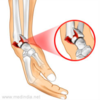
Name the fx:
Tx?

Olecranon Fracture
- ORIF
- Plate and screws
- Tension band wire
Why are pts unable to extend / straighten elbow when they have an olrcranon fx?
site of tricep tendon attachment
How would we tx Both Bones Forearm Fracture in kids?
adults?
Non-displaced- LA cast
•Displaced/Adults- ORIF
define a colles fx and the MOI
- Fx of Distal radial metaphyseal region with Dorsal angulation and impaction
- FOOSH in Dorsiflexion (palm side down)
- Extra-articular

Pt arrives with a “dinner fork deformity” what are we suspicious of?
Colles fx
define Smith fx and MOI:
- Fx of the distal radius with associated volar angulation of distal fracture fragment
- Fall on a flexed wrist
- Direct blow to back of wrist
How would we tx a nondisplaced smiths fx?
cast
How would we tc a displaced smith fx?
plates and screws
splint post-op
Identify fx
and the usual MOI

5th Metacarpal Fracture “Boxer’s Fracture”
- Fracture through 5th Metacarpal neck
- Occurs when a closed fist strikes a hard surface
Tx for boxer’s fx
- If >45 deg volar angulation, reduce fx to anatomic position
- Ulnar Gutter Cast Immobilization for 3-4 weeks, then transition to splint
- Surgical repair
- Unstable, or reduction fails
- Followed by cast immobilization for 3-6 weeks
What fx is this???
Smith’s
Pt arrives to ER after a FOOSH. They tell you they are experiencing arm pain.
Upon examination you notice a dinner fok deformity and the X-rays show a fx of distal radial metaphyseal region with dorsal angulation and impaction.
Dx?
Colles Fx
MOI of both Smith’s and Colles fx:
Colles: FOOSH, palmar surface of hand hits floor
Smith: dorsal surface (back of hand) hits the ground

Pt presents to clinic with a Boxer’s Fx after failing the first clin med exam and punching the wall. The fx has a 50 volar degree angulation:
How do we tx?
what type of cast do we use?
reduce fx to anatomic position
Ulnar Gutter Cast Immobilization for 3-4 weeks, then transition to splint
Pt arrives to ED with an intra-articular fx at the base of the 1st carpometacarpal joint. On X-rays you note a 2-piece fracture dislocation of base of 1st metacarpal with Intra-articular extension.
The Fracture has 2 mm displacement.
Tx?

CRPP fixation
IF >3mm ORIF w/ cortical screw
(this was a bennett fx)
What is the name of the fx that shows an intra-articular fracture of the 1st metacarpal that is comminuted?
Does this have a better/worse prognosis?
Rolando fracture
worse! :(
What type of case do we use on a Bennet Fx?
Thumb Spica 4-6 wks
What is the most frequently fractured carpal bone?
Scaphoid
Pt arrives to ED holding wrist. She compains of pain on the radial side of wrist and with forced dorsiflexion.
They tell you they had a FOOSH, upon examination you notice tenderness w/ axial compression of thumb toward the snuff box.
Likely dx?
What cast are we using?
scaphoid fx
thumb spica 6-8 wks
Why is there such a high incidence of AVN with a proximal 5th metacarpal fx?
- Blood Supply:
- Major source: dorsal carpal branch of radial artery
- 80% of scaphoid via retrograde blood flow

What is arthritis of the 1st CMC joint called?
Basal Joint arthritis
65 year old woman arrives to clinic complaining of opposition pain. She says writting has become very difficult nd she can no longer open jars.
Upon examination she reports pain at both the trapeziometacarpal joint and the scaphotrapezial joint.
Dx?
FIRST line tx?
Basal joint arthritis
NSAIDs / Tylenol
A 70 year old pt arrives to clinic with a hx of Basal Joint Arthritis. She has recieved 2 intraarticular cortisone injections and has no relief when taking tylenol and NSAIDs.
What is our next step in tx?
- Joint Arthroplasty
- Using half of the Flexor Carpi Radialis (FCR tendon) for ligament reconstruction combined with excision of trapezium
- “anchovy technique”
Artery that supplies 80% of scaphoid via retrograde blood flow??
Radial!
Pt with a history of RA presents w/ flexion contracture of (PIP) joint extension of (DIP) joint.
what is this deformity called?
Boutonniere Deformity
Manifestation of RA!!
Swan neck deformit shows _________ of proximal interphalangeal (PIP) joints and compensatory ________ of distal interphalangeal (DIP) joints.
hyperextension / flexion
boutonniere is opposite
When tx a Boutonniere deformity it is IMPORTANT to tell the pt they must wear the splint CONTINUOUSLY:
__ wks for a young pt
___ wks for an adult pt
6 wks child
3 wks elderly
What deformity is repaired with silver or other Ring-type splints (Oval 8)?
Swan neck deformity
what deformity is tx w/ with K-wire?
Boutonniere Deformity
what is the most common compressive neuropathy of upper extremity??
carpal tunnel
pt presents w a positive Tineal sign and positive Phalen’s sign.
Dx?
carpal tunnel
what is a positive phalens sign??
the occurrence of pain or paresthesias in at least one finger innervated by the median nerve
index finger, thumb, middle finger, and half the ring finger, and the nail bed
Stenosing tenosynovitis of 1st dorsal compartment of wrist??
Dx?
deQuervain’s Tenosynovitis
what are the 2 tendons that are affected in deQuervain’s Tenosynovitis?
- Abductor pollicis longus (APL)
- Extensor pollicus brevis (EPB)
define Gamekeepers thumb:
- Injury to ulnar collateral ligament of thumb at MCP joint
- Results in instability of MCP joint and decreased thumb grip strength
- AKA Skier’s Thumb
Pt arrives to ED after an injury resulting from falling on outstretched hand while gripping ski pole. You identify a partial tear.
Dx?
Tx?
Gamekeepers thumb
- Conservative (partial tear)
- Thumb spica cast/splint immobilization for 4-6 weeks
Tx for finger dislocations:
- Recheck NV status
- Volar Alumafoam splint
- Buddy-tape
- Tylenol, NSAIDs, and Ice
- Follow up with Hand/Ortho
A baseball player arrives to clinic after an injury to his finger. He reports most of his pain is located distally. he is unable to extend DIP joint.
You get an X-ray:
Dx?

Mallet finger
when would we use a DIP joint splint?
and what is most important to educate pt about this spint?
- Used for Mallet Finger
- Puts DIP in approx 10 degrees of hyperextension for tissue contact
- Explain to patient that this is NOT TO BE REMOVED for approx. 8 weeks
MOI of Bennett Fracture-Dislocation
Forced abduction of 1st metacarpal
Compare clinical presentation of Mallet vs Jersey finger in regards to MOI and position of affected finger.
Jersey: MOI- hyperextension –> Inability to flex finger at DIP joint
Mallet: MOI struck (trauma) Inability to extend DIP joint
What is injured in Jersey finger?
Which finger is almost ALWAYS affected?
- Avulsion injury of Flexor Digitorum Profundus tendon from insertion at base of distal phalanx
- Ring finger involved 75% of cases
- During grip, ring fingertip is 5 mm more prominent than other digits in ~90% of patients
- FDP insertion into ring finger anatomically weaker than middle finger
Can you dx Jersey Finger via X-ray?
No - usually appear normal
MRI: will show disruption of FDP at volar base of distal phalanx ± avulsion fragment
Pt arrives at clinic and reports of frequent hand pain and she is having a difficult time straightening or bending her finger, she describes this as a “locking” feeling.
Dx?
Tx?
Trigger finger
NSAID
Splinting
Glucocorticoid injection
Trigger finger results in the thickening of the ___ ______ ____ _______. In serious cases this must be treated surgically where we would release the ___ _____
- Flexor tendon and sheath
- A1 pulley
Pt complains of inability to extend hands and fingers. Upon examination you see this.

You also note a firm nodularity on volar surface of hand with coalescing cords of soft tissue on the webs and digits.
Dx?
Tx?
Duytren’s Contracture (•Palmar Fibromatosis)
- Cortisone injections into sheath
- Collagenase injections- reduces contractures improves ROM
Which fingers are affected earliest in Dupuytren’s Contracture?
Fourth and fifth fingers affected earliest
what is the most common soft tissue tumor of hand?
ganglion cyst
Pt presents to clinic with joint pain in her wrist. Upon examination you note a fluid filled swelling overlying a joint or tendon sheath.
Dx?
what is this fluid most likely (2 options)
Ganglion Cyst
•Contains mucinous or gelatinous fluid
Pt w/ a hx of OA presents with visible swelling on the dorsal side of finger. You also note a groove in the fingernail?
Dx?
Tx?

Mucous Cyst (remember usually assoc. w/ OA!)
- Intralesional corticosteroid injectionst riamcinolone
- Surgical excision—Hand surgeon
where do mucous cysts normally appear?
- Benign cysts usually at DIP
- Dorsal surface
what is an Infection of fingertip pulp called?
felon
Tx for felon?
I&D
Antibiotics directed at Staph and MRSA
- Dicloxacillin
- Cephalexin
- Bactrim
- Clindamycin
- Nafcillin
- Doxycycline
Pt presents with a nail laceration, in order to fix you must provide a finger block.
When choosing your local anesthsia would you grab:
Lidocaine
Lidocaine + epi 1%
Lidocaine
NO EPI W FINGER LACS
Pt presents to clinic w what they believe is a rash. upon examination you note grouped vesicles on an intensely erythematous base.
You ask them if it is itchy and they report that it is much more painful then itchy. You also note axillary lymphadenopathy.
Dx?
What fingers are most common to find this dx?
Herpetic Whitlow
thumb and index
Pt presents to clinic w/ a painful, edematous fingertip with vesicular lesions.
What tests would you ude to confirm your dx?
(there are 3)
Herpetic whitlow
Tzank smear
- Viral culture
- Serum antibody titers
Tx for herpetic whitlow?
- Self-limited
- Symptomatic relief
- Acyclovir may be beneficial- PO and Topical
- Famciclovir
- Valacyclovir
unroof tense vesicles
When assessing for a boxer’s fx what degree of volar angulation is acceptable?
up to 40 degrees
