MSK Flashcards

1
Q

Classic association with lupus in the knne

A

Patellar tendon tear (with patella Alta)

2
Q

Where does tibialis posterior insert?

A

Navicular bone and medial cuneiform.

3
Q

Tarsal tunnel contents

A

Tom, Dick, Harry, PT artery and nerve. Cover by flexor retinaculum.

4
Q

What other injury associated with plantaris rupture?

A

ACL tear.

5
Q

Avulsion of calcaneal tuberosity seen in which group of patients?

A

Seen in diabetes

6
Q

Loosers zones seen with

A

Osteomalacia and rickets

7
Q

What’s Panner’s disease

A

Diffuse abnormal signal in the capitellum. Seen in young throwers (5-10 years)

8
Q

First RA spot in the foot?

A

5th metatarsal HEAD.

9
Q

What’s Jaffe-Campanacci

A
  • Multiple NOFs,
  • Cafe au lait spots,
  • mental retardation,
  • cardiac malformations
10
Q

lucent skull lesion with beveled edges

A

EG

11
Q

MM in the spine

A

lytic lesions sparing the posterior element

12
Q

“mini brain” appearance in spine

A

Plasmacytoma

13
Q

Which ligament is involved in supracondylar spur “avian spur”

A

Ligament of Struthers can compress median nerve

14
Q

NOF-like lesion in anterior tibia with bowing in a kid

A

Osteofibrous Dysplasia

15
Q

H-shaped vertebrae seen with

A

Gaucher, SCA

16
Q

1st CMC arthritis?

A

Classically involved in OA Spared in RA

17
Q

most common malignancy in teens in lower extremity

A

Synovial Sarcoma

18
Q

Differences of synovial sarcoma from other ST sarcomas

A
  • can involve bones
  • can be painful
  • Triple sign on MRI (T2 with all intensities)
19
Q

Destructive mass in a leukemia patient

A

Granulocytic Sarcoma (chloroma)

20
Q

Reduced Boehler angle

A

indicates intra-articular calcaneal fracture. Lessn than 20 degress

21
Q

Chalk stick spine fracture

A

seen in ankylosing spondylitis.

22
Q

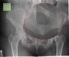
Malignant appearing tumor is the pelvis in adult

A

Chondrosarcoma is the most common.

23
Q

“Cotton wool” appearance of the skull

A

seen in osteoblastic phase of Paget’s.

24
Q

Mastocytosis in xray

A

gives diffuse sclerotic osseous replacement which starts axially. It’s due to release of histamine and prostaglandins.

25
Q

Primary bone lymphoma

A

can present as Normal xray with medullary changes in cross-sectional studies. Can show sequestrum (normal piece of bone in midst of pathological process).

26
Q

MARGINAL erosions.

A

Both RA and psoriatic arthritis

27
Q

Features of Gout touphi

A

don’t usually get calcified. Toughi enhance with Gd.

28
Q

What’s Thyroid acropachy

A

an extreme manifestation of Grave’s appears as smooth peri-osteal reaction.

29
Q

What’s the most medial head of the Tricep tendons

A

The most medial head of triceps is the LONG head, the medial head is located in the middle.

30
Q

When there’s rice bodies in a bursa

A

ddx would include RA, TB and coccidiomycosis.

31
Q

Os acromiale may cause

A

higher rates of rotator cuff impingement.

32
Q

What’s pilon fractures

A

characterized by intraarticular comminution, whether they are the result of rotational injury or axial load injury.

33
Q

PVNS on xray

A

normal bone density with prominent subchondral cysts on xray.

34
Q

How is Grashey view obtained?

A

90 degrees angled at the glenoheumeral joint.

35
Q

What’s the relationship between the humeral head and glenoid in external view of the shoulder

A

there’s always overlap between the humeral head and the glenoid.

36
Q

What’s the position of the greater tuberosity in external view of the shoulder?

A

the greater tuberosity is lateral.

37
Q

Position of tuberosities in internal view of the shoulder?

A

the greater tuberosity should not be seen on x-ray as it overlays the humeral head.

The red arrow points to the lesser tubercle of the humerus in profile.

38
Q

In axillary view

A

anterior part of the shoulder is located north in the image (superiorly).

39
Q

In which view Hill sachs is seen

A

seen only in internal rotation view.

40
Q

What’s the arm position in posterior shoulder dislocation

A

the arm is locked in internal rotation position.

41
Q

Brown tumors (osteoclastoma) seen in?

A

seen in both primary and secondary hyperparaPTH. More in primary.

42
Q

Subchondral sclerosis seen with

A

either CPPD or OA.

43
Q

Hook-like osteophytes

A

classically seen in hemochromatosis and CPPD. CPPD involves index and middle, hemochromatosis involves 4 MCP.

44
Q

most commonly fractures part of the humerus

A

Surgical neck

45
Q

Ganglion cyst

A

is NOT lined by synovial fluid.

46
Q

Morton neuroma

A

is peri-neural fibrosis around the nerve, it’s NOT a real tumor. Only fibrous reaction from wearing high heels for a long time.

47
Q

The fovea of the femur

A

is where the ligamentum teres attaches to the head of the femur. It’s the only part of the head that Is not covered by cartilage.

48
Q

How AVN occurs with hip joint effusion

A

distension of the joint causes compression of the vascular supply; which may lead to avascular necrosis.

49
Q

Isolated degenerative changes of the patellofemoral joint

A

are highly suggestive of CPPD.

50
Q

teres minor has fatty atrophy

A

quadrilateral space syndrome.

51
Q

Contrast mixture in Ct Arthrogram

A

The most appropriate choice of contrast mixture is iodinated contrast 1:1 with saline and/or anesthetic

52
Q

in DEXA

A

the lower of the T-scores between the PA spine and the hip is used.

53
Q

Location of the meniscal ossicle

A

most commonly seen in the posterior horn of the medial meniscus.

54
Q

pseudolesion of the humeral head seen in which view?

A

is typically noted on internal rotation views of the shoulder. Additional radiographic views, such as an external rotation view, will not demonstrate a similar finding

55
Q

What is the most common accessory muscle of the ankle?

A

Peroneus quartus

56
Q

What is the most common carpal coalition?

A

Lunotriquetral

57
Q

Bipartite patella

A

Can have anterior knee pain. Can have T2 bright signal

58
Q

If you see iliac horns

A

nail-patella syndrome. This autosomal dominant disorder has a clinical tetrad that includes the development of iliac horns, in conjunction with involvement of the nails, patella, and elbows.

59
Q

Bridging ossification between osseous structures in fibrodysplasia ossificans progressiva typically occurs where initially?

A

Sternocleidomastoid. it’s MO.

60
Q

Most lethal type in OI

A

Type II

61
Q

Movement of os odontoideum

A

It moves with the atlas on flexion and extension.

62
Q

What is a limbus vertebra.

A

typically results from an intraosseous herniation in the growing spine that may separate the ring apophysis from the vertebral body.

63
Q

The most common cause of scoliosis

A

idiopathic, accounting for approximately 85% of cases. Other causes of scoliosis include leg length discrepancy, congenital causes, neuromuscular causes, neurofibromatosis, connective tissue disorders, trauma, tumors, and radiation therapy.

64
Q

Which group has higher incidence of subperiosteal abscess?

A

Kids. the periosteum is more loosely attached to the cortex. As pus develops in the medullary space it eventually breaks through the cortex and fills the subperiosteal space, creating a subperiosteal abscess.

65
Q

well-defined, lytic lesion without a defined sclerotic margin, eccentric in location, and extending to the subarticular margin of the tibia.

A

GCT

66
Q

when do you treat NOF

A

if associated with a pathologic fracture, curettage and bone grafting may be indicated.

67
Q

MRI appearance of synovial chondromatosis

A

The chondromatosis bodies are commonly low on T1 and high on T2 nd typically enhances. There can be bone erosions.

68
Q

Most common location for osteoid osteoma

A

Femur

69
Q

Myxomas have a predication for which of the following tissues?

A

Muscles.

70
Q

Extensive bone marrow edema surrounding an otherwise well-circumscribed epiphyseal lesion in a skeletally immature patient

A

Chondroblstoma

71
Q

Which of the following paraneoplastic syndromes is associated with hemangiopericytomas (solitary fibrous tumor)?

A

Hypoglycemia

72
Q

surface soft tissue mass with mature bone extending from the center of the lesion to the periphery with the marrow nearly always involved.

A

parosteal osteosarcomas

73
Q

hamartomatous disorder composed of hemangiomatous and lymphangiomatous lesions in the axial and appendicular skeletal and visceral organ involvement.

A

Cystic angiomatosis

74
Q

Content of the rotator interval ?

A
  • superior glenohumeral ligament (SGHL)
  • coracohumeral ligament,
  • long head of the biceps tendon.
75
Q

the most common proximal fifth metatarsal fracture.

A

avulsion fracture (peroneus brevis)

76
Q

In Bennett fracture, The unopposed action of which tendon causes proximal retraction of the larger fracture fragment?

A

Abductor pollicis

77
Q

first radiographic manifestation of fracture healing?

A

Widening of the fracture line with blurring of fracture margins

78
Q

5th metatarsal avulsion fracture could be from either:

A

avulsion of the peroneus brevis or avulsion of the lateral component of the plantar aponeurosis

79
Q

which ligament is avulsed in Segond fracture

A

Lateral Capsular Ligament

80
Q

Posterior interosseous nerve syndrome.

A
  • Radial nerve injury/ entrapment.
  • forearm pain and weakness of the extensor muscles.
  • Sensation is usually preserved.
  • MRI typically shows denervation edema or atrophy of the involved muscles with sparing of the extensor carpi radialis longus.
81
Q

The most common type of perilunate injury

A

transscaphoid perilunate dislocation

82
Q

The most frequently involve tendon in quadriceps tendon tear?

A

Vastus intermedialis.

83
Q

The pes anserine bursa

A

separates the sartorius, gracilis, and semitendinosus tendons from the medial tibia and MCL.

84
Q

T-sign in elbow arthrogram

A

contrast extending between the deep fibers of the distal ulnar collateral ligament and sublime tubercle of the ulna. This finding is diagnostic of a partial-thickness, articular side tear of the distal ulnar collateral ligament. This injury is seen as a result of repetitive or excessive valgus force upon the elbow.

85
Q

What are Looser zones?

A

They represent focal deposition of uncalcified osteoid, seen as an ill-defined horizontal linear lucency in the cortex that does not span the complete diameter of the bone.

86
Q

where do erosions and sclerosis typically present in the sacroiliac joints?

A

Iliac side of the inferior sacroiliac joint. (I with I; iliac with inferior)

87
Q

What is the only discriminating feature of hemochromatosis, that is not present in CPPD arthropathy?

A

Osteoporosis

88
Q

Which part of the lumbar facet joint first shows changes of degeneration?

A

Superior articulating process

89
Q

When do you consider cement loosening?

A

if lucency is more than 2 mm.

90
Q

How does amyloid look in MR?

A

the tendons, capsule, and synovium will be low in signal intensity on all sequences. Erosions are typically filled with low signal intensity material.

91
Q

Productive arthoropaty

A

Gout and Psoriatic.

92
Q

first sign of collapse in osteonecrosis.

A

Crescent sign

93
Q

Expansile posterior element of spine mass

A

ABC or OSTEOBLASTOMA.

94
Q

DDX for bones lesion with MARROW EDEMA:

A
  • Osteoid osteoma,
  • osteoblastoma,
  • chondroblastoma,
  • EG, or
  • lymphoma
95
Q

disc spaces widening:

A

Langherhans or Sickle cell.

96
Q

In the wrist, the magic angle effect

A

seen in the extensor carpi ulnaris (ECU) on axial sections.

97
Q

the 2nd MCC of soft tissue mass in the HAND following a ganglion cyst.

A

Giant cell tumor of tendon sheath, can have bony erosions

98
Q

entrapment or irritation of the sciatic nerve within the buttocks.

A

Piriformis syndrome

99
Q

Hyperostosis frontalis

A

usually involves the inner table of the skull and may show some MDP uptake.

100
Q

Vessel and nervevinjured in posterior elbow dislocation.

A

Ulnar nerve and brachial artery

101
Q

Reiter’s and psoriasis

A

are IDENTICAL! Should go together in DDx. Reiter’s however DOES NOT occur in women.

102
Q

What’s ivory phalanx

A

represents osteosclerosis and is relatively specific for psoriatic arthritis.

103
Q

The Hawkins sign

A

subchondral lucent band seen on the frontal ankle radiograph 6–8 weeks after ankle immobilization. The lucent band represents increased bone reabsorption from active hyperemia. The presence of the Hawkins sign implies an intact blood supply to the talar dome and is a good prognostic indicator in talar neck fractures. Absence of the Hawkins sign suggests avascular necrosis.

104
Q

What maintains the arch of the foot?

A

the posterior tibial tendon and the spring ligament complex. Tearing could lead to acquired flat foot.

105
Q

Guyons canal formed by

A

pisirom and hamate. Contains Ulnar artery and nerve.

106
Q

Where does the plantaris tendon sit?

A

Between medial head of gastrocnemius and soleus. Example of ruptured tendon

107
Q

Standard views in tarsal coalition evaluation,

A
  • 45 degrees internal oblique.
  • AP
  • Lateral
108
Q

Madelung deformity is most associated with

A

Ulnocarpal impaction (positive ulnar variance).

109
Q

What’s the diagnosis?

A

osteopoikilosis

  • Autosomal dominant
  • Bone islands near the joints
  • Can have mild arthritis.
  • Associated with keloids
  • No malignant potential.
110
Q

What’s the most common indication for THA revision?

A

Mechanical loosening.

111
Q

What other fracture most commonly associated with Colle’s fracture?

A

Ulnar styloid fracture.

112
Q

Posterior impingement of the ankle

A
  • It is classically described in ballet dancers.
  • It is usually a unilateral phenomenon.
  • from repetitive ankle plantar flexion
  • Can be related to Os trigonum.
113
Q

Where does osseous lymphoma arise from?

A

Areas of persistent red marrow.