MSK Flashcards
What are some of the complications of a fracture?
Early - Intermediate: Haemorrhage, Neurological injury, compartment syndrome, infection, DVT and PE, dislocations.
Late - malunion, AVN, athropathy, contractures
What are the 4 main stages of fx healing?
- Haematoma formation 2. fibrocartilaginous callus formation 3. bony callus formation 4. bone remodelling
What are some factors that can interrupt or delay healing? think fracture characteristics and patient factors.
Fracture characteristics: movement, misalignment, infection, malignancy, impaired blood supply, severe damage
Patient factors: Vit D deficiency, malignancy, diabetes mellitus, drug therapy (eg: corticosteroids), smoking.
Describe the Gustilo-Anderson Classification of Open Fx
Type I: Wound <1cm
Type II: Wound 1-10cm
Type IIIA: Wound >10cm, Adequate tissue for coverage, any segmental/comminuted fx (even if <10cm), farm injuries
Type IIIB: extensive periosteal stripping, soft tissue inadequate for coverage - requires soft tissue transfer
Type IIIC: Vascular injury
What is the emergency management of Open Fx?
- A to E according to ATLS
- Stop haemorrhage
- Assess neurovascular status
- Analgesia
- Straighten and align limb - repeat neurovascular
- Remove gross contaminants and Photograph wound
- Dress wound with sterile dressing
- Splint fracture - repeat neurovascular
- IV antibiotics - Co-amoxiclav (1.2g IV 8hrly)/Clindamycin (600mg) if allergic to penicillin
- Check Tetanus status and administer prophylaxis
- X-ray the limb
- Refer to surgery
What is the surgical management of Open Fx
Wound debridement + washout, Ex-fix, may need soft tissue grafts
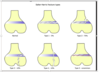
Describe Salter Harris Classification
See image
What is Beighton’s score for?
Assesing joint hypermobility - score of >/=5 out of 9 defines joint hypermobility
Define neuropraxia based on Seddon’s classificaiton of nerve injuries.
Reversible block to nerve conduction. Nerve is intact but mechanical pressure caused demyelination of axons.
Define Axonotmesis based on Seddon’s Classification
Interruption of axons in segment of nerve. Loss of conduction but endoneural tubes are intact.
Wallerian degeneration occurs distal to lesion. Axonal regeneration starts where proximal stump sprout numerous unmyelinated tendrils where one will find its way to the endoneural tube
Define Neurotmesis based on Seddon’s
Complete division of nerve usually occuring in oen fx.
There is Wallerian Degeneration but endoneural tubes are destroyed. Axon regeneration cannot occur without surgical intervention.
Neuroma may form
What are some risk factors for DVT/PE
Cancer, Age >60yrs, personal hx/family hx of VTE, Obesity, COCP/OCP, HRT, Recent period of immobilisaiton, recent surgery (esp hip fx),
Describe the diagnosis of DVT
- Use DVT Well’s Score to identify likelihood of DVT - if 2 points or more is likely. If 1 or less is unlikely.
- If DVT likely: Do proximal leg vein US within 4 hrs
- If negative, do D-dimer test
- If US/D-Dimer positive -> start LMWH
- If US cannot be done within 4 hrs, start LMWH anyways.
- If DVT unlikely: arrange a d-dimer test
- If d-dimer positive, arrange 4hr proximal leg vein US
Describe diagnosis of PE
- Do PE Wells Score - if >4, PE is likely. If = 4 PE is unlikely.
- If PE likely - Immediate CTPA, if there is delay in CTPA then give LMWH first.
- If PE unlikely - arrange d-dimer. If D-dimer positive, CTPA. If there is delay in CTPA, give LMWH.
What is the management of DVT/PE?
- LMWH or Fondaparinux should be given initially after DVT/PE diagnosed.
- This should be continued for at least 5 days or until INR >/=2.0 for at least 24hrs.
- Exceptions are those with renal impairment - use UFH
- Warfarin should be given within 24hrs of diagnosis (start same time as LMWH).
- Warfarin should be continued for:
- 3 months if DVT/PE is provoked
- 6 months if DVT/PE is unprovoked/or if patient has active cancer.
- Target INR - 2-3
Where does the clavicle usually fracture?
Between the middle and distal thirds
What is the conservative management of Clavicle fx?
Immobilisation: Broad arm sling, analgesia, physiotherapy
What are the complications of clavicle fracture?
Acute: pneumothorax, haemothorax, brachial plexus injury, blood vessel injury.
Late: Non-union, malunion, deformity, thoracic outlet syndrome.
In which direction does the shoulder commonly dislocates?
Anterior-inferior direction
What nerve is can be damaged from an anterior dislocation of the shoulder
Axillary nerve
Describe the management of anterior dislocation of shoulder?
Commonly conservative:
- neurovascular assessment before reduction
- closed reduction under anaesthesia - methods: external rotation, stimson maneuvre, milch technique
- Reassess neurovascular status and x-ray
- early physiotherapy
What are some complications of anterior shoulder dislocation?
Bankart lesions - soft bankart (detachment of anterior-inferior labrum) and bony bankart (involvement of the glenoid margin)
What is the blood supply to the humeral head?
Anterior humeral circumflex artery.
Where does the proximal humerus commonly fracture?
surgical neck
Desribe the management of proximal humerus fracture
Conservative: Collar and Cuff immobilisation + early physio
Surgical: Closed reduction percutaneous pinning or ORIF with plate or IM nail.
Complications of Proximal Humerus Fractures?
Axillary nerve injury, avascular necrosis of head of humerus, malunion, rotator cuff injuries, adhesive capsulitis, arthritis.
Why can someone with a midshaft fractre of humerus present with wrist drop
Because of radial nerve injury as radial nerve courses around the shaft of humerus.
