Molecular chaperones Flashcards

1
Q

How does helix helix packing work?

(to form supersecondary structures)

A

Ridges/pegs made by side chains on every 3rd (3n) or 4th (4n) residue (or all (n) residues) of an alpha helix slot into corresponding troughs on neighbouring helix.

Ω-angle between helices determined by whether each is n/3n/4n and whether parallel or antiparallel

2
Q

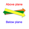
Why do beta sheets pack on other beta sheets at an angle (Ω between -20 and -50deg)?

A

Because each ß-sheet is twisted. (right handed twist)

the side chains on each beta strand form ridges and the ridges of other sheets can fit into the troughs between the first sheet’s ridges.
3
Q

Which type of alpha helix ridges can typically associate with beta sheets?

A

4n

4
Q

5 conditions required for thermodynamic, spontaneous folding model of protein formation to work?

(suggesting the polypeptide contains all information required for folding into its final native state)

A

1) unique native state, no other possible folds with similar free energy
2) stability against small changes in environment
3) **kinetical accessibility: **flat easy route through free energy surface (no humps requiring energy input to overcome)
4) independent of covalent modifications (like phosphorylation or glycosylation)
5) not limited by cis-trans proline isomerisation which is very slow!

5
Q

Why must proteins fold in pre-defined pathway? (i.e. kinetic model)

(levinthal’s paradox)

A

because so many possible ways of folding for each protein that a 100 residue protein could take longer than the age of the universe to find the right one.

6
Q

How does secondary structure of proteins form?

A

Nucleation and cooperation (zipper model)

Early interactions (H-bonds) between neighbouring residues act as nucleation centres that bring other residues closer together in a cooperative manner.

7
Q

Why is a hydrophobic molecule in water entropically unfavourable?

A

Hydrophobic molecules repel water molecules and in doing so make them more ordered!

Therefore folding a hydrophobic peptide reduces its surface area and so interactions with water, so making hydrophobic interactions entropically favourable! despite ordering the protein itself!

8
Q

Define a molecular chaperone:

A

One of a diverse group of proteins that assist in the folding/unfolding and assembly/diassembly of other macromolecular structures.

They are not a permanent part of the folded structure

9
Q

Why do some proteins need chaperones to fold?

A

To overcome high energy barriers or escape troughs in their folding pathways

Or if they need covalent modifications to fold:

like phosphorylation, disulfide bridges, glycosylation, acetylation

10
Q

3 types/functions of chaperones?

A

Holders, hold unfolded proteins to prevent their hydrophobic regions from aggregating, or unfolding further! (e.g. Hsp27, prefoldin, trigger factor)

Un-folders: require ATP to unfold proteins, and prevent their aggregation. (e.g. Hsp 60[GroEL], hsp 70, hsp 100, CCT and TriC

**Folders: **guides protein folding, ATP independent

(e.g. in the ER: Calnexin, calreticulin, and protein disulphide isomerase- [which catalyses disulphide bond rearrangements])

<!--EndFragment-->

11
Q

Bacterial cytosol chaperone system?

A

70% small protein nascent chains only stabilised by Trigger Factor (a ribosome associated chaperone), then fold with no further assistance

20% TF then Hsp70 family (DnaK and DnaJ), undergo ATP-dependent cycles

10% then transit to Hsp60 (GroEL) system

12
Q

Archaea cytosol chaperone system?

A

1) Ribosome associated chaperone: NAC (nascent polypeptide associated complex)
2) Hsp70 system (DnaK/J) + Prefoldin
3) Hsp 60 system: known as Thermosome in archaea

13
Q

Eukaryotic cytosol chaperone system?

A

1) NAC (nascent chain associated complex)
2) Rac (ribosomal associated complex): Hsp70 and its J domain protein Hsp 40 (which recruits it)
3) Some passed to Hsp90 by HOP

(Or some chains recognised (like actin and tubulin) by Prefoldin brought to Hsp60 system: chaperonin TriC)

14
Q

What recognises substrates for eukaryotic Hsp60 system, chaperonin TriC? (substrates like actin and tubulin)

A

Prefoldin, PFD. (after processing by Hsp70 in RAC, which also interacts with Hsp60, prefoldin TriC)

15
Q

What happens in response to heat or other stress in cells?

A

Protein unfolding and aggregation, but also the release of small Hsp dimers from sHsp complexes.

These sHsp dimers bind to hydrophobic regions of unfolding proteins to stabilise them against further unfolding to await later recovery.

16
Q

What happens to proteins aggregated beyond repair?

A

Hsp100 and proteases degrade them

17
Q

What system repairs and refolds aggregated proteins?

A

Hsp 100 along with Hsp40,Hsp70,NEF (nucleotide exchange factor for Hsp70)

18
Q

Role of intramolecular chaperones? pro-sequences

A

Pro-sequences are N-terminal sequences of a protein necessary for its correct folding

but which are removed subsequently by autoproteolysis

and so are not part of the final protein structure

19
Q

3 domains of Trigger Factor? (bacterial ribosomal associated holder chaperone)

A

RBD (ribosomal binding domain)

SBD (substrate binding domain holds non-specific hydrophobic regions of nascent peptide, with own 4 hydrophobic patches A-D)

PPD (peptidyl-prolyl isomerase domain changes proline from cis to trans form)

20
Q

General 3 constituents of Hsp70 complex?

A

Hsp70 (chaperone) (in Rac or free in cytosol)

Hsp40 J-domain protein (ATPase activating)

NEF (prokaryotic GrpE)(mammalian HspBP1 [hsp70bindingprotein]) (releases ADP from hsp70)

21
Q

Hsp70 structure and function?

A

N-terminal ATPase domain allosterically linked to SBD (substrate binding domain)

When ATP bound, lid region in open conformation, associated with ATPase domain.

After ATP hydrolysed to ADP (courtesy of hsp40 activation). LID CLOSES tightly binding hydrophobic regions of substrate (like leucine residues).

(NEF (GrpE or Bag1) releases ADP and so substrate.)

22
Q

How does NEF cause ADP release from Hsp70 to allow ATP to bind?

(like GrpE in prokaryotes, and Bag1 in humans)

A

NEF Binds over top of ADP bound ATPase domain, forces it open, releasing ADP.

23
Q

How do Hsp40 J-domain proteins deliver substrate and activate ATPase activity of Hsp70? (like DnaJ, or human DnaJ protein 1, hDj-1)

A

Zinc finger domain binds and delivers hydrophobic regions of substrate,

and J-domain activates ATPase activity of Hsp70 (DnaK/Hsc70/Hsp70).

24
Q

What chaperone that looks like a jellyfish is missing in prokaryotes?

and what is the jellyfish specific for in eukaryotes?

A

Prefoldin, PFD a heterohexameric jellyfish shaped holder chaperone. (6 coiled coil tentacles with hydrophobic grooved ends for binding)

Transfers proteins to Hsp60 (GroEL in mitochondria, TriC/CCT in eukaryote cytosol)

PFD specific mostly to Actin and Tubulin folding in eukaryotes!

25
Q

Chaperonins?

A

include Hsp60 (mitochondrial chaperonin)

They are ATP-dependent unfolder chaperones

Group I: prokaryotic: GroEL/ES complex

Group II: Archaea/Eukaryotic. Thermosome in archaea /TriC/CCT in eukaryotes (which fold actin and tubulin, brought by prefoldin, PFD)

26
Q

How does GroEL (bacterial Hsp60 homolog) unfold proteins? (with the help of GroES (“hsp10”) and ATP)

A

Symmetrical tetradecamer, 2 rings of 7 subunits create enclosed space in which polypeptide substrate binds to apical hydrophobic regions on each monomer. (only one ring at a time)

Binding of ATP at equatorial region of monomers causes conformational change that expands GroEL ring and hides hydrophobic sites. (this stretches out and unfolds polypeptide)

Small GroES cap recognises this structure and binds, releasing the polypeptide, allowing it to fold.

ATP binding other side of protein releases ES cap, polypeptide and ADP from first side.)

27
Q

What is the role of the Hsp100 family of chaperones?

A

Processing aggregated proteins for degradation (by association with a peptidase like ClpP in the ClpA-ClpP complex)

Or for refolding.

(Clip thread proteins, using ATP, delivered by Hsp70 complex or thread them to Hsp70 complex for refolding.)

28
Q

Structure of Hsp100 family? (clip threaders)

A

Hexamer with central pore (of AAA+ superfamily of ATPases) through which substrate is threaded. (clip threading mechanism)

Hydrophobic Aromatic residues on loops bind substrate and change conformation pull it through (unfolding it), in response to ATP hydrolysis.

(Class I’s have 2 ATPase domains, e.g.Hsp104,ClpB,ClpA)

Class 2’s have 1 ATPase domain. ClpX)

29
Q

ClpA-ClpP complex role?

A

Protein degradation, ClpA is Hsp100 family chaperone (ATP dependent unfolder)

ClpP is protease/peptidase

30
Q

General role of Hsp90 chaperones?

A

**Interact with variety of folded proteins **to stabilise them, regulate them, perhaps aiding in their maturation or activation. (proteins such as steroid receptors and kinases)

Many co-chaperones involved.(like HOP)

Essential for eukaryote viability.

31
Q

Hsp90 structure?

A

3 domains: ATP binding, client binding, c-terminal dimerisation domain (with MEEVD-motif that binds stuff)

When ATP binds N-terminals these also dimerise!

32
Q

Which co-chaperones help Hsp90 to activate Steroid hormone receptors, and which for Kinase activation?

A

HOP co-chaperone helps Hsp90 activate SHRs

Cdc37 co-chaperone helps Hsp90 activate kinases.

33
Q

What are Hsp27 dimers used for?

A

They are a type of small Hsp, sHsp which are used to stabilise actin and to stabilise aggregating proteins in times of stress.

34
Q

Immunoglobulin domains

How do sHsp’s dimerise?

A

Beta strand exchange between Immunoglobulin domain beta sheet regions.

(Stabilised by phosphorylation.)

35
Q

What are the functions of the Pap proteins: PapD, PapC and PapG?

A

These Pap proteins are homologous to sHsp and function in pilus assembly.

PapD is a periplasmic chaperone

PapC is Usher pore through outer membrane

PapG is tip of pilus with adhesin tip for interactions

36
Q

What makes PapD (chaperone) different from PapG,F,E,K,A, and H?

And What is donor strand complementation?

A

It has an exposed n-terminal beta strand, **G1 ß-strand. **which has conserved P2-P4 residues.

Other Pap proteins lack this G1-strand, leaving them with a c-terminal F-strand with a groove with P2-**P5 **binding pockets.

The binding of P2-P4 residues of PapD into their respective pockets is donor strand complementation.

37
Q

Why is PapD donor strand complementation (involving its G1-ß-strand) necessary for correct order of pilus formation?

A

It binds to other Pap proteins’ F-strand P2-P4 pockets, preventing inappropriate interactions until the correct (most competitive) Pap protein outcompetes PapD for binding.

(attaching using empty P5 pocket, and outcompeting for P2-4 binding)

38
Q

What is the role of the n-terminal extension on the Pap proteins PapF,E,K,A,H?

A

Nte of the PapF/E/K/A/H proteins have P2-P5 residues:

Firstly their P5 residue binds in the unnocupied P5 pocket of the F-ßstrand groove of the preceding Pap protein, whilst it is bound to G1-ßstrand of PapD (chaperone).

The specific sequence of each Pap protein’s Nte determines whether it can outcompete the PapD G1-ßstrand for P2-P4 pocket binding on that particular preceding Pap’s F-strand groove.

PapD zips out, incoming Nte zips in.

39
Q

What is recognised by Signal Recognition Peptide?

A

N-terminal Signal sequence that targets proteins for membrane transport e.g. secretion in e.coli

Particularly its hydrophobic middle region!

(includes peptidase cleavage recognition site so it can be cut off later)

40
Q

Cotranslational secretion in e.coli?

A

Nascent polypeptide signal sequence recognised by SRP, this docks to SRP’s receptor: FtsY GTPase that transfers polypeptide to Sec trimeric channel complex, SecYEG.

Ribosome provides force for transport! (although PMF might be important)

41
Q

What happens if nascent polypeptide’s Signal sequence isn’t hydrophobic enough to be recognised by SRP and undergo cotranslational secretion? (in e.coli)

A

Post-translational secretion:

It is held by SecB, which docks with SecA, an ATPase motor that provides the force to translocate the polypeptide through the SecYEG trimeric channel in the inner membrane.

(PMF also provides some force again)

42
Q

What cleaves the off the signal sequence of a secreted polypeptide?

A

Signal peptidase 1. Spase-I.

43
Q

Signal recognition particle/protein **receptor **in eukaryotic ER import?

(contrasted with FtsY in prokaryotes)

A

SR alpha bound to SR beta in the membrane.

44
Q

What activates ATPase activity of Bip (eukaryotic Hsp70) following ER import?

And what removes Bip from polypeptides? (NEF)

A

A transmembrane J-domain protein complex called Sec62/63

Allows (ADP-)Bip (Hsp70) to bind to imported polypeptide for folding (closed lid domain conformation)

BAP (Bip associated protein) acts as NEF. (allowing BiP to bind ATP and release substrate)

45
Q

Describe the glycosylation process of polypeptides that are imported into the ER (through Sec61 pore):

A

First oligosaccharyltransferase transfers the tri-antennary structure from its dolichol carrier onto the first asparagine, N, through the Sec61 pore.

Then Glucosidase I and II remove glucose residues from the end of the structure.

The remaining structure acts as a signal for Calnexin, Calreticulin and PDI (protein disulphide isomerases like Erp57) to rearrange disulfide bonds so the protein can fold.

46
Q

What happens to proteins that are terminally misfolded following glycosylation and processing (by folders: Calnexin,Calreticulin and PDI,Erp57) in the ER?

A

(Having had all their glucose residues removed by glucosidases:)

Manosidase removes one of the mannose residues from the tri-antennary structure

This is recognised (by ER Degradation Enhancing Manosidases, EDEM) and the protein is sent for ER-associated degradation. ERAD quality control.

Or a glycoprotein glucosyltransferase may save them! by re-adding a glucose residue it targets them for further processing by the Calnexin, membrane bound calreticulin, PDI Erp57 complex.

47
Q

Mitochondrial targeting sequence?

A

Directs newly synthesised proteins to mitochondria.

Has an amphipathic helix structure!

(positive charge residues on one side, R and K)

(May or may not be cleavable)

48
Q

Structure of Mitochondrial Import system: Translocase of the Outer Membrane:

A

Receptor subunits (TOM20 and TOM70) recognise the positive charge of amphipathic MTS helix.

General Import Pore: GIP: formed by TOM40 pore, TOM22 and TOM5 receptors

(little TOM5 facilitates insertion of polypeptide into TOM40 pore)

(TOM22 binds protein again once its through the TOM40 pore.)

49
Q

Routes of transport to the general import pore, GIP, of the mitochondria.

A

All involve MSF (Mitochondrial import Stimulation Factor) that binds mitochondrial targeting sequence (ampipathic helix) (“presequence”)

Co-translationally coupled: (straight after NAC procesing) MSF to Tom20 (>tom22,tom5,tom40,tom22)

Post-translationally coupled: (after Rac [Hsp70L,Mpp11 J-protein] then free Hsp70,Hsp40 processing) To TOM70 etc

Or Targeted by internal targeting sequences! Not cleaved out like presequences.

50
Q

Translocase of the Inner Membrane: TIM structure

(inner mitochondrial membrane)

A

TIM23 recognises presquence (amphipathic helix) and acts as channel into mitochondrial matrix.

TIM17 is a channel into the inner membrane itself!

TIM44 attaches mtHsp70 complex to TIM23.

51
Q

Role of mitochondrial processing peptidase? MPP?

A

Removes presquence mitochondrial targeting sequences (amphipathic helices) after import into mitochondrial matrix.