Module Two Flashcards

Late 4th Week
Arrow points to optic placode
Mx=maxillary Process
Mnd=mandibular process
X=forelimb bud
Neural folds have not fused in area of PN

Early 5th week
Nasal pit deeply invaginated and surrounded by maxillary process laterally, dorsally by lateral nasal process and medio-ventrally by medial nasal process.
2nd arch is much enlarged and obscures rostral margin of 3rd arch.
Bulging abdomen reflects rapid growth of liver and cardiopulmonary system.
Square=forelimb
Circle=hindlimb
X=somites
Arrow=open neural groove
Star=residual yolk sac

Early 5th week
Mandibular process removed so roof of stomodeum can be viewed directly.
Arrow points to Bathke’s pouch.
Circle=anterior nares
Triangle=lateral nasal process
X=medial nasal process
Star=maxilary process
Square=Developing secondary palatal shelves

Mid 6th week
Oronasal development
X=stomodeum boredered inferiorly by mandibular process (star), laterally by maxillary process (square) and superiorly by medial nasal process (circle).
Straight arrow=external nares which are bordered by maxillary processes laterally, medial nasal processes medially, and lateral nasal processes (triangle) dorsolaterally.
Oronasal groove is groove between maxillary process and medial nasal process.
Nasolacrimal groove=junction between maxillary process and lateral nasal process. Later turns into nasolacrimal duct.
Curved arrow=primary choanae which forms inside the stomodeum at the junction of medial nasal and maxillary processes.

Showing the nasal fin (arrow) which is the epithelial seam between maxillary process and medial nasal process.
A small extension lies between maxillary process and lateral nasal process not shown in this micrograph.
This must be removed for normal fusion to take place.

Showing the area of fusion where nasal fin had been. Breakdown has allowed for mesenchyme in MNP and MxP to become confluent

6th week
Mandibular process removed to expose roof of stomodeum (star)
Circle=enlarged secondary palatal processes
Arrow=primary choanae which are now open

Mid 6th week
Mandibular process removed
X=developing tongue
Square=secondary palatal shelves growing vertically from maxillary processes
Star=lip emerging from maxillary process
Arrow=nasal canals which are now complete
Triangle=primary palate

Mid 6th week
Looking into oral cavity from front
Tongue separates two secondary palatal shelves (PS) that extend vertically from maxillary processes
Note nasal pit

8th week
Tongue has retracted so shelves of secondary palate (X) have elevated and extend toward eachother
Posterior nasal chamber and oral cavity still confluent
Triangle=primary palate
Nasopalatine duct becomes incisive foramen

8th week
Star=medial edge of left palatal shelf
X=right side of rudimentary nasal septum
Arrow=opening of left primary choana

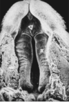
Late 9th week
Palatal shelves have joined but fusion is incomplete.
Marked grooves along midline
Arrow points to opening of nasopalatine duct behind incisive papilla.

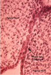
Late 9th Week
Initial fusion of palatal shelves after their elevation
MEE=medial edge epithelium

Formation of the secondary palate
A=7 weeks, B=8 weeks, C=9 weeks
Initial disposition of palatine shelves on each side of the tongue shown in A
Elevation coincident with depression of the tongue in B
Fusion with eachother and nasal septum in C

9th week
View from posterior to anterior
Palatal shelves are joined but still separated by a continous layer of epithelium (medial edge epithelium)
Fusion will only be complete when they are transformed into mesenchymal cells.


