Module 9.0 - Osteoarthritis and Rheumatoid Arthritis Flashcards
(18 cards)
What is osteoarthritis?
- It is a progressive joint disorder characterized by the slow erosion of protective cartilage on joint surfaces.
- Most common form of arthritis
- The distal interphalangeal joint (DIP) is more likely to be affected by OA.
- Marked disease expression variability
- Classified as inflammatory in nature due to the synovial response to reactive new bone formation
- Classified as primary and secondary OA

Describe the etiology/predisposing factors associated with osteoarthritis
A. Thought to be a ‘wear and tear’ syndrome where risks of developing OA increases with age
B. African American women are twice as likely to be affected as Caucasian women
C. Men are more likely to have OA in the hips; whereas women are more likely to suffer from OA in hands and fingers
D. Genetics- evidence suggests OA is an autosomal recessive trait
E. Metabolic abnormalities such as Paget’s disease
F. Obesity predisposes and causes mechanical stress on joints
G. Prior trauma- strains, sprains, dislocations, fractures
H. Hematologic/endocrine disorders: hemochromatosis, hemophilia and acromegaly
What are some subjective findings associated with osteoarthritis?
A. Pain in 1 or more joints, mostly weight-bearing joints, such as hips and knees
1. Stage 1: predictable sharp pain brought on by mechanical insult and can limit high impact activities with modest effect on function.
2. Stage 2: Pain becomes more constant and starts to affect ADLs, unpredictable episodes of stiffness.
3. Stage 3: Constant aching pain with episodes of intermittent intense, exhausting pain results in severe limitations in ADLs.
B. Stiffness and/or swelling in affected joints
C. Grating sensation (Crepitus) during range of motion
D. Instability of joint, buckling, locking when walking or climbing stairs
E. Fine motor deficits in hands- difficulty performing ADLs
What are the physical findings associated with osteoarthritis?
A. Tenderness along joint line (suggests articular pathology)
B. Limitation in range of motion - reduced; equal for both active and passive movements; results from marginal osteophytes and capsular thickening, synovial hyperplasia and effusion.
C. Bony swelling - remodeling of the bone and cartilage on either side of joint with marginal osteophytes evident in small and large joints.
D. Joint deformity - sign of advanced damage to joint
E. Instability - giving way or buckling common symptom in knee OA; can cause falls; sign of muscle weakness with subsequent altered patellar tracking (lateral patellar subluxation).
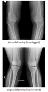
F. Angular deformities at knees (valgus and varus)

What are the two types of osteoarthritis and what joints does each type effect?
- Single- or multiple-joint OA – common areas are knees, hips, interphalangeal joints, first carpometacarpal joint, first MTP joint and apophyseal (facet) joints of lower cervical and lower lumbar spine.
- Generalized OA – implies a polyarticular subset of OA; slow accumulation of multiple joint involvement over several years; clinical marker for generalized OA is the presence of multiple Heberden’s nodes (posterolateral hard swellings of the distal interphalangeal (DIP) joints and Bouchard’s nodules (enlargement on proximal interphalangeal (PIP) joints). There is no set number of affected joints for diagnosis of generalized OA, but American College of Rheumatology (ACR) suggests OA at spinal, or hand joints plus at least 2 other joint regions for diagnostic criteria.

What are some laboratory/diagnostic tests used to diagnose osteoarthritis?
A. Plain film x-rays – narrowing of joint space with cyst formations, bone spurs and thickened synovial membranes; also wearing away of articular cartilage.
B. Magnetic resonance imaging (MRI) – not required for most patients unless radiographic changes are not evident or need for visualization of other structures in joint, such as effusions, synovium and ligaments.
C. Ultrasonography (U/S) – can identify OA associated structural changes such as synovial inflammation, effusion and osteophytosis; operator dependent limitations
D. Synovial Fluid Analysis – clear, yellow fluid with a normal WBC count (less than 1000/mm and glucose levels that approximate the patient’s serum glucose)
E. Sedimentation rate – may be elevated due to inflammation from free-floating intra-articular particles of cartilage
F. No laboratory test is specific for OA – complete blood count and chemistry panel should be obtained to detect any hematologic or renal impairment when treating with NSAIDs.
What are the specific joints involved in Osteoarthritis?
- Hand: Symptoms usually bilateral and joint involvement is usually symmetrical
- Knee: most common single cause of lower limb disability in adults over age 50. Usually bilateral with the patellofemoral joint and/or medial tibiofemoral joint most commonly affected.
- Hip: presents with pain, aching, stiffness and restricted movement; pain usually felt deep in anterior groin but may involve anteromedial or upper lateral thigh; pain is exacerbated with rising from a seated position
- Facet Joint: usually coexists with intervertebral disc degeneration, often termed ‘spondylosis’; Osteoarthritis to isolate symptoms specifically to facet joint; Lumbar facet pain is usually localized, unilaterally or bilaterally, which may radiated to buttocks, groin and thighs, typically ending ABOVE the knees; Cervical facet OA presents with ipsilateral neck pain, which does NOT radiate past shoulder and is aggravated by neck rotation or lateral flexion
- First metatarsophalangeal joint: usually bilateral and when symptomatic leads to localized big-toe pain on standing and during ambulation
How do you diagnose osteoarthritis?
The differential diagnosis of osteoarthritis depends largely on the location of the affected site as well as the presence, or absence of additional systemic symptoms. OA may be diagnosed without the use of radiography and/or laboratory testing in the presence of typical symptoms and signs in the at-risk age-group:
Persistent usage-related joint pain in one or few joints
- Age > or equal to 45 years
- Morning stiffness < 30 minutes
- Slow onset of pain over years, increased by joint use and relieved by rest
- Absence of constitutional symptoms
Consider additional testing and/or referral to a specialist for:
- Younger individuals with joint symptoms/signs of OA
- Presence of atypical symptoms
- Present of weight loss or other constitutional symptoms
- Knee pain with true ‘locking’ which suggests additional mechanical derangement

What are the overall goals for the management of osteoarthritis?
relieve symptoms, maintain/improve function, limit disability and avoid drug toxicity
How would you manage a patient with osteoarthritis of the hand?
- Rest and joint protection; OT consult for splinting as warranted
- Heat and cold therapy; PT consult as warranted
- Topical capsaicin
- Topical NSAID’s
- Oral NSAID’s, including COX-2 selective inhibitors- 1st line pharmacologic therapy
- Tramadol
- recommends the use of intra-articular therapies and opioids
- Recommends patients > 75 years of age use only topical NSAIDs, no oral agents
How would you manage a patient with osteoarthritis of the hip and knee?
- Weight reduction, if applicable
- Rest and elevation (knee) with the application of cold packs intermittently for 1-2 days
- Cardiovascular aerobic exercises within their functional capabilities
- Alternative therapies such as tai chi and acupuncture
- PT referral for quadriceps strengthening exercises (knee only), multimodal orthotics and assistive devices
- Acetaminophen
- Topical NSAIDs (knee only)
- Tramadol
- Intraarticular corticosteroid injections
- Recommends against use of chondroitin sulfate and glucosamine as well as topical capsaicin. ** Glucosamine can cause bronchospasm and should be avoided in patients with asthma or chronic lung disease.
- Surgical options, refer as warranted
What is rheumatoid arthritis?
Chronic, systemic autoimmune disease characterized by inflammation of connective tissue, causing thickened synovium and resulting in pain in and around the joints; while commonly noted intra-articularly, can spread to cardiovascular, hematopoietic and pulmonary structures.

What are the predisposing factors for rheumatoid arthritis?
A. Commonly diagnosed between 30-60 years of age, not inheritable
B. Gender: Women are 3 times more likely than men to be diagnosed
C. Specific genes: Variants of human leukocyte (HLA) antigen more likely to develop RA
D. Initiating factors: Many individuals who carry the HLA genes never develop RA, suggesting that additional factors must play a role in developing the condition.
- Infection: suspect that alterations in gut bacteria populations may initiate RA
- Cigarette smoking: is a recognized factor increasing risk of RA
- Stress – stressful life events (divorces, accidents, grief) common in people with RA within 6 months of diagnosis
What are the subjective findings associated with rheumatoid arthritis?
A. Symetric joint and muscle pain worse in a.m. and improves during day.
B. Weakness/fatigue
C. Anorexia
D. Weight loss
E. Generalized malaise- low grade fever
F. Numbness and tingling in hands

What are the intra-articular physical exam findings associated with rheumatoid arthritis?
- Rheumatoid Nodules: painless lumps that appears beneath the skin; mobile or fixed, commonly occurring on the underside of the forearm/elbow
- Swelling of joints, most commonly metacarpophalangeal (MCP) and PIP joints
- Multiple symmetric joint involvement
- Deformity of joints, commonly PIP, DIP and MCP of hands

What are the extra-articular physical exam findings associated with rheumatoid arthritis?
- Pleural effusions- may cause chestpain/shortness of breath
- Inflammation of the sclerae- may cause pain/vision problems
- Vasculitis- inflammation of the blood vessels
- Splenomegaly (Felty’s syndrome) – neutropenia/increased risk of infection
- Neuropathy- causing numbness, weakness and tingling
6. Sjogren’s syndrome: a systemic chronic autoimmune disorder; causing dry eyes/mouth; vaginal dryness plus more. Diagnosed by testing for the autoantibody SSA.
What laboratory/diagnostic tests are used to diagnose rheumatoid arthritis?
A. Rheumatoid factor (RF): antibody present in the blood of up to 80% of patients with RA; non-specific for RA
B. Anti-cyclic citrullinated peptide/protein antibody test (anti-CCP): more specific than RF in diagnosing RA
C. Granulocytopenia (Felty’s syndrome ) neutrophil count < 500
D. Anemia: hypochromic, microcytic – low hemoglobin, serum ferritin and low to normal total iron-binding capacity (TIBC)
E. Antinuclear antibody may be elevated
F. Erythrocyte sedimentation rate (ESR) is usually elevated
G. Radiographs: reveal joint swelling, joint erosion with progressive cortical thinning, osteopenia and joint space narrowing
H. Synovial fluid – yellow, turbid fluid, elevated WBC up to 100,000, normal glucose
I. RA is a clinical diagnosis- no single diagnostic test; based on characteristic signs and symptoms, laboratory & radiologic findings
How do you treat and manage a patient with rheumatoid arthritis?
- Referral to rheumatologist
-
Pharmacological: Disease-modifying anti-rheumatic drugs (DMARDs); usually includes combination drug therapy; DMARDs should only be ordered and monitored by a rheumatologist
- Methotrexate
- Cycylosporine (unlabeled use therapy)
- Gold preparations
- Hydroxychloroquine
- Sulfasalazine
- Leflunomide
- Etanercept
- Monitor liver function tests at onset of treatment with DMARDs and periodically thereafter
- PT and OT referrals for assistive devices and durable medical equipment
- Screen for Tb prior to starting DMARDs
- DMARDs not recommended in patients with Childs Pugh B cirrhosis or higher or in patients with untreated chronic hepatitis B or treated patients with chronic hepatitis b
- Refer to Orthopedist as warranted.
- Monitor for bone loss- use lowest dose of glucocorticoids as possible; supplement calcium and vitamin D, medications that reduce bone loss
- Pain Management: consider acetaminophen, tramadol and capsaicin cream; avoid narcotics except in severe cases; if long acting opioids used should be monitored by pain specialist


