Module 8.1 - CNS Disorders Flashcards
What is and what are the symptoms associated with a closed head injury?
- More common and involves the head striking a hard surface, a rapidly moving object striking the head or blast waves.
- The dura mater is intact and brain tissues are not exposed to the environment
- Blunt trauma can result in both focal brain and diffuse axonal injuries
- May be evidenced by immediate (generally no more than 5 minutes) loss of consciousness (LOC), loss of reflexes (fall to ground), transient cessation of respiration,briefperiod of bradycardia and decreased in blood pressure
- Increased cerebrospinal fluid (CSF) pressure and electrocardiogram (ECG) and electroencephalogram (EEG) changes occur on impact.
- Evaluation based on results of health history, LOC according to GCS, outcomes of imaging studies (CT or MRI) and assessment of vital parameters (ICP and EEG)

What are the goals of treatment for patients with closed head injuries?
Treatment is directed at controlled ICP, neuroprotection and managing symptoms
What is a contusion?
- A head injury produced by compression of the skull at the point of impact or, from blood leaking from an injured vessel.
- Injury may be coup (injury at site of injury) or contre-coup (injury from brain rebounding and hitting opposite side of skull) or both

How is the severity of a contusion determined?
- Severity varies with the amount of energy transmitted by the skull to underlying brain tissue
- The smaller the area of impact, the more severe the injury because of the concentration of the force.
- Brain edema forms around and in damaged neural tissues, contributing to increasing intracranial pressure (ICP).
- Multiple hemorrhages, edema, infarction and necrosis can occur within the contused areas.
- Tissue has a pulpy quality
- Maximal effects of these injuries peak 18-36 hours after severe head injury
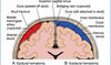
What is an epidural hematoma?
- Occurs when there is bleeding between the dura mater and the skull
- An artery is the source of bleeding in 85% of epidural hematomas
- Usually accompanied by a skull fracture

What is the most common site for an epidural hematoma?
- The temporal fossa is the MOST COMMON SITE of an epidural hematoma caused by injury to the middle meningeal artery or vein
- Individuals with a temporal epidural hematoma will suffer LOC immediately and become lucid within a few minutes

What are the subjective/physical exam findings associated with an epidural hematoma?
As hematoma accumulates, patient will complain of:
- Headache- increasing in severity
- Vomiting
- Drowsiness
- Confusion
- Seizure
- Hemiparesis may develop
Temporal lobe herniation can occur with decreasing LOC, ipsilateral pupillary dilatation and contralateral hemiparesis
How do you diagnose an epidural hematoma and how do you treat it?
- CT or MRI usually needed to diagnose epidural hematoma
- Medical emergency requiring surgical evacuation of hematoma
Prognosis good if treatment initiated prior to bilateral pupillary dilation occurs
What is a subdural hematoma?
- Bleeding between the dura mater and the brain
- Occurs in 10-20% of TBI
- Acute subdural hematomas occur RAPIDLY, usually within hours
- Commonly located at the top of the skull (at the cerebral convexities)
- As ICP increases, the bleeding veins are compressed (self-limiting)
- Cerebral compression and displacement of brain tissue causes temporal lobe herniation if not treated
What are the symptoms associated with a subdural hematoma?
- Headache
- Drowsiness
- Restlessness or agitation
- Slowed cognition and confusion
- Symptoms worsen over time and progress to LOC, respiratory pattern changes, and pupillary dilation
- Homonymous hemianopia (defective vision in either the right or the left field), dysconjugate gaze, and gaze palsies may occur.
What is a subacute subdural hematoma?
- Develop more slowly, often over 48 hours to a few weeks
- Occur more often in older adults and those that abuse alcohol due to brain atrophy with a subsequent increase in the size of the extradural space
- These subdural hematomas act like expanding masses, increasing ICP that eventually compresses the bleeding vessels
- Brain herniation can result
What is a chronic subdural hematoma?
- A subdural hematoma that develops over weeks to months
- Existing subdural space contains the liquefied clot from an acute bleed and/or accumulation of blood from a leaking vein
- A vascular membrane forms around the hematoma in approx. 2 weeks
- 80% will have chronic headaches and tenderness over hematoma on palpation
What are the symptoms associated with a chronic subdural hematoma and what is the treatment?
Symptoms:
- Confusion
- Memory loss
- Coma
- Difficulty speaking or swallowing
- Weakness with difficulty walking
- Loss of sensation
- Seizures
Require a craniotomy to evacuate gelatinous blood and to prevent brain herniation
What is an intracerebral hematoma?
- Occurs when there is bleeding within the brain
- Occur in 2-3% of head injuries, may be single or multiple and are associated with contusions
- MOST commonly located in the frontal and temporal lobes
- Penetrating or shearing forces traumatize small blood vessels

What are the signs/symptoms associated with an intracerebral hematoma?
This hematoma acts as an expanding mass, increasing ICP, compressing brain tissues causing edema and ischemia leading to:
- Decreased LOC, coma or confusional state from other injuries can make the cause of this increasing unresponsiveness difficult to detect
- Contralateral hemiplegia may occur and as ICP rises, temporal lobe herniation may appear
Delayed intracerebral hematomas may appear 3-10 days after head injury
How do you diagnose an intracerebral hematoma and how do you treat it?
- History and physical exam help to establish diagnosis
- CT/MRI and cerebral angiography confirm diagnosis
- Treatment is directed at reducing ICP to maintain cerebral perfusion pressure and allowing the hematoma to reabsorb slowly
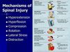
What are 4 different mechanisms of spinal cord injury?
- Hyperextension – Causes spinal cord to stretch (Ex: rear-end collisions, fall onto face, forehead or chin)
- Hyperflexion – greatest stress occurs at C5-6, causing bilateral facet dislocation
- Compression – Vertebral body is compressed and/or shattered, resulting in ‘burst’ fracture and bone fragments may become embedded into spinal cord (Ex: diving accidents or falls, when patient lands on their feet or buttocks)
- Whiplash – sudden hyperextension of the spine that stretches the ligaments as a result of the force of the lower body moving forward and the backward and downward movement of the head.
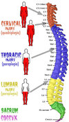
Describe the anatomy of the spinal cord
A. 33 vertebrae total
- Cervical spine (C1-C7)
Highly flexible, small in diameter
Easy to fracture secondary to above
- Thoracic spine (T1 – T 12)
Articulates with ribs
Not a common site for fracture due to its stability with ribs
- Lumbar spine (L1-L5)
Highly mobile, large in diameter
Requires greater amount of force to fracture
- Sacral spine (S1-S5)
- Coccygeal vertebrae (3-5 coccyges)
B. Spinal Cord
- Gray matter
- White matter
- Meningeal layer (pia mater, arachnoid and dura mater)

What are the 8 steps to conduct a physical assessment on a patient with a spinal cord injury?
1. Assess for airway, breathing and circulation and life-threatening injuries first
2. Assess respiratory ability: pulmonary complications main cause of early death in acute quadriplegia
- Chest excursion
- Use of intercostal muscles or diaphragm to breath
- Cervical cord injury at, or above, C3 results in respiratory arrest.
- Edema from upper cervical spine injury is considered life threatening secondary to breathing difficulty from impairment of diaphragm functioning.
- C5-C6 injuries spare the diaphragm, and diaphragmatic breathing can occur
- T1-L2 lesions can cause loss of intercostal muscle use
3. Assess need for Intubation if necessary
- Jaw thrust maneuver only
- Apnea
- Breathing difficulty
- Diaphragmatic fatigue exists
4. Monitor ABGs closely
5. Monitor for pneumonia, pulmonary edema and pulmonary emboli.
6. Motor assessment:
- Perform motor exam and grade strength on a 5 point scale
- Complete lesion: patient lacks sensory function, proprioception, and voluntary motor activity below level of injury; Worse prognosis for recovering neurologic function
- Incomplete lesion: parts of the spinal cord at the level of the lesion are intact; sacral sparing occurs; note sensory perception and voluntary contraction of the anus around the examiners finger
7. Assess Sensory function:
- Begin at the area of no feeling and proceed to the area of feeling
- Assess response to pain:
- Great toe L4
- Back of leg S1-S3
- Perianal area: S4-S5
- Umbilicus T10
- Nipple line T4
- Ring and little fingers C8
- Middle finger C7
- Thumb C6
- Top of shoulder C4
- If the patient is unable to feel pain the lesion is at or above the level spinal nerve level indicated above
8. Evaluate the patients back:
- Log roll patient- needs to be a coordinated maneuver
- Maintain in-line spinal stabilization
- Gently palpate spin for pain, tenderness, or gaps between spinous processes
- Observe for entrance or exit wounds, impaled objects or other injuries.
What are signs of C2-C3 spinal cord injury?
- Respirator paralysis (breathing center C3)
- Phrenic nerves exit C3-C5
- Flaccid paralysis
- Areflexia (DTRs)
- Loss of sensation below mandible

What are signs of C5-C6 spinal cord injury?
- Diaphragmatic breathing
- Paralysis of intercostal and abdominal muscles
- Quadriplegia
- Anesthesia below the clavicle and the ulnar half of the arm
- Areflexia (with possible exception of biceps reflex)
- Fecal and urinary retention
- Priapism (spontaneous erection)
What are signs of T12-L1 spinal cord injury?
- Paraplegia
- Anesthesia in the legs
- Areflexia in the legs
- Fecal and urinary retention
- Priapism

What are signs of L1-L5 spinal cord injury?
- Flaccid paralysis to partial flaccid paralysis
- Abdominal and cremasteric reflexes present
- Ankle and plantar reflexes absent

What is a primary spinal cord injury?
- Occurs with the initial mechanical trauma and immediate tissue destruction from shearing, compression, or penetration
- Can also occur if the injured spine is NOT adequately immobilized immediately following the injury
- Can also occur in the absence of vertebral fracture or dislocation from longitudinal stretching of the cord with or without flexion or extension of the vertebral column or both.
- The stretching causes altered axon transport, edema, myelin degeneration and retrograde degeneration