Misc Flashcards
What is the scoring system used to determine risk of stroke and whether anticoagulants are needed?
CHADS2
Congestive Heart failure 1
Hypertension 1
Age >75 1
Diabetes 1
Hx of ischaemia 2
Give some contraindications for anticoagulants
Bleeding disorder
thrombocytopaenia
haemorrhage
GI bleed
head injury
severe liver disease
Give examples of thigs that enhance the effect of Warfarin
Alcohol
Aspirin
clopiogrel
SSRIs
NSAIDs
Metronidazole
What food/vitamin should be avoided when on warfarin?
Vitamin K/greens
What clinical examination sign can be used for DVT?

What is the difference between a direct and indirect inguinal hernia?
Indirect - associated with a patent process vaginalis
Direct - associateed with a weakening of the inguinal floor
Diagnosis of swollen sac that is transilluminable
Hydrocoele
Diagnosis of swollen sac that feels like a bag of worms
Varicocoele
Anaphylaxysis algorhythm
ABCDE
Diagnose anaphylaxysis
(Acute onset, skin changes)
Help
Lie flat
Raise legs
Adrenaline
Pead BLS
D
R
“HELP”
A
B (up to 10 sec)
5 rescue breaths
C
CPR
4-5cm
100-120BPM
15:2
Commonest cause of radial nerve compression
humerus fracture
radial nerve compression symptoms
Wrist drop; weak extensors
Ulnar nerve compression commonest cause
fracture/trauma to elbow
Ulnar nerve compression symptoms
Claw hand
wasting of muscles apart from thenar eminence (median innervation)
loss of sensation in ring and little finger
What can cause hypereflexia?
UMN pathology
(stroke, MND, cord compression)
Think - loss of motor innervation, but reflex remains.
What can cause hyporeflexia?
Myelopathies
What is this called?

Snellen chart
read from 6 metres away.

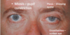
Ptosis.
eg in Horner’s syndrome,
myasthenia gravis (poss bilateral)
Name the muscles causing these movements in the the RIGHT eye


“Learn this diagram”
- Quote from eye week ICM handout…………

Switching light test -
one pupil remains dilated regardless of which eye is illuminated with the torch.
What can be deduced?
Efferent pupillary defect
The lesion is in the efferent limb of the pupillary light reflex
Switching light test -
both pupils dilate when the light is swung across from illuminating the normal eye and shone into the affected eye
What can be deduced?
A relative afferent pupillary defect
This indicates a lesion anterior to the chiasm on the affected side
Describe where you would place ECG leads V1-6
V1 - Right sternal border, 4th intercostal space
V2 - Left sternal border, 4th intercostal space
V3 - Midway between V2 and V4
V4 - Left mid clavicular line, 5th intercostal space
V5 - Left anterior axillary line, same level as V4
V6 - Left mid axillary line, same level as V4
What is the heart rate is on an ECG, the QRS complexes are:
3
4
5
large sqaures apart?
large sqaures divided by 300…….
3 = 100 bmp
4 = 75 bmp
5 = 60 bmp
How long is 1 small sqaure on an ECG?
How long is a normal PR interval in sqaures and time?
1 small square = 0.2ms
Normal PR is 120-200ms
3-5 small squares
If ECG abnormality is seen in
I, II and aVF
where is the pathology?
Inferior - Ventricular
Heart block or right coronory artery problem
If ECG abnormality is seen in
V1, V2, V3
where is the pathology?
Anterior
mainly Left ventricle
If ECG abnormality is seen in
V4, V5, V6
where is the pathology?
Lateral
Mainly left ventricle

Leuconychia
hypoalbuminaemia

Koilonychia
Iron deficiency anaemia

Dupuytrens contracture

Palmar erythema
liver disease
diabetes
Wilson’s disease

Liver flap
encephalitis

Kayser Fleischer rings
Copper deposits
Wilson’s disease

Ascites
Cirrhosis

Spider Naevi
High oestrogen

Caput medusa
Portal hypertension

Gyneacomastia
Increased oestrogen

Virchow’s Node
Abdo cancer
Name 4 LL pulses

CV Auscaltation “opening click”
indicative of?
Mitral stenosis
Cv auscaltation “ejection click”
indicative of?
aortic stenosis
What myotome is (mainily) being tested when elicited the following reflexes?
Biceps
Supinator
Triceps
Biceps - C5
Supinator - C6
Triceps - C7
What’s the scar from?

Pneumonectomy
What’s the clinical sign?

Central cyanosis
Clincal signs and what is it indicative of?

Horners syndrome.
Interruption of SNS to face poss due to tumour invading cervical plexus
What is this, and what clinical signs would you get?

Massive pleural effusion
Reduced L chest expansion
Deviated trachea away from affected side
Stony dull percussion
Reduced breath sounds
reduced vocal fremitis
reduced vocal resonance
What’s this!? and what are the clinical signs?

Pneumonectomy
traceha deviated towards affected side
reduced chest expansion on affected side
breath sounds diminished on affected side
hyperesonant
vocal fremitis and resonance reduced
What is this and what are the clinical signs?

Tension Pneumothorax
Trachea deviated away from affeted side
reduce chest expansion on affected side
hyperesonant on affected side
reduced breath sounds on affected side
reduced vocal fremitis and resonance on affected side


Whats this?
What’s it used for?

Nasal cannulae
Low flow oxygen for domicillary patients
What’s this?
What’s it used for?

Venturi mask
LTOT
What’s this?
What’s it used for?

Simple mask/medium concentration mask
STOT
What’s this?
What’s it used for?

Non-rebreathing mask
Emergency oxygen therapy
What’s this?
What’s it used for?

Bag mask ventilator BMV
CPR

Syaghorn calculus L kidney

Gallstone

Batteries!

Ureteric calculus

Gas in the biliary tree
Colecystectomy clips
What part of bowel is affected - how do you know what part it is?

Dilated colon - Large bowel
Haustra visible - absence of lines all the way accross the bowel (as in plicae circulares in small bowel)
‘coffee bean’ appearance

Dilated colon (sigmoid volvulus)
What’s wrong? how can you tell what part of bowel?
what can cause it?

Small bowel obstruction
Plicae circulares are visible all the way accross the bowel.
often caused by adhesions, malignancies

Perforated bowel - pneumoperitoneum
What’s wrong? how can you tell?

Perforated bowel
pneumoperitoneum
Riglers sign - you can see inside and outside of bowel wall
triangles/sharp corners where gas is outside the bowel.
What’s wrong? how can you tell?

Pneumoperitoneum
Riglers sign (see both sides of bowel wall in R upper quadrant)
What’s wrong? how can you tell?

Small bowel dilatation
plicae circulares
central
multiple loops
Whats’ wrong? How can you tell?

Small bowel dilatation.
plicae circulares
central
multiple loops
What’s wrong? How can you tell?

Large bowel obstruction
peripheral
fewer loops
haustra
What causes small bowel obstruction?
Adhesions
Hernia,
Malignancy
Causes of large bowel obstruction
Cancer
Diverticulitis
What’s wrong, how can you tell? What can cause it?

Large bowel obstruction
Peripheral
fewer loops
haustra
Caused by cancer, diverticulitis, hernias.
What’s wrong? How can you tell? What causes it? What else can you see?

Small bowel obstruction.
Plicae circulares
central
loops
Adhesions, hernias, cancer.
