Metabolic bone disease flashcards
Osteoporosis pathogenesis
rate of bone reabsorption > bone formation
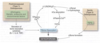
Bone remodelling unit
BRU Osteoblasts, osteoclasts
At what age does complete bone replacement cease to occur?
Unit 35-40 years old
How much bone is lost every reabsorption cycle?
Loss of bone mass is incremental with every reabsorption cycle (0.7%/year)
Classification of osteoporosis
Localised: following limb disuse Systemic
Primary systemic osteoporosis
Post-menopausal (type I), age related (type II - both sexes)
Secondary systemic osteoporosis
Endocrinopathies Neoplasias Nutritional Drugs Miscellaneous
Pathogenesis of primary osteoporosis
Contributing factors to primary osteoporosis
Genetic factors Nutritional status (calcium, vitamin D, high quality protein) Physical activity Environment
Osseous morphology post menopausal
Increased osteoclast activity Thinning out of trabeculae Loss of inter-connections
Osseous morphology of age related osteoporosis
Subperiosteal and endosteal resorption Thinning out of compact bone and trabeculae
Diagnosis of osteoporosis
Bone mineral density testing (i) Dual-emission X-ray absorptiometry (DEXA) (ii) Quantitative computered tomography (QCT) Biopsy: not used for routine diagnosis
Bone mineral dentidy T-score criteria for osteopenia and osteoporosis
Normal = >-1.0 Low bone mass (osteopenia) = between -1.0 and -2.5 Osteoporosis = <-2.5 and frigility A Z score is needed to avoid overestimation of bone mineral deficits.
Osteomalacia
Inadequate mineralisation of skeletal matrix. Outcome is defective osseous structure. In children there is also defective mineralisation of the cartilaginous matrix of the growth plate, and it is known as rickets.
Types of osteomalacia (according to causes)
Vitamin D-related Malabsorption syndromes Renal diseases Drug induced Others (poisons, tumours).
Pathogenesis of osteomalacia
(1) Deficiency of vitamin D causes hypocalcaemia
(2) Parathyroid hormone (PTH) secretion is activated, renal alpha1-hydroxylase is activated, Ca2+ is mobilised from bone, renal excretion of Ca2+ is decreased, HPO34 renal excretion increases.
(3) Serum levels of Ca2+ are restored nearly to normal.
(4) Hypophosphatemia persists with impairment of osseous mineralisation.
Paget’s disease of bone.
Osteitis deformans Bone resorption, intense bone formation, skeletal deformity. Uncommon before 40y. Male<female></female>
Developmental stages of Paget’s disease
- Osteoclastic activity 2. Osteoclastic x osteoblastic proliferation 3. Osteosclerotic phase
Pathogenesis of Paget’s disease
Obscure cause (environmental? Genetic?) Increased osteoclast recruitment
Pathology of Paget’s disease
In Paget’s disease large, abnormal, multinucleated osteoclasts cause excessive bone erosion with destruction of trabeculae and cortical bone. Each wave of bone destruction is followed by a vigorous but uncoordinated osteoblastic response, producing new osteoid to fill the defects left by the osteoclasts. The result is greatly disordered bone architecture.
There are three pathological phases: (1) Lytic phase (2) Mixed phase (3) Sclerotic phase
Lytic phase of Paget’s
Large osteoclasts with many nuclei (up to 100) Woven aspect with resorption pits
Mixed phase of Paget’s
Presence of osteoclasts Prominent osteoblasts Marrow replaced by LCT Osteoprogenitor cells Richness in blood vessels Woven and lamellar pattern
Sclerotic phase of Paget’s
Reduction in fibro-vascular pattern Normal marrow Coarse trabeculae (thick) Cortical fragility
Diagnosis of Paget’s disease
(1) X-ray: thick, coarse cortex and cancellous bone; wedge-shaped lytic edge (2) Serum alkaline phosphatase (ALP): is elevated >5x upper limit than normal (ULN) in association with osteoblastic activity. (3) Hydroxyproline: increased urinary excretion (4) Osteocalcin: serum elevation when osteoblastic activity is increased.
