MCB - Exam 3 Flashcards

1
Q

What differenciates cell types?

A

Cell - Structure and Function

  • All cells contain the same genetic material.
  • Differentiation depends on differences of gene expression.
2
Q

What are some differences in the ways cells express proteins?

A

Different Cell Types - Different Proteins

  1. Common proteins (housekeeping proteins), i.e. glucose metabolism.
  2. Specifically limited proteins, i.e. hemoglobin.
  3. Typical human cell expresses 30-60% of its 21,000 structural genes (protein coding) but level of gene expression varies - fingerprint expression profiles e.g. microarrays or RNA Seq.
  4. Other factors post transcription include: alternative splicing (dystrophin gene), post translational modification.
3
Q

What are the 6 locations where gene expression is controlled?

A

Gene Expression - Locations of Control

  1. Transcriptional control
  2. RNA processing control
  3. RNA transport and localization control
  4. Translation control
  5. mRNA degradation control
  6. Protein activity control
4
Q

Describe DNA binding motifs.

A

DNA Binding Motifs

  • Gene regulation requires:
    • Short stretches of DNA of defined sequence - recognition sites for DNA binding proteins.
    • Gene regulatory proteins - transcription factors that will bind and activate gene.
  • Recognition sequences for regulatory proteins e.g. GATA1: TGATAG
  • Logo: ex. TAATTGC
  • Recognition sequences can be proximal or distal to first exon (few base pairs - 50kb away).
5
Q

Describe DNA motif recognition.

A

DNA Motif - Recognition

  • Regulatory proteins associate with, recognize specific DNA sequence, and bind to bases in the major groove.
  • Major groove presents a specific face for each of the specific base pairs.
  • Surface of protein is extensively complementary to the surface of the DNA region to which it binds.
  • Contacts made with the DNA involve 4 possible configurations:
    • Possible H-bond donors.
    • Possible H-bond acceptors.
    • Methyl groups.
    • H atom.
6
Q

How many interactions are involved in a typical gene regulatory protein - DNA interaction?

A

10-20 interactions.

7
Q

Describe how sequence specific transcription factors are modular.

A

Sequence Specific Transcription Factors - Modularity

  • May have different modules such as:
    • DNA-binding module
    • Dimerization module - forms dimer with other subunits.
    • Activation module - turns on gene.
    • Regulatory module - regulates transcription factor.
8
Q

How was the modular nature of transcription factors proven?

A

Evidence for Modular Transcription Factors

  • Used a series of plasmids making mutant GAL4 protein and measured binding to UAS (upstream activation sequence) and expression activity.
9
Q

What are the various DNA-binding domain structural motifs?

A

DNA-Binding Domain Structural Motifs

  • Helix-turn-helix
  • Zinc finger motif
  • Leucine zipper
  • Helix-loop-helix
  • Homeodomain
  • Beta-sheet
10
Q

Describe the helix-turn-helix DNA binding motif.

A

DNA-Binding Motifs - Helix-Turn-Helix

  • Most simple and common DNA-binding motif.
  • 2 alpa-helices connected by short chain of AA that make the “turn” (turned at fixed angle).
  • Longer helix = recognition module - DNA-binding module - fits in major groove.
  • Side chains of AA recognize DNA motif.
  • Symmetric dimers: bind DNA as dimers.
11
Q

Describe the zinc finger domain DNA-binding motif.

A

DNA-Binding Motifs - Zinc Finger Module

  • DNA-binding motif includes Zn atom.
  • Binds to major groove.
  • Zn finger domains found in tandem clusters.
  • Multiple contact points.
12
Q

Describe the Leucine zipper DNA-binding motif.

A

DNA-Binding Motif - Leucine Zipper

  • 2 motifs:
    • Type I:
      • 2 alpha helical DNA binding domain.
      • Grabs DNA like clothes pin.
      • Activation domain overlaps dimer domain.
      • Interactions between hydrophobic AA side chains (leucines)
    • Type II:
      • Dimerizes through leucine zipper region (homo- / hetero-).
      • Interactions between hydrophobic AA side chains (leucines).
      • Leucine residue every 7 AA down one side of alpha-helix in dimerization domain: forms zipper structure.
13
Q

Describe the helix-loop-helix DNA-binding motif.

A

DNA-Binding Motifs - Helix-Loop-Helix

  • Consists of a short and a long alpha-chain connected by a loop.
  • Can occur as homodimers or heterodimers.
  • Three domains or modules to this protein:
    • DNA binding
    • Dimerization
    • Activation
14
Q

What is a homeodomain protein?

A

Homeodomain Protein

  • Homeodomain - 3 alpha helices.
  • Helix-turn-helix motif.
15
Q

Describe beta-sheet DNA recognition proteins.

A

Beta-Sheet DNA Recognition Proteins

  • 2 stranded beta-sheet
  • Beta-sheets consist of beta-strands.
  • Connected laterally by 2-3 backbone H-bonds.
  • Forms twisted, pleated sheet.
  • Binds to major groove of DNA.
16
Q

What is an example of a mutation in a Zn finger transcription factor leading to disease?

A

Zn Finger Transcription Factor Mutation

  • Hereditary spherocytosis.
  • Klf1 is a Zn finger protein that binds to promotor for all EMS proteins.
  • Mutation in Klf1 = no EMS proteins.
  • HS mutation: GAA to GAT or Glu to Asp in exon 3 (Zn finger domain 2).
17
Q

Describe the use of an electrophoretic mobility shift assay to identify transcription factors.

A

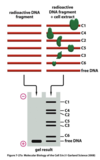
Transcription Factor Identification - Electrophoretic Mobility Shift Assay (EMSA)

  • Detection of sequence-specific DNA-binding proteins.
  • Uses radioactive DNA fragment (regulatory DNA sequence) with protein extract from cell.
  • DNA with proteins attached migrate according to size of protein.
  • Isolate protein to identify.
18
Q

Describe the use of affinity chromatography for the identification of transcription factors.

A

Transcription Factor Identification - Affinity Chromatography

  • 2 Steps:
    • Isolate DNA-binding protein.
    • Purification of sequence-specific binding proteins.
19
Q

How is chromatin immuno-precipitation used to identify DNA-binding sequences?

A

DNA-Binding Sequence Identificaiton - Chromatin Immuno-Precipitation (CHIP)

  • Allows identification of the sites in the genome that a known regulatory protein binds to.
  • Done in living cells.
  • End product can be run through PCR and used to identify sequence.
20
Q

Describe the gene control region of a typical eukaryotic gene.

A

Gene Control Region - Eukaryote

  • DNA region involved in regulating and initiating transcription of a gene.
  • Includes:
    • Promoter (where transcription factors and RNA polymerase II assembles).
    • Regulatory sequences to which regulatory proteins bind to control rate of assembly process at the promotor.
  • 9% of the 21,000 human genes (2,000) encode gene regulatory proteins.
  • Expression of mammalian genes is governed by an extremeley complex network of controls (very few generalities can be made).
  • RNA polymerase and general transcription factors assemble at the promoter.
  • Other gene regulatory proteins (activators or repressors) bind to regulatory sequences which can be adjacent, far upstream or in introns downstream of the promotor.
21
Q

How does DNA looping and a mediator complex affect activation?

A

Transcriptional Activation

  • DNA looping and a mediator complex allow the gene regulatory proteins to interact with the proteins that assemble at the promoter.
  • Mediator serves as an intermediary between gene regulatory proteins and RNA polymerase II.
22
Q

What effects the affinity between gene activator proteins and naked DNA vs DNA in nucleosomes and how can it be overcome?

A

Gene Activator Proteins

  • Binds with lower affinity to DNA in nucleosomes.
  • Binds with higher affinity to naked DNA.
  • Possibly due to the surface of the nucleotide recognition sequence facing inward.
  • 4 methods to overcome difference in affinity:
    • Nucleosome remodeling
    • Nucleosome removal
    • Histone replacement.
    • Histone modification (e.g. histone acetylation)
23
Q

How do gene repressor proteins inhibit transcription?

A

Gene Repressor Proteins - Inhibiting Transcription

  • Competitive DNA binding.
  • Masking the activation surface.
  • Direct interaction with the general transcription factors.
  • Recruitment of chromatin remodeling complexes.
  • Recruitment of histone deacetylases.
  • Recruitment of histone methyl transferase.
24
Q

Describe transcription inhibition by competitive DNA binding.

A

Transcription Inhibition - Competitive DNA Binding

  • Binding sites for receptor and activator overlap or are the same site.
25
Q

Describe transcription inhibition by gene repressor proteins masking the activation surface.

A

Transcription Inhibition - Masking Activation Surface

  • Both proteins bind to DNA, but repressor binds to the activation domain of the activator protein.
26
Q

Describe transcription inhibition by direct interaction of gene repressor proteins with the general transcription factors.

A

Transcription Inhibition - Direct Interaction with General Transcription Factors

  • Repressor binds to DNA and blocks assembly of general transcription factors.
27
Q

Describe transcription inhibition through the recruitment of chromatin remodeling complexes.

A

Transcription Inhibition - Recruitment of Chromatin Remodeling Complexes

  • Repressor recruits a chromatin remodeling complex which returns the promoter to the pretranscriptional nucleosome state.
28
Q

Describe transcription inhibition by recruitment of histone deacetylases.

A

Transcription Inhibition - Recruitment of Histone Deacetylases

  • Repressor attracts a histone deacetylase to the promotor - harder to remove deacetylated histones and open up DNA.
29
Q

Describe transcription inhibition through the recruitment of histone methyl transferase.

A

Transcription Inhibition - Recruitment of Histone Methyl Transferase

  • Repressor attracts a histone methyl transferase which methylates histones.
  • Methylated histones are bound to proteins which act to maintain chromatin in a transcriptionally silent form.
30
Q

Describe the assembly of gene regulatory proteins into complexes.

A

Gene Regulatory Proteins - Assembly into Complexes on DNA

  • Depending on the composition of complexes, proteins can be either activating or repressing.
  • The same protein can be part of an activating or repressing complex.
  • Regulation by “committee.”
31
Q

How are gene regulatory proteins controlled?

A

Gene Regulatory Proteins - Control

  • Synthesis
  • Ligand binding
  • Covalent modification - phosphorylation
  • Addition of subunit
  • Unmasking
  • Nuclear entry
  • Proteolysis
32
Q

How is mammalian gene regulation displayed in hemoglobin?

A

Mammalian Gene Regulation - Hemoglobin

  • alpha-globin like chains:
    • zeta
    • alpha
  • beta-globin like chains:
    • epsilon
    • gamma
    • delta
    • beta
33
Q

Describe hemoglobin switching.

A

Hemoglobin Switching

34
Q

How are the globin genes arranged?

A

Beta-Globin Genes

  • Arranged in linear fashion.
  • Ordered in 5’ to 3’ direction in same sequence of activation and expression during embryonic, fetal and adult development.
35
Q

Describe beta-globin gene regulation.

A

Beta-Globin Regulation

  • 100kb region containing 5 beta-globin genes and locus control region (LCR).
    • Far upstream but required for transcription.
  • Regulatory proteins bind to LCR.
36
Q

What is sickle cell anemia and what can cure it?

A

Sickle Cell Anemia

  • Sickle beta-globin.
  • If you can switch from adult Hb to fetal Hb, very little needed to cure sickle cell disease.
    • Understanding globin gene regulation may allow the induction of fetal Hb in sickle cell anemia and lead to a cure.
37
Q

What is post-transcriptional regulation of gene expression.

A

Post-Transcriptional Regulation of Gene Expression

  • Any change to the RNA after transcription that changes protein production.
    • Alternative splicing.
    • Spatial localization of mRNA.
    • RNA stability.
    • mRNA regulation.
      • IREs and IRPs
    • miRNAs
    • Post-translational processing and modification of proteins.
    • Degradation:
      • Proteasomes
      • Ubiquitin
38
Q

How can alternative splicing produce different forms of proteins from the same gene?

A

Post-Transcriptional Regulation of Gene Expression - Alternative Splicing

  • 75% of genes in humans undergo alternative RNA splicing.
  • Splice RNA transcripts differently producing different mRNAs.
  • Negative control of alternative splicing:
    • Repressor molecules prevent splicing machinery access to splice site.
  • Positive control of alternative splicing:
    • Activating molecules recruit and help direct splicing machinery.
39
Q

How can spatial localization of mRNA regulate gene expression?

A

Post-Transcriptional Regulation of Gene Expression - Spatial Localization

  • mRNAs leave nucleus through pores and either:
    • Travel to destination using cytoskeletal motors.
      • Anchor proteins hold mRNA in place.
    • Random movement leading to trapping.
    • Random movement & degeneration (RNA that is not trapped is degraded).
40
Q

How can RNA stability regulate gene expression?

A

Post-Transcriptional Regulation of Gene Expression - RNA Stability

  • Most mRNAs have 1/2-life 30 mins Globin 1/2-life is 10 hours).
  • Poly-A tail confers stability and gradual shortening by an exonuclease acts as a timer.
  • Once reduced to 25 nt, 2 pathways converge leading to degradation.
    1. Decapping - 5’ cap removed and exposed mRNA degraded from 5’ end.
    2. mRNA degraded from 3’ end through poly-A tail and into coding region.
41
Q

Describe how mRNA regulation plays a role in controlling iron absorption.

A

Iron Absorption - mRNA Regulation

  • Ferritin mRNA - storage of iron.
  • TfR mRNA - iron absorbance.
42
Q

Describe the iron cycle.

A

Iron Cycle

43
Q

Describe ferritin’s role in the iron cycle.

A

Iron Cycle - Ferritin

  • Intracellular protein
  • Binds 1,000’s of Fe3+ molecules.
  • Found in most cells.
  • Hemosiderin - granules of ferritin.
  • Excess iron is mainly stored in:
    • Liver
    • Lungs
    • Pancreas
44
Q

Describe the role of the transferrin-receptor (TfR) in the iron cycle.

A

Iron Cycle - Transferrin-Receptor

  • Transports dimeric transferrin into cell.
  • Erythroid precursors in bone marrow have 800,000 TfR molecules per cell.
45
Q

Describe transferrin receptor regulation.

A

Transferrin Receptor Regulation

  • Involves:
    • Iron responsive elements (IREs) - recognition sites.
    • Iron responsive regulatory protein (IRP) - aconitase.
  • Iron starvation:
    • No need to store Fe - decrease ferritin mRNA - IRP binds to IRE at 5’ ferritin mRNA - ferritin translation is blocked.
    • Cells must transport Fe into cells - make more TfR mRNA - IRP binds to IRE at 3’ transferrin receptor mRNA - transferrin made.
  • Iron excess:
    • Need to store Fe - make more ferritin mRNA - IRP does not bind to IRE at 5’ ferritin mRNA - ferritin made.
    • Decrease transport into cell - make less TfR mRNA - IRP does not bind to IRE at 3’ transferrin receptor mRNA - RNA degrades and no transferrin receptor is made.
46
Q

How can miRNAs regulate gene expression?

A

Post-Transcriptional Regulation of Gene Expression - miRNAs

  • miRNAs are regulatory RNAs that regulate mRNAs.
  • Noncoding RNAs, 22nt long, that silence expression of specific mRNA targets.
  • miRNAs bind to complementary sequences in the 3’ UT end of mRNA.
  • Degrade RNA or block translation.
47
Q

What are miRNAs?

A

miRNAs

  • Repressors of gene activity.
  • Development:
    • Primary miRNA - 100nt precursors with hairpin loop.
    • Processed into pre-miRNA.
      • Cropped in nucleus to form double stranded loop structure.
      • Cleaved by Dicer.
      • Joins witrh Argonaute and other proteins to form RISC: RNA-induced silencing complex.
    • Mature miRNA base pairs with mRNA & cleaves it - shutting down expression.
  • About 1,000 miRNAs in the human genome.
  • Occur in clusters.
  • A miRNA can regulate more than 1 mRNA and can repress 100’s of mRNAs.
  • May target 60% of mammalian genes.
  • Binding sites are wide-spread.
  • Can change expression profile in disease states, such as stroke, cardiovascular disease (decreased miR-29 in heart disease), or CA (increased miR-141 in prostate cancer).
48
Q

Describe the relationship of miRNA expression and disease.

A

miRNA Expression in Disease

  • Causative - miRNAs likely have mutations that cause disease.
    • Example - Tourette’s:
      • miRNA involvement was found with 1 form of Tourette’s.
      • Variant of SLITRK1 mRNA leads to increased miR-189 binding and is shown to be associated with Tourette’s.
  • Responsive - increased miRNA expression down-regulates genes in response to disease to limit severity.
49
Q

How can protein activity control regulate gene expression?

A

Post-Transcriptional Regulation of Gene Expression - Protein Activity Control

  • Post translational modifications required by proteins to be functional.
    • Phosphorylated
    • Glycosylated
    • Bind to other subunits or partners.
    • Modified by enzymes (i.e. thrombin cuts fibrinogen to form fibrin).
  • Proteins must fold into their 3-D conformations.
  • Molecular chaperones help proteins fold.
    • Heat shock proteins (Hsp60 & Hsp70)
  • Bind co-factors.
50
Q

How can protein degradation regulate gene expression?

A

Post-Transcriptional Regulation of Gene Expression - Protein Degradation

  • Ubiquitin identifies unfolded or abnormal proteins for destruction.
  • Ubiquitin binding process:
    • E1 ubiquitin activating enzyme links ubiquitin to cysteine side-chain.
    • Transfered to E2 ubiquitin conjugating enzyme (with accessory protein E3 ubiquitin ligase).
      • E3 activated by phosphorylation, ligand binding, or protein subunit addition.
    • Transferred to lysine side chain of proteins with degradation signal.
      • Degradation signal activated by phosphorylation, unmasking of signal by protein dissociation, or creation of destabilizing N-terminus.
  • Ubiquitin chain is recognized by proteasome.
  • Aberrant protein destroyed.
51
Q

Describe the relationship of proteosomes and disease therapy.

A

Proteasomes and Therapy

  • Proteasome inhibitors used to treat multiple myelomas (CA of plasma cells).
    • Abnormal cells accumulate in bone marrow and interfere with RBC production.
    • Incurable but treatable.
  • Bortezomid interacts with 1 proteolytic site on proteasome, reversibly inhibiting it.
  • May prevent degradation of pro-apoptotic factors triggering programmed cell death in neoplastic CA cells.
  • Potential future use for proteasomes: activating them to enhance clearance of misfolded proteins such as beta-amyloid in Alzheimer’s.
52
Q

What are 4 types of gene expression controls, other than post-transcriptional and post-translational?

A

Other Controls of Gene Expression

  • Coordinated expression of genes.
  • Decision for specialization.
  • Methylation and genomic imprinting (which genes get expressed or repressed from mom and dad).
  • X-chromosome inactivation.
53
Q

Describe coordinated gene expression.

A

Coordinated Gene Expression

  • Expression of critical regulatory protein can trigger battery of downstream genes.
  • Coordinated gene expression in response to need.
  • Example: glucocorticoid cortisol (response to stress) - increase BGL - aid in fat, protein, carb metabolism - diurnal (high at 8am vs. low at 12am).
54
Q

Describe the specialization in controlling gene expression.

A

Controlling Gene Expression - Specialization

  • Combinations of gene control can produce many types of cells.
  • Decision at each step (1 or none, 2 or 3, 4 or 5)
55
Q

How does methylation regulate gene expression?

A

Regulating Gene Expression - Methylation

  • DNA can be regulated by proteins and can be covalently modified.
  • Methylation shuts down gene expression.
  • DNA methylation can be inherited:
    • Methylated parent strand serves as template for daughter strand.
    • Maintenance methyltransferase methylates cytosine.
    • Basis for genomic imprinting.
56
Q

What is genomic imprinting?

A

Genomic Imprinting

  • Differential expression of genetic material depending on the parent of origin.
57
Q

What is epigenetics?

A

Epigenetics

  • Regulation of expression of gene activity without altering gene structure (e.g. methylation).
58
Q

Describe Prader Willi syndrome (PWS).

A

Prader Willi Syndrome

  • Genomic imprinting disorder.
  • Caused by paternal deletion on chromosome 15 in the region 15q11-q13.
  • Patients inherit gene deletion from father, genes in this range only expressed from paternal origin so deletion means that neither the maternal or paternal genes are expressed.
  • Presentation:
    • Stage 1: Infantile hypotonia; poor suck; feeding difficulties - failure to thrive.
    • Stage 2: Hyperphagia (uncontrollable eating); onset of early childhood obesity (cardinal feature and most significant health problem).
  • Avg age of onset - 2y (range 1y-6y).
  • Greater than 40% body fat or 2-3x higher than general population.
  • Hypogonadism.
  • Short stature; small hands and feet; hypopigmentation.
  • Mental deficiency; behavior problems (skin picking, OCD)
59
Q

How does X-chromosome inactivation regulate gene expression?

A

Regulation of Gene Expression - X-Chromosome Inactivation

  • F = XX; M = XY
  • Because females have 2 X-chromosomes and humans do not deal well with duplicate genes, dosage compensation occurs and 1 X chromosome is inactivated.
  • X-chromosome is inactivated by becoming highly condensed heterochromatin (Barr body).
    • Inactivation is random.
    • Maintained post cell divisions.
    • Gamete cell formation resets inactivation.
    • Inactivation starts and spreads from X-inactivation center (XIC) by formation of XIST RNA which coats entire X-chromosome.
  • Females are mosaics of 2 types of cells - either the paternal or maternal x is inactivated.
60
Q

What is the dogma of life?

A

Dogma of Life

  • Must find food.
  • Must not become food.
  • Must reproduce.
61
Q

What is cytokinesis?

A

Cytokinesis

  • Cell division into two cells.
62
Q

What is the goal of the cell cycle?

A

Cell Cycle - Goal

  • Produce 2 genetically identical daughter cells.
  • DNA in each chromosome must be faithfully replicated into 2 copies.
  • Precise replication of 6.4 x 10⁹ base pairs in the diploid human genome.
63
Q

What is the challenge of the cell cycle?

A

Cell Cycle - Challenge

  • Mistakes occur at rate of 1 x 10⁻⁹ per replication (6.4 X 10⁹ base pairs)
  • About 6 mistakes occur in one cell division.
  • The replicated chromosomes must be accurately distributed in daughter cells (segregation).
64
Q

What is the importance of the cell cycle?

A

Cell Cycle - Importance

  • 3x10¹³ cells in body, ~ 1016 cell divisions in one lifetime.
  • Some cells replaced constantly (intestinal cells 3-4 days), some not (liver cell 1 year).
  • If system malfunctions - cancer can result.
  • Cancer is a disease of excess cell proliferation.
65
Q

How is the cell cycle controlled?

A

Cell Cycle - Control

  • Complex network of regulatory proteins (ordered series of biochemical switches) that govern progression through the cell cycle.
    • Initiate main events such as chromosome duplication and segregation.
    • Responds to signals inside and outside the cell.
  • Coordinates growth of cell (chromosomes and organelles) with cell division.
  • Proteins coordinate events so they occur at the appropriate time.
    • Prevents preparation for segregation of chromosomes until DNA replication is complete.
  • Regulates cell #’s in a multi-cellular organism.
66
Q

What are the major chromosomal events in the cell cycle?

A

Cell Cycle - Major Chromosomal Events

  • Chromosomal duplication.
  • Chromosomal segregation.
  • Cytokinesis.
67
Q

What are the 4 phases of eukaryotic cell division?

A

Cell Division - 4 Phases

  1. Prophase - chromosomes condense into rigid rods called sister chromatids (become attached to mitotic spindle - a bipolar array of microtubules).
  2. Metaphase - sister-chromatids line up at equator of cell attached to opposite poles of spindle.
  3. Anaphase - sister chromatids become daughter chromosomes and are pulled to opposite poles of spindle.
  4. Telophase - spindle disassembles, chromosomes packaged into separate nuclei, cytokinesis occurs.
68
Q

What are the 4 phases of the cell cycle?

A

Cell Cycle - 4 Phases

  • S phase - DNA synthesis
  • M phase - separate chromosomes and divide cells.
  • GAP phases - allow more time for growth:
    • G1 - between M & S
    • G2 - between S & M
69
Q

What is interphase?

A

Cell Cycle - Interphase

  • G1, S, G2
70
Q

How long does the M phase last?

A

Cell Cycle - M Phase

  • 1 hour
71
Q

What are the 3 major transition checkpoints in the cell cycle?

A

Cell Cycle - Transition Check Points

  • Checkpoint I: START - cell commits to cell cycle entry and chromosome duplication (also called restriction point).
    • If conditions are not conducive to mitosis, cell cycle will not proceed.
    • Once started, cells will continue cell cycle even if conditions change.
  • Checkpoint II: G2/M - chromosome alignment on spindle in metaphase.
  • Checkpoint III: metaphase-to-anaphase transition - trigger sister chromatid separation and cytokinesis.
72
Q

What models are used for studying the cell cycle?

A

Cell Cycle - Studying

  • Yeast
  • Animal embryos:
    • Frog embryos - initially has no detectable G1 or G2 with S and M lasting about 15 minutes.
  • Cell lines:
    • Fibroblasts are a mammalian cell line, however they stop dividing in culture after a certain # of cycles (25-40).
    • Immortalized cell lines:
      • Murine erythroleukemia cells (MEL)
        • Useful for studying erythroid cell development and RBC generation.
        • Can induce erythropoeisis using growth factors.
      • Human erythroleukemia cells (HEL)
73
Q

Describe Cdks.

A

Cell Cycle - Cdks

  • Cyclin dependent kinases (Cdks) govern cell cycle, phosphorylate proteins downstream to activate them and regulate cell cycle events.
  • Causes cyclical changes in phosphorylation of substrates that regulate cell cycle events.
  • Activities of Cdks rise and fall during cell cycle.
  • Levels of Cdks remains constant.
74
Q

Describe cyclins.

A

Cell Cycle - Cyclins

  • Proteins that regulate Cdks.
  • Levels vary and cycles during the cell cycle (hence name ‘cyclins’).
    • Expression controls what step of the cell cycle the cell is in (cyclin present -> cyclin-Cdk complexes formed triggering cell cycle events).
  • Cdks are dependent on cyclins - must be bound to cyclin to have protein kinase activity.
  • Direct Cdks to their specific target.
75
Q

What are the 4 classes of cyclins?

A

Cyclins - 4 Classes

  1. G1/S cyclins:
    • Start cell cycle
    • Activates Cdks in late G1, helping trigger progression through START.
    • Levels drop in S phase.
  2. S cyclins:
    • Stimulates DNA duplication by binding Cdks after progression through START.
    • Levels remain high until mitosis.
  3. M cyclins:
    • Initiate mitosis by binding to Cdks that stimulate entry into mitosis at G2/M checkpoint.
    • Removed at mid-mitosis.
  4. G1 cyclins:
    • Govern activity of G1/S cyclins, controlling progression through START checkpoint.
76
Q

What are the 4 Cdks?

A

Cdk - 4 Types

  • 4 different Cdks that form cyclin-Cdk complexes:
    • G1/S-Cdk
    • S-Cdk
    • M-Cdk
    • G1-Cdk
77
Q

Describe the formation and activation of the cyclin-Cdk complex.

A

Cyclin-Cdk Complex - Formation & Activation

  • Cdk is inactive without cyclin being bound. The active site is blocked by a region of the protein called the T-loop.
  • Binding of cyclin causes T-loop to move out of active site, partially activating Cdk.
  • Phosphorylation of Cdk at T-loop, by Cdk activating kinase (CAK), fully activates enzyme (“cave site”).
78
Q

Describe the regulation of Cdk activity by an inhibitor.

A

Cdk - Regulation by Inhibitor

  • Wee1 - a kinase inhibits M-Cdk by phosphorylating the “roof” site, inhibiting the M-Cdk complex (ensures lots of primed M-Cdk by end of G2).
    • Cdc25 - a phosphotase that dephosphorylates the “roof” site of M-Cdk, reactivating the M-Cdk complex to initiate mitosis.
    • Double-circuit positive feedback ensures fast M-Cdk activation.
      • Activated M-Cdk activates more Cdc25.
      • Cdc25 inhibits Wee1.
  • p27 - a Cdk Inhibitory Protein (CKI) that binds to both Cdk and cyclin to inactivate complex.
    • Primarily used for control of G1/S-Cdks & S-Cdks early in cell cycle.
79
Q

Describe the link between CKIs and disease.

A

CKI - Disease

  • Hereditary melanoma:
    • Mutation in INK4A, a CKI involved in the G1 phase, causes loss of activity.
    • Leads to uncontrolled cell cycle (cancer).
  • p53 & p21
    • p53 - CKI that is a major tumor suppressor.
    • p21 - CKI to stop cell division.
      • p21 transcription is a target of p53.
      • If p53 fails, p21 expression is reduced, leading to uncontrolled cell division (cancer).
80
Q

Describe the regulation of Cdk activity by proteolysis.

A

Cdk - Regulation by Proteolysis

  • SCF:
    • Used to activate S-Cdk at S phase by removing CKI.
    • SCF-ubiquitin ligase adds ubiquitin to CKIs, marking them for destruction.
    • SCF activity depends on F-Box subunit, which helps SCF recognize target protein.
    • Adds ubiquitin to CKI on S-Cdk in G1, activating S-Cdk for S phase.
  • APC/C (anaphase-promoting complex (or cyclosome)):
    • Ubiquitin ligase that initiates progression from metaphase to anaphase in mitosis.
    • Adds ubiquitin to:
      • Securin - protects cohesin protein linkages that hold sister chromatid pairs together in early mitosis.
      • S- and M-cyclins in order to inactivate most Cdks in the cell.
81
Q

Describe cohesins.

A

Cohesins

  • Members of SMC proteins (structural maintenance of chromosomes)
  • Form rings around sister chromatids.
82
Q

Describe securin.

A

Securin

  • Protects cohesins.
  • Inhibits protein called separase, which cleaves cohesin.
83
Q

Describe separase.

A

Separase

  • Protein that cleaves cohesin.
  • Necessary for sister chromatids to become daughter chromosomes.
84
Q

Describe APC/C and its role in the transition from metaphase to anaphase.

A

APC/C - Transition from Metaphase to Anaphase

  • APC/C is a ubiquitin ligase that initiates progression from metaphase to anaphase in mitosis.
  • Levels rise in mid-mitosis.
  • Adds ubiquitin to:
    • Securin - protects cohesin protein linkages that hold sister chromatid pairs together in early mitosis by inhibiting separase.
  • Cyclins
    • Activated by Cdc20.
    • Targets S-cyclins and M-cyclins.
    • Leads to addition of polyubiquitin to M-cyclin in M-Cdk complex.
      • Cyclins destroyed, Cdks dephosphorylated.
    • In anaphase S-cyclins destroyed.
85
Q

Describe the cell cycle control system.

A

Cell Cycle - Control System

  1. Signals cause activation of G1-Cdk
  2. G1-Cdk stimulates genes making G1/S-cyclin & S-cyclin. Proceeds through START checkpoint.
  3. G1S-Cdk activity induces S-Cdk activity causing DNA replication.
  4. M-Cdk drives expression through G2/M checkpoint.
  5. Apc/C + Cdc20 triggers destruction of securin and cyclins at metaphase-to-anaphase transition. Anaphase occurs, mitosis completes.
86
Q

What are the 2 parts of cell cycle regulation in mitosis?

A

Cell Cycle Regulation - Mitosis

  1. Increase of M-Cdk activity at G2/M triggers prophase, prometaphase and metaphase.
    • Assembly of mitotic spindle.
    • Attachment to sister chromatids.
  2. Metaphase-to-anaphase transition: APC/C triggers destruction of securin, cohesins and cyclins.
87
Q

Describe chromosome duplication control.

A

Cell Cycle Regulation - Chromosome Duplication

  • Promotes complete and accurate replication of DNA.
  • Prevents more than one cycle of duplication per cell cycle.
  • 2 steps:
  1. At G1 phase, prereplicative complexes or PRE-RC assembles at origins of replication.
  2. At S phase replication forks are created.
  • Chromosome duplication followed by mitosis.
  • No new PRE-RCs made in mitosis, assembly is inhibited by Cdk activity.
88
Q

What is condensin?

A

Condensin

  • Five subunit protein complex.
    • Related to cohesin.
    • Contains 2 SMC subunits (structural maintenance of chromosomes).
    • Contains 3 non-SMC subunits.
  • Forms a ring-like structure and uses ATP to promote compaction and resolution of sister chromatids.
89
Q

What is the mitotic spindle?

A

Mitotic Spindle

  • A bipolar array of microtubule proteins that pulls sister chromatids apart at anaphase.
  • M-Cdk triggers assembly of spindle.
  • Microtubule organization begins at mitosis stage.
  • All spindle microtubules bind to centrosome.
  • Consists of 3 types of microtubules:
    • Astral microtubules - interact with cell cortex.
      • Radiate outward from the poles and contact the cell cortex helping to position the spindle.
    • Kinetochore microtubules - attach each chromosome to spindle pole.
      • Plus ends attached to sister chromatid pairs at large protein structures called kinetochores (located at centromere)
    • Interpolar microtubules - hold two halves of spindle together.
      • Plus ends from one pole interact with plus ends from other pole.
90
Q

Describe gamma-TuRC.

A

Gamma-TuRC

  • Involved with nucleation of microtubule at the MTOC.
  • Binds negative end of microtubule.
91
Q

Describe centrosomes.

A

Centrosomes

  • Protein organelles.
  • Consist of:
    • Matrix
    • Pair of centrioles.
    • Gamma-TuRC (more than 50 copies)
  • Replicate with 2 pairs of centrioles per centrosome.
  • Migrate to poles to form mitotic spindle.
92
Q

Describe the motor proteins of the mitotic spindle.

A

Mitotic Spindle - Motor Proteins

  • 2 major types of proteins:
    • Dyneins:
      • Tend to move to center of cell.
      • Minus-end directed.
    • Kinesins:
      • 3 classes:
        • kinesin-5
        • kinesin-4,10
        • kinesin-14
      • Tend to move to periphery of cell.
      • Typically plus-end directed with acception of kinesin-14.
      • 2 globular heads and elongated coil-coil tails.
      • Plays important role in chromosome separation.
93
Q

Describe kinesin-5

A

Mitotic Spindle Motor Proteins - Kinesin-5

  • 2 motor domains that interact with plus end of anti-parallel microtubules.
  • Move anti-parallel microtubules past each other to force or push the spindle poles apart.
94
Q

Describe kinesin-14

A

Mitotic Spindle Motor Proteins - Kinesin-14

  • Minus end oriented.
  • Single motor domain.
  • Pulls poles together.
  • Opposes kinesin-5 (no kinesin-5 spindle collapses).
95
Q

Describe kinesin-4,10.

A

Mitotic Spindle Motor Proteins - Kinesin-4,10

  • AKA chromokinesins.
  • Plus end directed.
  • Push attached chromosomes away from pole.
  • Oppose movement of kinetochore microtubules, which pull chromosome from kinetochore toward pole.
96
Q

Describe dyneins with respect to the mitotic spindle.

A

Mitotic Spindle Motor Proteins - Dyneins

  • Minus end directed.
  • Link plus ends of astral microtubules to actin skeleton at cell cortex.
  • Pull spindle poles away from each other.
97
Q

Describe kinetochores.

A

Kinetochores

  • Giant multilayered protein structure built on the chromosome.
  • Multiple microtubules attach to kinetochore (Ndc80 complex)
  • Exposed open end for addition and removal of tubulin subunits.
    • Removal of subunits leads to force on kinetochore pulling chromosomes to pole of cell.
  • Spindle microtubules attached to each sister chromatid at the kinetochore.
98
Q

Describe microtubule binding to the kinetochore.

A

Kinetochore - Microtubule Binding

  • Bipolar attachment.
  • Sister chromatids must attach to opposite poles of mitotic spindle (biorientation).
  • Formation of unstable connections is not allowed.
  • Stable attachment is detected by tension on kinetochore.
99
Q

What are the three forces involved in chromosome movement?

A

Chromosome Movement - 3 Forces

  • Depolymerization:
    • Depolymerization of plus end of microtubule pulls the kinetochore and chromosome toward the pole.
  • Microtubule flux:
    • Pulled toward spindle poles while being dismantled at minus ends.
    • Tubulin added at plus end while being removed at minus end - interpolar microtubules.
  • Polar ejection force:
    • Kinesin-4,10 motors on chromosomes interact with microtubules and transport chromosomes from poles.
    • Results in push-pull phenomenon.
100
Q

Describe anaphase A and B.

A

Anaphase A & B

  • Anaphase A - chromosomes move apart due to microtubule depolymerization at kinetochore.
  • Anaphase B - separation of spindle poles by kinesin-5 motor proteins (also dynein).
101
Q

Describe cytokinesis.

A

Cytokinesis

  • Final step of cell cycle.
  • First visible change is the cleavage furrow.
  • Contractile ring underlying the cleavage furrow.
    • Composed of actin and myosin filaments.
    • Assembly of ring results from local formation of new actin filaments (depends on formin).
  • Rings contract, vesicles fuse with membrane to create new membrane.
  • 4 stages:
    • Initiation
    • Contraction
    • Membrane insertion.
    • Completion
102
Q

How is cell division controlled?

A

Cell Division - Control

  • Extracellular signaling molecules regulate cell size and number.
  • 3 classes of signaling molecules:
    • Class 1: mitogens - stimulate cell division by triggering G1/S-Cdk activity.
    • Class 2: growth factors - stimulate cell growth.
    • Class 3: survival factors - suppress form of programmed cell death known as apoptosis.
  • IF ANY OF THESE IS ABERRANT - CANCER RESULTS!
103
Q

Describe mitogens.

A

Mitogens

  • Ensures that cells only divide when more cells are needed.
  • Example: PDGF, EGF, EPO
  • Activate the Ras-MAPK pathway:
    • Mitogen binds to receptor.
    • Ras causes activation of MAPK (mitogen activated protein kinase) cascade.
    • Leads to increase of gene regulatory proteins including Myc.
    • Myc promotes entry into cell cycle by increasing expression of G1 cyclins.
104
Q

Describe the role of Myc and E2F I in the cell cycle.

A

Cell Cycle - Myc and E2F I

  • G1-Cdk activates group of gene regulatory factors called E2F proteins.
  • E2F binds to promoters of G1/S cyclin and S cyclin genes (leads to DNA transcription).
  • Enter into S phase of cell cycle.
  • DNA synthesis begins.
105
Q

Describe the role of Rb protein in the cell cycle.

A

Cell Cycle - Rb Protein

  • Functions as a regulator:
    • E2F protein is inhibited by interaction with Rb protein (retinoblastoma protein).
    • Shuts down entry into S phase.
    • Active G1-Cdk phosphorylates Rb to reduce binding to E2F.
  • No regulation of entry into cell cycle = cancer.
106
Q

Describe Rb protein.

A

Retinoblastoma Protein Family

  • Rb identified through studies of inherited eye cancer (retinoblastoma) in children.
  • Rare - 4 per million.
  • Occurs before age 2.
  • Rb is tumor supressor that prevents over-proliferation of cells.
  • Loss of both copies of Rb genes leads to cell and tumor proliferation of retina.
  • No inhibition = retinoblastoma.
107
Q

Describe the role of ATM/ATR in cancer.

A

ATM/ATR

  • DNA damage activates ATM and ATR protein kinases.
    • Blocks division by stopping progression through cell cycle.
  • ATM/ATR kinases associate with site of damage and phosphorylate Chk1 and Chk2 proteins (checkpoint kinase 1 & 2).
    • Major target of Chk1/Chk2 is p53 which stimulates transcription of p21.
    • p21 CKI binds to G1/S-Cdk and S-Cdk to inhibit activity - no cell division- damaged DNA must be repaired.
  • ATM/ATR not working = Cancer
108
Q

Describe Ataxia Telangiectasia.

A

Ataxia Telangiectasia (AT)

  • ATM protein dysfunction leads to AT.
  • AT patients exhibit higher incidence of lymphoma and leukemia.
  • Ataxia - poor coordination
  • Telangiectasia - small dilated blood vessels.
109
Q

Review

A
110
Q

Describe relationship of Ras and cancer.

A

Ras is mutated in 30% of cancers.

111
Q

Describe relationship of p53 and cancer.

A

p53 mutations occur in 50% of human cancers.

112
Q

Describe the relationship of mitogen activating genes and cancer.

A

Many mitogen activating genes identified as cancer-promoting genes or oncogenes.

113
Q

Describe the PI-3 kinase pathway.

A

Pi-3 Kinase Pathway

  • Most important growth signaling pathway.
  • PI-3 kinase adds ATP to PIP2 (inositol phospholipids).
  • Activated TOR which activates many factors for cell growth.
  • PI-3 kinase phosphorylates PIP2 which activates TOR and downstream factors for cell growth.
114
Q

What are the three mechanisms that coordinate cell growth with division?

A

Cell Growth and Division - Coordination

  • 3 mechanisms:
    1. Rate of cell division determined by extracellular factor leading to cell growth.
    2. Cell growth and division controlled separately by growth factors and mitogens.
    3. Cell growth and division both stimulated by extracellular factor.
115
Q

What effect does mitogen depletion have on cell division?

A

Cell Division - Limited by Mitogen

  • Cells in culture exhibit “density-dependent inhibition of cell division.”
  • Depletion of mitogens and addition of fresh serum leads to cell proliferation.
116
Q

Describe the effect myostatin has on muscle cells.

A

Myostatin

  • Inhibits muscle cell growth.
  • Knockout myostatin leads to large muscles.
  • Myostatin inhibitors used to treat DMD.
117
Q

What is apoptosis?

A

Apoptosis

  • Programmed cell death, or cell death under physiological conditions.
    • Used to eliminate unwanted cells.
    • Important for removal of abnormal, non-functional, potentially dangerous cells.
      • Lymphocytes after destroying and ingesting microbes.
      • Cells with DNA damage that cannot be repaired.
    • Used to maintain correct organ size (cell division = cell death).
  • Clean form of cell death:
    • Tightly controlled and regulated.
    • No spread of damage or initiation of inflammatory process.
  • Most common cell death.
118
Q

What are the two types of cell death?

A

Cell Death - Types

  • Apoptosis
  • Necrosis
119
Q

What is necrosis?

A

Cell Death - Necrosis

  • Accidental cell death or cell death by injury.
  • Dirty way of dying - cells may rupture, releasing contents and initiating an inflammatory response.
120
Q

How is apoptosis critical for development?

A

Apoptosis - Critical for Development

  • Removes unwanted cells.
  • Sculpts hands, feet, etc, during embryonic development.
  • Examples:
    • Tail of tadpole.
    • Development of mouse paw.
121
Q

Describe the phenotype of apoptosis.

A

Apoptosis - Phenotype

  • Overall shrinkage of cell volume and nucleus.
  • Loss of adhesion to neighboring cells.
  • Formation of blebs on surface.
  • DNA fragmentation.
  • Cytoskeleton collapses.
  • Nuclear envelope disassembles.
  • Rapid engulfment of dying cell by phagocytosis.
122
Q

What are the biochemical characteristics of apoptosis?

A

Apoptosis - Biochemical Cahracteristics

  • DNA fragmentation visible on agarose gel.
    • Endonucleases cleave DNA in linker regions of nucleosomes.
  • Cytochrome C released from mitochondria is a marker of apoptosis.
123
Q

What are caspases?

A

Apoptosis - Caspases

  • Caspase = cysteine aspartyl specific protease.
    • Cysteine in active site.
    • Cleaves proteins at aspartic AA residues.
  • Proteases that mediate intracellular proteolytic cascade of apoptosis.
  • Activation of caspases is key event.
  • 2 Classes:
    • Initiater caspases - activates multiple procaspases into executioner caspases.
    • Executioner caspsases - destroys actual targets:
      • Cleaves downstream proteins.
      • Cleaves inactive endonuclease.
      • Targets cytoskeleton.
      • Attacks cell adhesion proteins
      • Cells roll up into ball.
124
Q

What are procaspases?

A

Apoptosis - Procaspases

  • Initial, inactive, precursor of caspases.
  • Activated by protease cleavage.
  • Procaspases cleaved at specific sites to form a large and small subunit which form a heterodimer.
125
Q

Describe the caspase cascade.

A

Apoptosis - Caspase Cascade

  • Machinery always in place.
  • Irreversible.
  • 2 pathways:
    • Internal:
      • Stimuli - DNA abnormalities.
      • Mitochondrial dependent.
    • External:
      • Stimuli - removal of survival factors and proteins of tumor necrosis factor family.
      • Mitochondrial independent.
126
Q

Describe the extrinsic apoptotic pathway.

A

Apoptosis - Extrinsic Pathway

  1. Extracellular signals (Fas ligand) bind to cell surface death receptors (Fas) and trigger pathway.
    • Receptors are transmembrane proteins with 3 domains:
      • Extracellular binding domain.
      • Single transmembrane domain.
      • Intracellular death domain.
    • Homotrimers
    • Members of TNF family.
  2. Adaptor proteins recruited:
    • FADD adapter (Fas associated death domain) & procaspase-8, both with death effector domain.
  3. Death inducing signal complex (DISC) formed by Fas, FADD, and procaspase-8 or -10.
  4. Caspase-8 or -10 activated.
  5. Downstream executioner caspases activated - caspase-3.
  • There are inhibitory proteins that restrain the extrinsic pathway.
    • Decoy receptors - receptors with ligand binding domain but no death domain, binds death ligand but does not activate apoptosis.
    • FLIP - competitive inhibitor against procaspase-8 and procaspase-10 (resembles procaspase with no proteolytic domain).
    • Decoy and FLIP act as sponges absorbing ligand.
127
Q

Describe the intrinsic apoptotic pathway.

A

Apoptosis - Intrinsic Pathway

  • Cells activate apoptosis from inside cell in response to injury, DNA damage and lack of oxygen, nutrients, or extracellular survival signals.
  1. Translocation of cytochrome c from the intermediate space of mitochondria is key event.
  2. Released into cytosol and will bind to Apaf1 (procaspase-activating adaptor protein).
  3. Apaf1 forms apoptosome which actiates caspase-9.
  4. Caspase-9 activates downstream exectutioner caspases - caspase-3 (common to both pathways).
128
Q

Describe the Bcl2 family of proteins.

A

Bcl2 Family of Proteins

  • Regulate intrinsic pathway.
  • Bcl2 controls release of cytochrome c into cytosol (Bcl = B cell lymphoma)
    • 2 types:
      • Anti-apoptic (pro-survival) - blocks release of cytochrome c (example: Bcl2)
      • Pro-apoptotic - promotes release of cytochrome c.
    • 4 distinct Bcl homology domains (BH1-4)
      • Proapoptotic proteins include BH-123 or BH-3 only.
129
Q

Describe the BH123 protein.

A

Apoptosis - BH123

  • Pro-apoptotic - triggers the intrinsic pathway.
  • Become activated.
  • Form aggregation in mitochondrial outer membrane.
  • Induce release of cytochrome c.
  • Apoptosome formed by binding to Apaf1
130
Q

Describe Bcl2.

A

Apoptosis - Bcl2 Proteins

  • Anti-apoptotic proteins.
  • Includes Bcl2 and Bcl-XL (Both BH1234)
  • Mainly located on cytosolic surface of outer mitochondrial membrane.
  • Prevent apoptosis by binding to pro-apoptotic proteins (e.g. BH123) and prevent aggregation into active form.
  • Can be inhibited by BH3-only protein.
131
Q

Describe the BH3-only protein.

A

Apoptosis - BH3-Only Protein

  • Pro-apoptotic.
  • Activated BH3-only protein is cytosolic.
  • Translocates to mitochondria after apoptotic signal activates it.
  • Inhibits anti-apoptotic Bcl2 protein from inhibiting aggregation to release cytochrome c.
132
Q

Describe IAPs.

A

Inhibitors of Apoptosis (IAPs)

  • Bind and inhibit caspases.
  • Some IAPs add ubiquitin to caspases to mark them for destruction by proteasome.
  • IAPs block apoptosis by binding to caspases.
133
Q

What are anti-IAPs?

A

Anti-IAPs

  • Pro-apoptotic
  • If there is apoptotic stimuli or apoptosis signals, this triggers the release of anti-IAPs from mitochondria to block the activity of IAPs.
  • Allowing executioner caspases to be activated.
134
Q

How can insufficient apoptosis contribute to disease?

A

Apoptosis - Insufficient

  • Insufficient apoptosis leads to excessive #’s of cells and cancer.
  • Excessive Bcl2 (inhibits apoptosis) promotes the development of cancer by inhibiting apoptosis of DNA-damaged cells allowing them to proliferate.
  • Mutated p53 can no longer cause cell cycle arrest and no longer promotes apoptosis allowing DNA damaged cells to continue to divide promoting cancer.
  • Excessive apoptosis can also be a problem (i.e. AMI & Stroke).
135
Q

What are some of the statistics and basic characteristics of cancer?

A

Cancer

  • 1/5 will die of cancer.
  • 1/3 will contract cancer.
  • Top 2 causes of death - MI and CA.
  • Lung CA is most deadly. primarily due to inability to remove tumor in many cases.
  • Disease of age.
    • Takes time to accumulate mutations necessary to develop.
136
Q

What is cancer?

A

Cancer

  • A disease in which an individual mutant clone of cells begin by prospering at the expense of its neighbor cells.
  • Descendants of these clones can destroy the whole cell society in your body.
  • Pathways are affected by cancer, which means we can come up with ways to combat it.
137
Q

Describe the properties of CA cells.

A

Cancer - Cell Properties

  • 2 heritable properties:
    1. Reproduce in defiance of normal restraints on cell division and cell growth.
      • Become self-sustaining, do not need signals to grow.
        • Normal cells have strong requirement for growth factors.
      • Release autocrine growth factor signals.
      • Do not respond to anti-growth signals.
      • Do not require signals to proliferate.
      • Do not respond to apoptosis signals.
    2. Invade areas normally reserved for other cells.
  • Get help from stromal cells.
  • Induce angiogenesis.
  • Do not display replicative senescence - immortal.
138
Q

How does cancer kill?

A

Cancer - How it Kills

  • As a tumor spreads, it squeezes or destroys blood vessels and nerves until organ can no longer do its job.
139
Q

Describe the pathology of cancer.

A

Cancer - Pathology

  • An abnormal cell that grows (increases in mass) and proliferates (divides) out of control will give rise to a tumor or neoplastic growth.
  • If neoplastic cells do not become invasive, tumor is benign (not cancer) and can be surgically removed as cure.
  • Tumor is malignant (cancer) if cells have ability to invade surrounding tissues.
140
Q

How are cancers classified?

A

Cancer - Classification

  • Carcinoma: from epithelial cells (most common).
  • Sarcoma: from connective or muscle tissue.
  • Leukemias and lymphomas - from WBCs and their precursors.
141
Q

What is an adenoma?

A

Adenoma - benign epithelial tumor with glandular organization (adenocarcinoma if malignant).

142
Q

What is an adenocarcinoma?

A

Adenocarcinoma - malignant epithelial tumor with glandular organization.

143
Q

How are cancers named?

A

Cancer - Naming

  • Have names reflecting tissue of origin.
  • Examples:
    • Basal-cell carcinoma - keratinocyte stem cell in skin.
      • Rarely metastasize.
    • Melanoma - pigment cells in skin.
      • Often widely metastasize.
144
Q

What is metastases?

A

Metastases

  • Cells break loose, enter into blood or lymph, travel to new areas and form secondary tumors.
  • Metastases kills patients.
145
Q

Describe tumor development.

A

Tumor Development

  • Represents tumor progression.
  • Involves multiple mutational events that:
    • Confer a proliferative advantage allowing cells to grow more rapidly than normal.
  • All tumors from single ancestor.
    • Single cell that outgrows, out-divides and out-lives neighboring cells.
  • Evolve from benign growth to invasive cancers and ultimately metastasize.
  • Develop slowly (smoking in 20’s = potential for lung CA in 50’s and 60’s)
146
Q

Describe colon cancer tumor development.

A

Tumor Development - Colon Cancer

  • Mutation in APC gene (tumor supressor).
  • Cells with APC mutation gain an advantage in growth.
  • Form polyps - benign tumor.
  • Mutation in Ras - becomes “cancer” gene.
  • Loss of other tumor suppressor (p53) - carcinoma.
  • Tumor moves out into bloodstream.
  • Gains capacity to invade = now malignant.
147
Q

Describe how chronic myelogenous leukemia develops from a single heritable cell.

A

CML - Dervied from Single Cell

  • Philadelphia chromosome:
    • Responsible for CML
    • Translocation between 9 & 22.
    • Smaller chromosome.
  • All CML cells have same chromosomal aberration, no spectrum of different defects.
  • Must come from a single cell.
148
Q

Describe tumor progression in cancer.

A

Cancer - Tumor Progression

  • Example: cervical cancer.
    • Cervix is a stratified epithelia.
    • Cell proliferations begins in basal epithelia.
    • Low grade or high grade intraepithelial neoplasia.
      • Surgical removal can cure at this stage.
    • Can develop into carcinoma.
    • When cells move through basal lamina, invade surrounding tissues, and can enter blood - metastases can happen.
    • Pap smear is early detection technique (scraping of cells from surface of cervix) - examine for invasive carcinoma.
    • Best weapon against CA is early detection.
149
Q

What is carcinogenesis?

A

Carcinogenesis

  • Generation of cancer.
  • 2 types of carcinogens:
    • Chemical
    • Radiation (x-rays, UV)
  • Leads to DNA damage, that if unrepaired, leads to cancer.
  • DNA repair mechanisms, cell cycle controls and apoptosis does an incredible job preventing CA.
  • Ames test:
    • Uses culture of histidine-dependent Salmonella
    • Mixed with potential mutagen and liver extract.
    • Plated on histidine difficient agar.
    • If growth observed - assumed that suspected mutagen caused mutation that allows bacteria to grow without histadine.
150
Q

What is mutagenesis?

A

Mutagenesis

  • Mutant change in DNA.
  • ~ 1016 cell divisions in a person’s lifetime, 10-6 mutations per cell division = 1010 incidents of mutations in lifetime.
  • Given enough time, some mutations could lead to cancer.
  • DNA repair mechanisms, cell cycle controls and apoptosis does an incredible job preventing CA.
151
Q

What does cancerous growth depend on?

A

Cancer - Growth

  • Depends on defective apoptosis or defective growth.
  • Obesity related to incidence of CA - more cells/more chance of mutations.
  • Loss of programmed cell death or DNA repair leads to progeny cells that continue to accumulate mutations = CA.
152
Q

Describe the steps in the process of metastasis.

A

Metastasis - Steps

  • Metastasis is the most deadly aspect of cancer - can’t eradicate by surgery or irradiation.
  • Cells must become invasive and form new colonies - hard to do - 1/1,000 invading cells survive.
153
Q

What is angiogenesis / neovascularization?

A

Angiogenesis/Neovascularization

  • Formation of new blood vessels.
  • Tumors must get O2 and nutrients like normal cells.
  • Release factor to induce new blood vessel formation.
  • Target of CA therapy (Folkman).
  • Neovascularization - the formation of new blood vessels basically from scratch.
  • Angiogenesis - sprouting of blood vessels from pre-existing blood vessels.
154
Q

Describe the composition of tumors.

A

Tumors - Cell Composition

  • Contain:
    • Cancer cells
    • Fibroblasts
    • Stroma (supportive connective tissue).
  • Needs communication between these cells.
155
Q

Why is cancer considered an environmental disease?

A

Cancer - Environmental Disease

  • In US and Europe 1/5 die of CA.
  • Environment and life decisions lead to CA.
  • Tobacco accounts for 24% of lung, kidney, and bladder cancer.
  • Diet: high fat/low fiber: accounts for 37% of bowel, pancreas, prostate and breast CA.
  • In third world countries infectious diseases kill more people - lucky if get CA because lived long enough to get it.
156
Q

Describe the link between smoking and CA.

A

Cancer - Smoking

  • 90% of lung CA cases = smokers.
  • 13% of lung CA victims survive - not operable.
  • Dogs with lung CA - 100% lived with smokers (second-hand smoke).
157
Q

Describe the genetic causes of cancer.

A

Genetic Causes of Cancer

  • Cancer critical genes - genes whose alteration frequently results in CA.
  • Two types of mutations in genes that regulate cell proliferation:
    • Overactivity mutations - gain of function - oncogenes - involves single mutation event and activation of gene causing proliferation (dominant).
      • Oncogenes = gas pedal.
      • Mutation of a single copy of proto-oncogene converts it to an oncogene and has dominant effect.
    • Underactivity mutations - loss of function - tumor supressor genes - involve genes that inhibit growth.
      • Tumor-suppressor genes = brakes.
      • Mutation events: one gene = no effect, second mutation = causes problems (recessive).
158
Q

Describe the role of DNA maintenance genes in cancer.

A

DNA Maintenance Genes

  • Subset of tumor-supressor genes.
  • Mutations involve inactivation of caretaker genes that create genomic stability.
  • Include DNA repair genes & checkpoint genes.
159
Q

Describe the discovery and roles of retroviruses and cancer.

A

Retroviruses

  • 1911 Rous discovered that a virus (Rous sarcoma virus (RSV)) could cause cancer in chickens.
  • Virus was shown later to be a retrovirus.
  • Retrovirus contains an RNA genome, which gets reverse-transcribed into DNA, that contains v-src oncogene and is integrated into host genome.
  • Transformed cells form small colonies of proliferating cells caused by v-src oncogene.
  • Viral oncogenes have closely related counterparts in normal cells that are involved in regulation of cell growth and proliferation (v-src/c-src).
  • Virus causes c-src to be constantly activated.
160
Q

Describe Ras.

A

Ras

  • First human oncogene discovered.
  • Monomeric GTPase for signal transduction.
  • Ras oncogenes, when mutated cannot shut off by hydrolyzing GTP to GDP (cholera).
161
Q

Describe the activation of oncogenes.

A

Oncogenes - Activation

  • 4 different dominant mutations, only one allele needs to be present for activity.
  1. Deletion or point mutation in coding sequence: makes hyperactive protein.
  2. Regulatory mutation: produce more normal protein (promoter mutation).
  3. Gene amplification: several copies instead of 1 - normal protein overproduced.
  4. Chromosomal rearrangement: brings new regulatory sequence that causes overproduction OR creates overactive fusion protein.
    • Example: EGF receptor
      • Rearrangement removes extracellular domain, causing receptor to become active without ligand.
      • Receptors dimerize to fool cell to produce signal to divide.
      • Associated with glioblastoma.
162
Q

How can ligands contribute to cancer?

A

Ligands

  • If produced constitutively, they cause continuous proliferation and growth (CA cells produce own ligand - autocrine signaling).
163
Q

How can receptors contribute to cancer?

A

Receptors

  • Tyrosine kinase receptors - when constitutively produced, no ligand needed.
164
Q

How can transcription factors contribute to cancer?

A

Transcription Factors

  • In nucleus, these proteins constantly induce transcription - e.g. fos and jun activate gene expression including those important for cell cycle progression - overproduction can lead these to act as oncogenes.
165
Q

How can cell cycle proteins contribute to cancer?

A

Cell Cycle Proteins

  • Anything that can cause cell proliferation - overproduction of these proteins leads to cancer.
166
Q

Describe the role of Bcl2 in cancer.

A

Cancer - Bcl2

  • Inhibits apoptosis - overproduction allows cancerous cells to survive and grow.
  • Example of rearrangement mutation.
  • B cell lymphoma identified as resulting from translocated gene.
167
Q

What are tumor suppressor genes.

A

Tumor Suppressor Genes

  • Generally encode proteins that inhibit cell proliferation, if lost cancer can occur.
  • Can be thought of as the “brake pedal.”
  • 2 major categories:
    1. Proteins that normally restrict cell growth and proliferation.
      • Inhibit progression through G1/S in cell cycle (Rb, CKI).
      • Receptors or components of a signaling pathway that inhibit cell proliferation.
      • Proteins that promote apoptosis (caspases).
    2. Proteins that maintain integrity of the genome.
      • Checkpoint control proteins (ATM, ATR - detect DNA damage, stop cell cyle).
      • Remember Ataxia Telangiectasia.
      • DNA repair enzymes or pathways.
168
Q

What are oncogenes?

A

Oncogenes

  • Genes that have a growth promoting effect on cells.
  • Mutations that lead to overactivity, gain of function, cause proliferation, cancer results.
  • Mutations causing proliferation are dominant, can be thought of as the “gas pedal.”
169
Q

Describe retinoblastoma.

A

Retinoblastoma

  • Two forms:
    • Hereditary (40%)
      • Effects both eyes.
      • One defective gene inherited, predisposing to cancer.
      • Somatic event occurs - eliminating one good copy leads to loss of heterozygosity (LOH) and cancer.
    • Sporadic (60%)
      • Typically one tumor in one eye.
      • Cells start off normal (no mutation of Rb).
      • Two hit hypothesis - 2 separate mutations to Rb lead to cancer.
  • Rare childhood tumor (1/20,000)
    • Occurs before 2y.
    • 95% diagnosed before 5y.
  • Rb is tumor suppressor (prevents over-proliferation of cells by inhibiting cell division).
    • Loss of both copies of Rb gene leads to cell and tumor proliferation.
170
Q

Describe the Rb protein and its role in controlling cell proliferation.

A

Rb Protein

  • E2F binds to promoters of G1/S cyclin and S cyclin genes.
  • E2F binds to DNA synthesis protein genes - induces gene expression - drives cell cycle.
  • E2F inhibited by interaction with Rb protein.
  • Rb inhibits cell division - can be inactivated by phosphorylation.
  • Loss of Rb and no control over cell proliferation leads to CA.
  • Rb pathway incldues both tumor supressors or oncogenes:
    • Cdk or cyclin overproduced (oncogene), leads to excessive phosphorylation of Rb, preventing Rb from stopping E2F, leading to cell proliferation (CA).
    • CKI (tumor supressor) if lost or inactive, Cdk not inactivated, leads to cell proliferation.
171
Q

Describe p53.

A

p53

  • Huge tumor supressor gene.
  • Involved in:
    • Cell cycle arrest
    • DNA repair
    • Apoptosis
    • Block of angiogenesis.
  • Majority of CA have p53 mutation.
  • Loss of p53 = loss of several functions:
    • Loss of checkpoint control in cell cycle.
    • Loss of cell cycle arrest in response to DNA damage.
    • Loss of DNA repair activities.
    • Loss of apoptosis in response to DNA damage.
  • p53 (gene regulatory protein):
    • Stimulates transcription of gene encoding CKI called p21.
      • p21 binds to G1/S-Cdk and S-Cdk and will stop cell cycle.
    • Activates expression of pro-apoptotic proteins BH123 and BH-3 only.
172
Q

Describe the relationship between viruses and cancer.

A

Viruses and Cancer

  • Papilloma viruses - cause warts and cervical CA.
  • Viral DNA exists as extrachromosomal material (like plasmid in bacteria).
  • Normally replication of the viral DNA coincides with the replication of chromosomes.
  • However, if viral DNA integrates with host DNA - interferes with control of cell division in basal cells - malignant tumor develops.
    • Caused by viral proteins E6 and E7.
      • Bind to 2 tumor suppressor genes: Rb & p53.
      • Allowing cells to replicate uncontrolled.
173
Q

How is cell proliferation activated by DNA tumor viruses?

A

DNA Tumor Viruses - Activation of Proliferation

  • Caused by viral proteins E6 and E7.
  • E7 binds to Rb, preventing it from inhibiting E2F (gene regulatory protein), allowing overexpression of G1/S-Cdk and S-Cdk, which leads to uncontrolled cell growth and division.
  • E6 binds to p53, inactivating it, preventing CKI production, allowing Cdks to act uncontrollably leading to uncontrolled proliferation.
174
Q

What is a pro-oncogene?

A

Pro-Oncogene

  • Normal gene, usually involved in regulation of cell proliferation, that can be converted to an oncogene by a mutation.
175
Q

Describe oncogene collaboration.

A

Oncogene Collaboration

  • Often mutation leading to one oncogene does not result in tumor or CA.
  • Additional mutations leading to more than one oncogene, leads to earlier and higher rate of tumor development.
  • Example:
    • Myc Tg mouse - most cells do not give rise to CA.
    • Ras Tg mouse - more severe than Myc Tg - tumors occur earlier.
    • Myc Tg + Ras Tg mouse - tumors develop earlier and at higher rate.
176
Q

How can cancer be stopped?

A

Cancer - Stopping

  • Barriers to metastasis.
  • Screening and prevention
  • Treatment
177
Q

Describe cancer cell metastasis and barriers.

A

Barriers to Metastasis

  • Metastasis:
    • First step often local invasion, only a few cells will pass this barrier.
    • Initial entry into blood or lymphatic vessels is facilitated by angiogenesis of new blood vessels.
      • Most cancers have acquired this ability, through mutations in genes that control apoptosis, before becoming invasive.
    • Many cancer cells survive in circulation and exit at remote sites.
    • Few successful in colonization and forming metastatic tumors. Most die after entering a foreign tissue, fail to proliferate or only form micrometastasis.
178
Q

Describe colorectal cancer prevention.

A

Colorectal Cancer Prevention

  • One of the most preventable cancers.
  • Colonoscopy allows early detection.
  • Takes 10 years for tumor progression so have time to check.
  • Usually start colonoscopies at 50.
179
Q

Describe colorectal cancer.

A

Colorectal Cancer

  • Arises from epithelial lining of large intestine.
  • Gut is highly mitotic and renewed at a rapid rate (approximately 1 week to replace all cells).
    • Renewed from stem cells.
  • Mutations disrupt organization signals and begin tumor progression.
  • Colonoscopy can detect small bengin tumor called a polyp (adenoma).
    • Polyp can be removed - prevents tumor from becoming malignant.
  • Stats:
    • 10% of CA deaths from colorectal CA.
    • Age > 55y
180
Q

What mutations are common in colorectal cancer?

A

Colorectal Cancer - Common Mutations

  • 40% of colorectal CAs have point mutation in K-Ras.
  • 60% inactivating mutation of p53.
  • Loss of APC leads to familial adenomatous polyposis coli (FAP).
    • Hundreds of polyps, many opportunities for malignancy.
    • 80% of colorectal cancers show inactivation of both APC genes.
    • Hereditary - have one copy of APC inactivated or deleted, LOH leads to loss of APC gene and cancer.
      • Most not hereditary.
181
Q

What is hereditary non-polyposis colorectal cancer?

A

HPNCC

  • Unusual cancer cells - look almost normal.
  • Most have normal # or near-normal # of chromosomes.
  • Usually colorectal cancer cells have multiple copies of chromosomes with abnormalities such as translocations and deletions.
182
Q

Describe cancer treatment.

A

Cancer Treatment - Chemotherapy

  • Drugs that treat cancer.
  • Stops cell division - impact rapidly dividing cells such as CA cells.
    • Can also effect hair follicles, stomach lining cells, and blood-producing cells.
    • Can lead to numerous side effects.
  • Cancer strategy - give as strong a dose as possible to kill tumor without killing the patient.
183
Q

What is targeted cancer therapy?

A

Targeted Cancer Therapy

  • Uses knowledge of the process that has been mutated in the cancer cell in order to locate an appropriate treatment that will have the greatest effect on the cancer.
  • Example: the Philadelphia Chromosome in CML:
    • Abl (on chromosome 9) is a tyrosine kinase for cell signaling.
    • When Bcr (chromosome 22) is fused to N-terminus of Abl through translocation, Abl becomes highly active and highly expressed.
    • Leads to cell proliferation - CML.
    • Gleevec (treatment) inhibits tyrosine kinase activity and causes disappearance of Philadelphia Chromosome in >80% of patients.
184
Q

Describe the theory of combination therapy.

A

Combination Therapy

  • If patients are treated with one treatment, may not be completely effective, leading to incomplete treatment of CA and possible resistance to treatment or possibly multiple treatments.
  • Treating patients with multiple drugs simultaneously is an advantage for CA therapy.
185
Q

Describe personalized medicine in cancer treatment.

A

Personalized Medicine - Cancer Treatment

  • Cancers can be extremely heterogenous.
  • Identifying gene expression profile of a particular CA can identify the dysregulated cancer-critical genes.
  • This allows custom-driven treatments to be selected to target specific proteins.
186
Q

Describe anti-angiogenesis therapy.

A

Anti-Angiogenesis

  • Tumors require formation of new blood vessels to grow.
  • Preventing the formation of new blood vessels is believed to starve the tumor.
187
Q

Describe mitochondria.

A

Mitochondria

  • Maternally inherited - mitochondria in eggs.
  • Self replicate, segregate during cell division by chance.
  • Rate of mtDNA mutation is higher than nuclear genes.
  • Provide cellular energy in the form of ATP for the cell by:
    • ETC
    • Oxidative phosphorylation.
  • Have their own DNA / genome.
    • Contains 37 genes for 13 proteins and 24 parts of the machinery to make proteins (rRNA and tRNA).
    • Replicates and gets passed on to new mitochondria.
  • If mtDNA mutation occurs, a mixture of normal mitochondria and mutant mitochondria occurs in one cell - called heteroplasmy.
    • Need certain level of aberrant mitochondria vs. normal mitochondria for disease to occur.
    • Threshold effect of mutant mitochondria are required for disease manifestation.
188
Q

Describe the ETC.

A

ETC

  • e- flow down ETC - pumping H+ into space between IM and OM.
  • H+ gradient formed - when H+ goes down gradient the energy is captured and used to form ATP.
189
Q

What are mitochondrial myopathies?

A

Mitochondrial Myopathies

  • A muscle disease caused by mitochondrial dysfunction.
  • Characteristics of mitochondrial disorders: clinical variability and age related progression of disease.
  • 4 major types of Mitochondrial Genetics Diseases:
    • MERRF: myoclonus epilepsy with ragged red fibers (tRNA mutation).
    • MELAS: mitochondrial encephalopathy, lactic acidosis and stroke-like episodes (tRNA mutation).
    • KSS: Kearns-Sayre Syndrome (mtDNA rearrangements).
    • CPEO: chronic progressive external opthalmoplegia (mtDNA rearrangements).
  • LHON - Leber Hereditary Optic Neuropathy - blindness in late adolescence, muscle not affected (SBP substitution in mtDNA coding for Complex I).
190
Q

What are the clinical characteristics of mitochondrial myopathies?

A

Mitochondrial Myopathies - Clinical Characteristics

  • Muscle weakness
  • Excercise intolerance
  • Lactic acidosis
  • Neurological signs: mitochondrial encephalopathies.
  • Other associated abnormalities:
    • Vomiting, seizures, dementia
    • Stroke-like episodes
    • Ptosis
    • Opthalmoplegia (paralysis of extraocular muscles)
    • Blindness
    • Cardiomyopathy
191
Q

What are mitochondrial myopathies with ragged red fibers?

A

Mitochondrial Myopathies - Ragged Red Fibers

  • Mitochondrial myopathy where aggregates of abnormal mitochondria form red sarcolemmal blotches called Ragged Red Fibers.
192
Q

Describe mutant segregation of mitochondria.

A

Mitochondria - Mutant Segregation

  • Miotochondria segregate, during cell division, by chance.
  • Ratio of mutant mitochondria in daughter cells is random and may or may not meet the threshold needed for disease.
193
Q

Describe mitochondrial genetics in disease.

A

Mitochondrial Genetics in Disease

  • Heteroplasmy: mixture of normal and abnormal mitochondria.
  • Threshold - if cells carry too many mutant mitochondria - disease results.
  • Need a mutation to occur AND a certain percentage of the mitochondria to be aberrant.
  • Some tissues require more energy than others - e.g. neural and muscle tissue.
  • Neurological and muscle tissues affected by mitochondrial genetic diseases.
    • Brain/CNS, heart, skeletal muscle.
194
Q

Describe mitochondrial inheritance.

A

Mitochondrial Inheritance

  • Passed from mother to offspring.
195
Q

Describe the mitochondrial respiratory chain.

A

Mitochondria - Respiratory Chain

  • Consists of 87 proteins.
  • Encoded by nuclear and mitochondrial genome.
  • Mitochondrial genome encodes 13 of these proteins.
  • 16,569 bp, ring-shaped, double stranded genome resembling bacterial genomes.
  • Mature egg contains >100,000 copies (1/3 of DNA of mature egg cells).
196
Q

Describe the role of mitochondrial genes in encoding for proteins in the mitochondrial respiratory chain.

A

Mitochondrial Genes - Respiratory Chain

  • 5 complexes in chain.
  • ETC = 4 enzyme complexes (I-IV) that oxidize NADH and FADH2.
  • Complex V is an ATP synthase - uses H+ gradient to alter ADP to ATP.
  • Complexes I-V encoded by nuclear and mitochondrial DNA.
  • mtDNA encodes 37 gene products: 13 protein (including some of the subunits of complexes, except for complex II), 22 tRNAs and 2 rRNAs.
  • Transcription/translation of mt mRNA occurs in the mitochondria.
197
Q

Describe mtDNA.

A

mtDNA

  • 16,569 pairs of DNA.
  • Most cells contain about 1,000 mtDNA molecules.
  • No DNA repair mechanism.
  • No recombination of the mtDNA.
  • 10x higher mutation rate than nuclear DNA.
  • Only inherited from mother.
198
Q

Describe mitochondrial mutations.

A

Mitochondria - Mutations

  • 2 major divisions:
    • Point mutations in mtDNA tRNA genes lead to MELAS and MERRF.
    • mtDNA genome deletions and rearrangements lead to KSS and CPEO.
199
Q

Describe MERRF.

A

MERRF - Myoclonus Epilepsy with Ragged Red Fibers

  • Clinical features:
    • Myoclonus - involuntary jerking of muscle, often first symptom.
    • Myoclonic epilepsy
    • Ataxia (lack of coordinated muscle movements).
    • Ragged Red Fibers (muscle tissue)
    • Seizures, dementia.
  • 90% caused by 2 mutations of tRNALys.
  • Mutations:
    • 85% due to A to G mutation in the mtDNA tRNALys gene at nucleotide position 8344.
    • 5% due to G to C at position 8356 in tRNALys mtDNA gene.
200
Q

Describe the role of heteroplasmy and MERRF.

A

Heteroplasmy - MERRF

  • Characteristics have clinical variability and age related progression of disease due to heteroplasmy.
  • Example: MERRF - there is a genotype/phenotype correlation.
    • 25yo - 95% mutant tRNALys has severe clinical presentation.
    • 25yo - 85% mutant tRNALys is normal and healthy, but develops disease later in life.
201
Q

Describe MELAS.

A

MELAS

  • Mitochondrial encephalopathy, lactic acidosis and stroke-like episodes.
  • Clinical presentation:
    • Seizures
    • Stroke-like episodes of hemiparesis (weakness on one side of body).
    • Blindness
    • Headaches
    • Anorexia
    • Recurrent vomiting
    • Lactic acidosis
    • Ragged Red Fibers
  • AOO: 2-10 years old (60%)
  • Mutation:
    • Typically caused by A3243G mutation in tRNALeu.
202
Q

Describe Kearns-Sayre Syndrome.

A

Kearns-Sayre Syndrome

  • Clinical presentation:
    • Retinitis pigmentosa (degenerative eye disease leading to blindness)
    • At least one of the following:
      • Cardiac conduction abnormality.
      • Cerebelar ataxia.
      • Cerebral spinal protein level above 100 mg/dL.
    • May present with optic atrophy, hearing loss, dementia, seizures, cardiomyopathy, lactic acidosis.
    • Red Ragged Fibers seen in skeletal muscle.
  • AOO: before 20 years old
  • Mutation:
    • 85% of KSS due to mtDNA rearrangements including duplicated mtDNA, deleted mtDNA and insertions.
    • 200 different deletions.
203
Q

Describe CPEO.

A

CPEO

  • Chronic Progressive External Opthalmoplegia
  • Mild to moderate mitochondrial myopathy.
  • Clinical presentation:
    • Ptosis
    • Red Ragged Fibers observed in skeletal muscle.
  • Mutation:
    • mtDNA rearrangements.
204
Q

Describe LHON.

A

LHON

  • Leber Hereditary Optic Neuropathy
  • Mitochondrial mutation that only affects optic nerve.
  • No muscle involvement.
  • Clinical presentation:
    • Acute or subacute, bilateral, central vision loss.
      • Onset and progression typically rapid.
      • Initially affects one eye, but eventually both eyes affected at same time.
    • Degeneration of the retinal ganglion cell layer and optic nerve.
  • AOO: 20-30 years old.
  • Mutation:
    • Single base pair substitutions of mtDNA coding genes of Complex I proteins.
    • No tRNA or mtDNA rearrangements.
205
Q

What is bioethics?

A

Bioethics

  • The study of ethical issues emerging from advances in biology and medicine.
  • A moral discernment as it relates to medical policy and practice. It includes the study of moral choices relating to medical and biological research.
  • Concerned with the ethical questions that arise in the relationships among life sciences, biotechnology, medicine, politics, law, and philosophy.
206
Q

What is the Institutional Review Board (IRB)?

A

Institutional Review Board (IRB)

  • An independent body made up of medical, scientific, and nonscientific members, whose responsibility it is to ensure the protection of the rights, safety, and well-being of human subjects involved in a trial by among other things, reviewing, approving, and providing continuing review of trials, of protocols and amendments, and of the methods and materials to be used in obtaining and documenting informed consent of the trial subjects.
  • Must be registered with and regularly inspected by the FDA.
207
Q

What is research misconduct?

A

Research Misconduct

  • A fabrication, falsification, or plagiarism in proposing, performing, or reviewing research, or in reporting research results.
208
Q

What is a serious adverse event (SAE)?

A

Serious Adverse Events (SAE)

  • Any experience that suggests a significant hazard, contraindication, side effect, or precaution that develops during a clinical trial.
209
Q

What is informed consent?

A

Informed Consent

  • Consent to participate in a medical experiment by a subject after achieving an understanding of what is involved. It is essential that participants understand that participating in a research study is completely voluntary; they can withdraw from the study at any time or choose not to participate without it having any impact on their clinical care.
210
Q

What is autonomy?

A

Autonomy

  • The capacity of an individual to make an informed, uncoerced decision.
211
Q

What is a clinical trial?

A

Clinical Trial

  • The systematic investigation of the effects of materials (e.g., investigational drugs, devices) or methods (e.g., surgery, radiation) on a disease state conducted according to a formal study plan (protocol).
212
Q

What is good clinical practice (GCP)?

A

Good Clinical Practice (GCP)

  • A standard by which clinical trials are designed, implemented, and reported to assure that the data are scientifically sound and that the rights of the subjects are protected.
213
Q

What is Dr. James Lind known for?

A

Research confirming citrus as cure, and preventative, for scurvy.

214
Q

What is Dr. John Haygarth known for?

A

Dr. John Haygarth - 1799

  • First demonstration of placebo effect.
  • Wanted to evaluate the effectiveness of “Metallic Tractors.”
  • Used:
    • True tractors.
    • Pieces of bone.
    • Slate pencils.
    • Painted tobacco pipes.
  • Found same effect for all instruments.
215
Q

What is the Biologics Control Act?

A

Biologics Control Act - 1902

  • Regulated the production of vaccines and antitoxins.
  • 1901 - 13 children died in St. Louis from diptheria antitoxin that was contaminated with tetanus spores.
216
Q

What is the Pure Food and Drug Act?

A

Pure Food and Drug Act - 1906

  • Required medications to report their ingredients.
  • Response to The Jungle, Upton Sinclair; banned the selling of impure food.
217
Q

What is the Therapeutics Trials Committee?

A

Therapeutics Trials Committee - 1931

  • Created by the Medical Research Council of Great Britain.
  • Considered applications by companies for trials of new products.
218
Q

What is the Tuskegee Syhilis Study?

A

Tuskegee Syphilis Study - 1932

  • 399 African-American men with syphilis told they were being treated for “bad blood.”
  • Initially given mercury mixture, which was the treatment of the day, but only in small amounts and were never treated with PCN.
  • Study continued until 1972 when study gained public attention - led to Belmont Report.
  • At end of study, 28 died of syphilis, 100 died of related complications, 40 of their wives had been infected, and 19 of their children born with congenital syphilis.
219
Q

What is the Nuremburg Code?

A

Nuremberg Code

  • Resulted from Military Tribunal’s decision on Nazi medical experiments (16 doctors guilty of war crimes and crimes against humanity, 7 doctors executed).
  • 10 Points:
    1. Consent must be voluntary.
    2. Possibility of important results, with no other means to get information.
    3. Trial must be justifiably based on past knowledge.
    4. Avoid all unnecessary physical and mental suffering.
    5. No trial can be conducted where reason to believe that death or disabling injury will occur (some exceptions).
    6. Trial must be of more benefit than risk.
    7. Provisions must be made to protect patients from injury or death.
    8. Trial conducted by scientifically qualified persons.
    9. Right of patient to withdraw from study.
    10. Doctor must discontinue trial if new information suggests probable cause that trial could result in injury, disability, or death.
220
Q

What is the Willowbrook Study?

A

The Willowbrook Study

  • New York Institution for Mentally Ill Children infected children with hepatitis (orally and injected), to study natural progression of hepatitis.
    • Nurses fed patients with Hepatitis infected feces in it.
  • Parents gave consent under duress. Told their children could not receive further treatment if they did not consent.
  • Study ended in 1976.
  • Benefits of study were greatly exagerated.
221
Q

What is the Jewish Chronic Disease Hospital Study?

A

Jewish Chronic Disease Hospital Study

  • Injection of live CA cells into patients with various chronic illnesses.
  • Doctors claimed to have received oral consent but pt’s not told it was CA cells, no documentation of consent done.
    • Doctors felt that telling patients that they were being injected with CA cells would frighten them unnecessarily.
  • Purpose of study was to see how quickly diseased persons could reject CA cells.
222
Q

What is the Declaration of Helsinki?

A

Declaration of Helsinki - 1964

  • Developed by The World Medical Association
  • Outlines the Ethical Principles of Medical Research.
  • Basic principles:
    • Doctor’s duty to protect the life, health, privacy, and dignity of patient.
    • Research protocol must be reviewed by an independent committee (IRB/IEC).
    • Benefit > Risk; must be stopped if risks > benefit.
    • Patients must volunteer and give consent.
    • Assent must be obtained from minors, if capable.
223
Q

What is the National Research Act?

A

National Research Act - 1974

  • Created the National Commission for the Protection of Human Subjects of Biomedical and Behavioral Research to write the basic ethical principles for research in the US.
  • Lead to the creation of the Belmont Report.
224
Q

What is the Belmont Report?

A

Belmont Report - Ethical Principles and Guidelines for the Protection of Human Subjects - 1979

  • 3 fundamental ethical principles identified for human subjects research:
    • Respect for Persons
      • Autonomy of individuals.
      • Persons with diminished autonomy are entitled to protection.
    • Beneficence
      • Do no harm.
      • Maximize benefit and minimize risk.
    • Justice
      • Benefits and risks must be distributed equally.
  • Made a distinction as to what the IRB needs to review:
    • Clinical practice: Interventions designed solely to enhance the well being of the individual and have reasonable expectation of success.
    • Research: activity designed to test a hypothesis, permit concliusions to be drawn, and thereby to develop or contribute to generalized knowledge.
225
Q

What are the levels of IRB review?

A

IRB Review - Levels

  • Full Board:
    • More than “minimal risk” to subjects.
    • Not covered under other review categories.
    • Example: intervention involving physical or emotional discomfort or sensitive data.
  • Expedited:
    • Not greater than minimal risk .
    • Fits one of the 9 Expedited Review Categories.
    • Example: Collection of biospecimens by noninvasive means.
  • Exempt:
    • Less than “minimal risk.”
    • Fits one of the 6 exempt categories.
    • Example: Research with de-identified records, anonymous surveys.
226
Q

Describe IRB members.

A

IRB Membership

  • Must have diverse membership including non-scientist and outside member (from outside institutions), physicians, scientists, ethics specialist and lawyer is helpful.
  • No alternatives, but can have video conference or phone in.
227
Q

Describe IRB member liability.

A

IRB Member Liability

  • Following FDA regulations does not protect committee members from a malpractice lawsuit, although it does minimize exposure to liability.
  • Member should be recused when reviewing a project that they are involved in, or during any other potential conflict of interest.
228
Q

What is compassionate approval?

A

IRB - Compassionate Approval

  • Emergency approval for use of test article (drug).
  • Example: patient is going to die, and this drug might help.
  • When it happens, the IRB must be informed within 5 days of occurence.
229
Q

What is differential gene expression?

A

Differential Gene Expression

  • Controls development.
  • All organisms begin life as a single cell that divide, proliferate, and differentiate into different cell types.
230
Q

In development of an embryo, what are the four phases of development?

A

Embryo Development - Phases

  1. Proliferation
  2. Specialization
  3. Interaction
  4. Movement
231
Q

How do cells display ‘memory’ in development?

A

Development - Cell Memory

  • Cells retain a record of signals their ancestors received during embryonic development.
  • Genes expressed by cell depend on environment, both present and past.
232
Q

What does it mean to say that homologous proteins are functionally interchangeable?

A

Homologous Proteins

  • Basic machinery for development is similar for all organisms.
  • Homologous proteins are functionally interchangeable.
233
Q

Describe the conserved mechanisms for development.

A

Development - Conserved Mechanisms

  • Conserved in most all animals.
  • After fertilization, the zygote divides rapidly.
  • Results in formation of many small cells.
  • These cells dependent on food stored in egg by the mother.
  • Genome is inactive.
  • Later, genome becomes activated and cells divide and cohere to form blastula (hollow fluid filled ball of cells).
  • Blastula then undergoes massive rearrangements to form gastrula (has 3 major layers).
234
Q

Describe the process of gastrulation.

A

Gastrulation

  • Blastula consists of a sheet of epithelial cells facing the external medium.
  • This sheet gives rise to ectoderm.
    • Ectoderm is precursor of nervous system and epidermis.
  • Part of epithelial sheet becomes tucked into the interior giving rise to endoderm.
    • ​Endoderm is the precursor of gut, lung and liver.
  • Group of cells move into the space between ectoderm and endoderm giving rise to mesoderm.
    • Mesoderm is precursor of muscles and connective tissue.
235
Q

What is gastrulation?

A

Gastrulation

  • Transformation of the blastula, a hollow sphere of cells, into a layered structure with a gut.
236
Q

What is ectoderm?

A

Ectoderm

  • Precursor of nervous system and epidermis.
  • Derived from outer sheet of epithelial cells of blastula.
237
Q

What is endoderm?

A

Endoderm

  • Precursor of gut, lung and liver.
  • Derived from invagination of epithelial sheet of blastula, during process of gastrulation.
238
Q

What is mesoderm?

A

Mesoderm

  • Precursor of muscles and CT.
  • Derived from cells migration to space between endoderm and ectoderm during the process of gastrulation.
239
Q

Describe the similarities in development between different organisms.

A

Similarities in Development

  • 50% of genes in a fruit fly, nematode worm, and human have recognizable homologs in the other species.
  • Higher organisms have several homologs of the same gene - gene duplication.
  • Gene regulatory proteins - most important for development.
240
Q

How is development controlled?

A

Development Program

  • Instructions for producing a multicellular animal is contained in non-coding regulatory DNA associated with each gene.
  • DNA contains regulatory elements that serve as binding sites for gene regulatory proteins.
  • Regulatory DNA defines the sequential program for development.
  • Coding sequences in DNA similar in most organisms but non-coding sequences make one organism different from another and provide uniqueness.
241
Q

Describe how development decisions impact cell fate.

A

Development Decisions and Cell Fate

  • Cells make developmental decisions long before they show any outward signs of differentiation.
  • “Determined” - cells that are fated to develop into a specialized cell type despite changes in environment.
  • “Completely undetermined” - cells that can change rapidly due to alterations in environment.
  • “Committed” - cells that have some attributes of a particular cell type but can change with environment.
242
Q

What are positional values?

A

Positional Values

  • Before acquiring a particular fate, cells express genes that are markers of their location. i.e. they are ‘regionally determined’.
  • Position specific character of cell called positional value.
  • Cells retain ‘memory’ of positional value.
243
Q

What are the mechanisms underlying cellular differentiation?

A

Mechanisms of Cellular Differentiation

  • Cells can become different due to asymmetric division (e.g., development of germ cells).
  • Significant sets of molecules distributed unequally between daughter cells.
  • Cells born the same can become different due to change in environment after birth (different molecules induced).
  • These molecules then directly or indirectly alter pattern of gene expression between the 2 cells.
244
Q

Describe inductive signaling.

A

Inductive Signaling

  • Most important environmental cues are signals from neighboring cells.
  • Induction of a different developmental program in select cells in a homogenous group leading to altered character - inductive signaling.
  • Few cells closest to the source take on induced character - signal is limited in time and space.
  • Types of signals:
    • Short range: cell-cell contacts.
    • Long range: substances that can diffuse through the extracellular medium.
245
Q

What are morphogens?

A

Morphogens

  • Morphogen - a long range inductive signal that imposes a pattern on a field of cells.
  • Exerts graded effects by forming gradients of different concentrations.
  • Each concentration can direct the target cells into a different developmental pathway.
  • Gradient formed by:
    • Localized production of an inducer that diffuses away from its source.
    • Localized production of an inhibitor that diffuses away from its source and blocks the action of a uniformly distributed inducer.
  • Morphogens need an ‘on’ and ‘off’ system.
  • Antagonists or extracellular inhibitors bind to the signal or its receptor and block interaction.
246
Q

Describe lateral inhibition and positive feedback.

A

Lateral Inhibition and Positive Feedback

  • System starts off homogenous and symmetrical.
  • Environment imposes weak asymmetry.
  • Positive feedback amplifies effect.
  • Broken asymmetry is ‘all or none’ phenomenon.
  • Irreversible - once achieved, external signal becomes irrelevant.
  • E.g., Delta - notch signaling.
247
Q

What are the factors underlying diversity in patterns.

A

Factors Underlying Diversity in Patterns

  • Combinatorial control
  • Cell memory
  • Sequential induction
248
Q

What is combinatorial control?

A

Combinatorial Control

  • Response of a cell to a given signal may differ based on the presence of other signals (combinations create variety).
249
Q

What is cell memory?

A

Cell Memory

  • Effect of a given signal depends on previous experiences of the cell (which may have altered its chromatin, regulatory proteins, transcription and RNA).
250
Q

What is sequential induction?

A

Sequential Induction

  • Different signals formed/secreted in a spatial and temporal manner.
251
Q

What are signaling pathways?

A

Signaling Pathways

  • Handful of conserved family of proteins.
  • Ultimate result of inductive events is change in DNA transcription.
  • Some genes turned ‘on’ others turned ‘off’.
  • Response depends on spatial and temporal expression of different sets of genes.
252
Q

Describe a typical neuron.

A
253
Q

What are the phases of neural development?

A

Phases of Neural Development

  • Phase 1 - different cell types (neurons, glial cells) develop independently at widely separate locations in embryo according to local program and are unconnected.
  • Phase 2 - axons and dendrites grow out along specific routes setting up a provisional but orderly network of connections between various parts of the nervous system.
  • Phase 3 - continues into adult life, connections are adjusted and refined through interactions with distant regions via electric signals.
254
Q

Describe the origin of the nervous system.

A

Nervous System - Origin

  • Neurons are produced in association with glial cells (provide supporting framework and nutrition).
  • Both cell types develop from ectoderm from a common precursor.
  • CNS (brain, spinal cord, and retina) derived from neural tube.
  • PNS (nerves, sensory neurons) derived from neural crest.
255
Q

Describe the formation of the neural tube.

A

Neural Tube - Formation

  • Neural tube derived from a single layered epithelium.
  • Starts with a neural grove on the dorsal side of embryo.
  • Gradually deepens as neural folds become elevated.
  • Ultimately the folds meet and coalesce in the middle line and convert the groove into a closed tube, the neural tube is the neural canal.
256
Q

Describe the development of the neural tube.

A

Neural Tube - Development

  • Delta notch signaling controls differentiation into neurons (lateral inhibition and positive feedback).
  • Signal proteins secreted from ventral and dorsal side of neural tube act as opposing morphogens, causing neurons at different dorso-ventral positions to express different gene regulatory proteins.
  • Neurons continue to be generated over days, weeks, or even months giving rise to greater diversity.
257
Q

What does delta notch signaling control in the development of the neural tube?

A

Neural Tube - Delta Notch Signaling

  • Delta notch signaling controls differentiation into neurons (lateral inhibition and positive feedback).
258
Q

Describe the effects of opposing morphogens on gene expression in neural tube development.

A

Effect of Opposing Morphogens on Gene Expression

  • Signal proteins secreted from ventral and dorsal side of neural tube act as opposing morphogens, causing neurons at different dorso-ventral positions to express different gene regulatory proteins.
259
Q

Describe the development of the neural crest.

A

Neural Crest - Development

  • Neural crest cells originate at the dorsal end of the neural tube.
  • Migrate extensively during or shortly after closure of the neural tube or neurulation.
  • Generate several differentiated cell types:
    • Neurons and glial cells of the PNS.
    • Epinephrine-producing cells of the adrenal gland.
    • Many of the skeletal and connective tissue components of the head.
  • Fate of the neural crest cells depends on where they migrate to and settle.
260
Q

Describe the molecular mechanisms of neuronal migration.

A

Neuronal Migration - Molecular Mechanisms

  • A typical immature neuron has a cell body, a long axon and several short dendrites.
  • Axon and dendrites not distiguishable at first.
  • Tip of axon/dendrite has an irregular, spiky enlargement called growth cone.
  • Growth cone crawls through surrounding tissue, trailing the axon or dendrite behind.
  • Growth cone has the engine and steering apparatus that directs the process along the right path.
  • One of the growth cones starts migrating fast, becomes dominant, and develops axon-specific proteins - this will form axon.
261
Q

Describe growth cones.

A

Growth Cones

  • Growth cone behavior is dictated by its cytoskeletal machinery.
  • Growth cone throws out filopodia and lamelopodia.
  • Monomeric GTPases Rho and Rac control the assembly/disassembly of actin filaments, which control movement of growth cone.
  • Growth cones withdraw cells from unfavorable surfaces and steer them towards favorable ones where they persist for longer time.
262
Q

Describe the migration of growth cones.

A

Growth Cones - Migration

  • Growth cones travel towards target cells along predictable routes.
  • Exploit two major cues to find their way:
    • Extracellular matrix environment - sensed by receptors present on membrane.
    • Chemotactic factors - released by neighboring cells. Attractive or repulsive.
263
Q

Describe how growth cones use the extracellular matrix to migrate.

A

Growth Cones - Migration Using Extracellular Matrix

  • Growth cones often follow a path taken by other cells - contact guidance (consequently, nerve fibers are usually found in bundles).
  • Mediated by homophilic cell adhesion molecules.
  • Two important classes:
    • Immunoglobulin superfamily
    • Cadherin family
  • Provide a mechanism for selective guidance and recognition.
  • Matrix molecules such as laminin favor axonal outgrowth.
  • Other such as chondroitin sulfate proteoglycans inhibit growth.
264
Q

Describe the development of the spinal cord.

A

Spinal Cord - Development

  • Differences in gene expression modulate the characteristics of neurons and the connections they make.
  • Dorsal neurons of spinal cord receive and relay sensory information from sensory neurons located in the periphery of the body.
  • Ventral clusters of spinal cord neurons develop as motor neurons send out long axons to connect with specific subset of muscles.
  • Intermediate location has inter-neurons that connect specific set of nerve cells to each other.
265
Q

Describe the use of chemotactic factors in the migration of growth cones.

A

Growth Cone Migration - Chemotactic Factors

  • Secreted by cells, act as guidance factors at strategic points along path.
  • May be attractive or repulsive.
  • Examples:
    • Netrin
    • Slit
    • Semaphorin
266
Q

Describe the mechanism of commissural neuron guidance.

A

Mechanism of Commissural Neuron Guidance

  • First stage depends on secretion of netrin by cells of the floor plate.
    • Binding of netrin to its receptor causes opening of TRPC (Transient receptor potential C) channels.
    • Allow entry of extracellular Ca2+.
    • Leads to activation of machinery for extension of filopodia and movement of growth cone.
    • Non-commissural neurons in neural tube do not have netrin receptors, so do not migrate towards floor plate.
  • Midline cells secrete Slit.
    • Slit receptor Roundabout present on commissural neurons.
    • Slit repels growth cones and blocks entry to the midline.
    • Growth cones become sensitive to another repulsive signal called semaphorin.
    • Trapped between 2 sets of repellants, growth cones travel in a narrow track.
267
Q

What regulates which growth cones synapse and where?

A

Neurotrophic Factors

  • Axonal growth cones reach eventual target cells.
  • Halt, communicate and make synapses with target cells.
  • Signal from target tissue regulate which growth cones synapse and where.
268
Q

Describe neurotrophic factors.

A

Neurotrophic Factors

  • Most neurons are made in excess and up to 50% die after they reach target cell.
  • Target cell produces limited amount of specific neurotrophic factors needed for survival.
  • Those that do not get enough, die by programmed cell death.
  • Can be reversed by increasing number of target cells and exacerbated by decreasing number of target cells.
269
Q

Describe nerve growth factor.

A

Nerve Growth Factor

  • First prototypical factor to be identified was nerve growth factor (NGF).
  • Belongs to the family of neurotrophins.
  • NGF receptor is tyrosine kinase (TrkA).
  • Promotes survival of specific sensory neurons and sympathetic neurons.
  • Effects:
    • Short term - effect on growth cone and neurite extension. Effect is local, direct, rapid and independent of communication with cell body.
    • Long term - effect on cell survival. Mediated by its receptor, uptake into cells via endocytosis and stimulation of dowstream signaling pathways.
270
Q

Describe activity-dependent synaptic remodeling such as in retinal/tectal neurons.

A

Activity-Dependent Synaptic Remodeling (Retinal/Tectal Neurons)

  • Each axon initially branches widely and makes multiple synapses with target cell.
  • Profusion of weak synapses.
  • Network subsequently trimmed by elimination of synapses and retraction of axon branches.
  • Accompanied by sprouting of axons to develop denser distribution of synapses that survive.
  • Synaptic remodeling dependent upon 2 rules that create spatial order:
    • Axons from cells in different regions of retina compete for tectal neurons.
    • Axons from neighboring sites which are excited at same time cooperate/collaborate to retain and strengthen synapses with tectal neurons.
  • Activity-dependent synaptic remodeling depends on electrical activity and synaptic signaling.
271
Q

Describe adult memory.

A

Adult Memory

  • The rule that ‘neurons that fire together wire together’ also applies to adult brain.
  • Synapses are strengthened by external events that cause 2 or more neurons to be activated at the same time.
  • Entry of Ca2+ through the glutamate receptor (NMDA receptor) triggers lasting change in synaptic strength.
  • Corresponding change in physical structure of synapse.
  • Individual dendritic spines remodeled, new spines appear.
272
Q

Describe the characteristics of stem cells.

A

Stem Cells - Characteristics

  • Not terminally differentiated.
  • Can divide without limit.
  • Ability to renew themselves.
  • Following division each new cell has ability to remain stem cell or become differentiated into a different cell type.
  • Undergo slow division.
273
Q

What does totipotency mean?

A

Developmental Capacity - Totipotency

  • Ability of cell to give rise to all cells of an organism, including embryonic and extra-embryonic tissues.
  • Example: zygote
274
Q

What does pluripotency mean?

A

Developmental Capacity - Pluripotency

  • Ability of a cell to give rise to all cells of the embryo and subsequently adult tissues.
  • Example: embryonic stem cells.
275
Q

What does multipotency mean?

A

Developmental Capacity - Multipotency

  • Ability of a cell to give rise to different cell types of a given lineage.
  • Example: adult stem cells.
276
Q

Describe the maintenance of stem cells.

A

Stem Cells - Maintenance

  • A steady pool of stem cell population maintained by:
    • Asymetric division - creates 2 cells, one with stem cell characteristics and another with the ability to differentiate.
    • Independent choice - Division makes 2 identical cells but the outcome is stochastic and/or influenced by environment.
277
Q

Describe asymmetric division of stem cells.

A

Stem Cells - Asymmetric Division

  • Creates 2 cells, one with stem cell characteristics and another with the ability to differentiate.
  • Has drawbacks: cannot explain how existing stem cells rapidly increase their numbers.
278
Q

Describe independent choice of stem cells.

A

Stem Cells - Independent Choice

  • Division makes 2 identical cells but the outcome is stochastic and/or influenced by environment.
  • More flexible than asymmetric division theory, explains the sharp increase in stem cell numbers when needed for repair.
  • Environment may influence batches of cells and does not have to be 50/50 for every division.
279
Q

Describe founder stem cells.

A

Founder Stem Cells

  • Proportions of the parts of a body are determined early in development.
  • Each organ/tissue has a fixed number of founder cell populations programmed to have a fixed number of divisions.
  • Controlled by short range signals that operate for a few hundred cell diameters.
  • Define the size of large final structures.
  • If the adult organ needs to be renewed, founder stem cells can divide giving rise to one daughter cell that remains a stem cell and a set of cells that have a set number of transit amplifying divisions.
280
Q

Describe transit amplifying cells.

A

Transit Amplifying Cells

  • Cells mixed with stem cells that divide frequently.
  • Transit from a cell with stem cell characteristics to a differentiated cell.
  • They are programmed to divide a limited number of times.
  • Part of strategy for growth control.
281
Q

Describe adult stem cells.

A

Adult Stem Cells

  • Tissue specific.
    • Examples: epidermal stem cells, intestinal stem cells, hematopoeitic stem cells, neural stem cells, etc.
  • Serve as an internal repair system in many tissues, dividing without limit to replenish cells that may be damaged.
  • In some tissues replace cells that have very rapid turnover.
  • Need a specialized microenvironment where they can stay as a stem cell (niche).
  • Both intrinsic and extrinsic factors regulate them.
282
Q

Describe the layers and cells of the epidermis.

A

Epidermis - Layers and Cells

  • Layers from deep to superficial:
    • Basal cell layer:
      • Attached to basal lamina and contains only dividing cells in epidermis.
    • Prickle cell layer:
      • Cells have numerous desmosomes that attach tufts of keratin filaments.
    • Granule cell layer:
      • Forms boundary between inner metabolically active strata and outer dead epidermis cells.
    • Squame layer:
      • Flattened dead cells, densely packed with keratin but no organelles.
283
Q

Describe the renewal of the epidermis.

A

Epidermis - Renewal

  • Continuous wear and tear needs constant repair.
  • Self-renewing process:
    • Basal cells divide with some maintaining the basal layer and others move to layers above.
    • Cells move through prickle cell layer, granule cell layer, etc.
    • Change in gene expression at each step of differentiation, acquiring phenotype appropriate for that layer.
    • Cells start undergoing partial degradation, losing nucleus and other organelles.
      • Depends on partial activation of apoptotic machinery.
    • Time from ‘birth’ of cell in basal layer to shedding from surface is 1 month.
284
Q

Describe the regulation of epidermal stem cells.

A

Epidermal Stem Cells - Regulation

  • Regulation helps to control size of stem cell population.
  • Contact with basal lamina controls numbers of stem cells (most important signal).
  • Maintenance of contact preserves stem cell potential.
  • Loss of contact triggers terminal differentiation.
  • Stem cell markers not well known.
  • Proliferative potential of stem cells directly correlates with expression of beta-1 subunit of integrin (helps mediate adhesion to basal lamina).
  • Clusters of cells with high levels of integrin found near the basal lamina and in bulge of hair follicle.
285
Q

What factors govern the renewal of the epidermis?

A

Renewal of Epidermis - Governing Factors

  • Rate of stem cell division.
  • Probability that one daughter cell will remain a stem cell.
  • Rate of division of transit amplifying cells.
  • Timing of exit from basal layer and the time the cell takes to differentiate and be sloughed away from surface.
286
Q

Describe the signaling pathways in epidermis renewal.

A

Epidermis Renewal - Signaling Pathways

  • Various signaling pathways involved - perturbations cause epidermal cancers.
  • Pathways:
    • Hedgehog
    • Wnt signaling
    • Notch signaling
    • TGFBeta
287
Q

Describe the hedgehog signaling pathway in epidermis renewal.

A

Signaling Pathways - Hedgehog

  • Overactivation of hedgehog pathway makes cell continue to divide even after exit from basal layer.
  • Deficit of hedgehog signal leads to loss of sebaceous glands.
288
Q

Describe the Wnt signaling pathway in epidermis renewal.

A

Signaling Pathways - Wnt Signaling

  • Up-regulation of Wnt signaling causes extra hair follicles to develop (gives rise to tumors).
  • Loss of Wnt signaling leads to failure of hair follicle development.
289
Q

Describe the notch signaling pathway in epidermis renewal.

A

Signaling Pathways - Notch Signaling

  • Notch signaling restricts size of stem cell population.
  • Lateral inhibition causes neighbors of stem cells to become transit amplifying cells.
290
Q

Describe the role of TGFBeta in epidermis renewal.

A

Signaling Pathways - TGFBeta

  • TGFBeta plays a key role in repair of skin wounds promoting formation of collagen rich scar tissue.
291
Q

What is sensory epithelia?

A

Sensory Epithelia

  • A specialized epithelium that covers certain parts of the body (sensory tissue of the nose, ears, and eyes).
  • Derived from ectoderm.
  • Contains elaborate devices that collect signals from the external environment and deliver them to the CNS.
  • Sensory cells present in the sensory epithelium act as transducers, converting signals from the environment into an electrical form that can be interpreted by the CNS.
  • Well conserved through evolution.
292
Q

What are sensory cells?

A

Sensory Cells

  • Photoreceptors - eyes.
  • Auditory hair cells - ears.
  • Olfactory sensory neurons - nose.
  • All are either neurons or neuron-like.
  • Each cell carries at its apical end a specialized structure that detects the external stimulus and converts it into a change in membrane potential.
  • At its basal end it makes a synapse with neurons that relay the sensory information to specific sites in the brain.
293
Q

Describe pohotoreceptors.

A

Photoreceptors

294
Q

Describe auditory receptors.

A

Auditory Receptors

295
Q

Describe olfactory neurons.

A

Olfactory Neurons

  • Humans ~40 million olfactory neurons.
  • Bipolar neurons with a dendrite facing the extracellular environment (interior space of the nasal cavity) and an axon that travels along the olfactory nerve to the olfactory bulb in the brain.
  • Many tiny hair-like cilia protrude from the dendrite.
  • Supporting cells present in between neurons - hold neurons in place and separate them from one another.
  • Basal cells in the epithelium - cells in contact with basal lamina.
  • Sensory surfaces of epithelium kept moist and protected by a layer of fluid (mucus).
296
Q

Describe olfactory receptors.

A

Olfactory Receptors

  • The free surfaces of cilia have odorant receptor proteins (olfactory receptors).
  • A type of G protein coupled receptor.
  • Odorant receptor genes - 1000 in dog, 350 in humans.
  • Each neuron expresses only one of these genes enabling the cell to respond to only one class of odorant (organic smell molecules).
  • Structural features of odorant recognized by the receptor.
  • All olfactory neurons respond by a common mechanism.
  • A given olfactory receptor can bind to a single class of odor molecules (may include a variety of odor molecules).
  • Affinity variable depending on how well the ligand binds to the receptor.
  • Activated olfactory receptor in turn activates an intracellular G-protein (Golf).
  • Activates adenylate cyclase in the plasma membrane which results in an influx of sodium and calcium into the cell.
  • This influx of positive ions causes the neuron to depolarize, generating an action potential.
297
Q

Describe the function of olfactory receptors.

A

Olfactory Receptors - Function

  • Action potentials relayed via the axon to the brain.
  • Relay stations in brain called glomeruli.
  • Located in olfactory bulbs one on each side of the brain.
  • 1800 glomeruli/bulb in mouse brain.
  • Although olfactory neurons expressing the same odorant receptor are located in different places on the olfactory epithelium, their axons converge on the same glomerulus.
298
Q

Describe the regeneration of olfactory neurons.

A

Olfactory Neurons - Regeneration

  • Individual olfactory neurons survive for only a month.
  • Neural stem cells residing among the basal cells in the olfactory epithelium generate replacements for the lost neurons.
  • Basal stem cells in contact with basal lamina divide and differentiate into olfactory neurons.
  • Odorant receptor proteins help in axonal guidance and allow the growth cone to migrate to and establish connection with correct glomerulus in olfactory bulb.
  • Regeneration of olfactory receptor cells is one of the only few instances of adult neurogenesis in the CNS.
  • Has raised considerable interest in dissecting the pathways for neural development and differentiation in adult organisms.
299
Q

Describe olfaction in neurodegenerative disorders.

A

Olfaction in Neurodegenerative Disorders

  • Reduced olfaction has been observed in aged individuals and in people with age-dependent neurodegenerative disorders.
  • In idiopathic Parkinson’s disease, reduced sense of smell precedes clinical symptoms of the disease by almost 10 years.
  • In Alzheimer’s disease severity of the disease correlates with the degree of loss of olfaction.
  • Mechanisms not understood. Could serve as biomarker for these diseases.
  • CNS is the most dificult to construct in adult life.
  • Adult brain and spinal cord have little capacity to self-repair or regenerate.
  • Once neurons degenerate, there is no replacement (e.g., AD, PD, stroke).
300
Q

Describe stem cells in the brain.

A

Stem Cells in Brain

  • Recently discovered that brain has stem cells, which can divide and produce neurons and glial cells.
  • Prevalent only in certain parts of the brain.
    • Ventricles of forebrain.
    • Hippocampus.
301
Q

Describe the stem cells in the ventricles.

A

Stem Cells - Ventricles

  • Stem cells present in the region lining the brain ventricles.
  • These continuously divide and produce new stem cells.
  • Migrate to olfactory bulb where they differentiate into olfactory neurons.
  • Linked to renewal of olfactory epithelium.
302
Q

Describe the stem cells in the hippocampus.

A

Stem Cells - Hippocampus

  • Involved in learning and memory formation.
  • Continuous turnover of cells in hippocampus.
  • Plasticity of adult brain associated with turnover of a specific subset of neurons.
  • About 1400 fresh neurons are generated every day, giving a turnover of 1.75% of the population per year.
303
Q

Describe the experimental evidence for neural stem cells.

A

Neural Stem Cells - Experimental Evidence

  • Cultures established from self renewing region of brain and fetal brain tissue show neurospheres.
  • Clusters of neural stem cells, neural cells and glial cells.
  • Neurospheres can be propogated through several generations.
  • Depending upon culture conditions they can stay as stem cells, differentiate into neurons or differentiate to produce both neurons and glial cells.
304
Q

Describe the applications for neural stem cells.

A

Neural Stem Cells - Applications

  • Neural stem cells can be grafted into adult brain.
  • Have remarkable ability to adjust their behavior to match new location.
  • Stem cells from hippocampus when implanted into olfactory bulb pathway give rise to neurons that become correctly incorporated into the olfactory bulb.
  • Suggests applications in the treatment if diseases involving neuronal degeneration.
305
Q

Describe neurogenesis in adult brain.

A

Neurogenesis in Adult Brain

306
Q

Describe regeneration and repair in non-mammalian organisms.

A

Regeneration and Repair in Non-Mammalian Organisms

  • Planaria - freshwater flatworm:
    • Has epidermis, gut, brain, eyes, peripheral nervous system, musculature, excretory and reproductive system.
    • Has extraordinary capacity to regenerate.
    • Small tissue fragment can form complete animal.
    • When starved it decreases size, maintaining proportions, by reducing cell numbers, and will grow back to normal size when food introduced.
    • Cycles can be repeated indefinitely without impairing survival or fertility.
307
Q

Describe the role of stem cells.

A

Stem Cells - Role

  • Neoblasts - stem cells, undifferentiated cells, found along with differentiated cells.
  • 20% of cells in body are neoblasts that are widely distributed.
  • Neoblasts continually differentiate into body cells which are dying via apoptosis.
  • Cells that die are phagocytosed by neighboring cells in process called “cell cannibalism”.
  • Process is well coordinated.
  • Upon X-ray iradiation, animal loses ability to undergo cell division and dies, injection of a single neoblast rescues this and helps develop the full organism with all its body parts.
308
Q

Describe regeneration in non-mammalian organisms.

A

Regeneration in Non-Mammalian Organisms

  • Transformation common in non-mammalian species which are able to regenerate lost tissues and organs.
  • E.g., regeneration of a newt limb following amputation.
  • Differentiated muscle cells in the stump reenter the cell cycle, de-differentiate and become embryonic cells and proliferate to form a limb bud similar to the embryo.
  • Regenerate the missing limb.
309
Q

What is the promise of stem cell research?

A

The Promise of Stem Cell Research

  • Identify drugs and test potential therapeutics.
  • Toxicity testing.
  • Tissues/cells for transplantation.
  • Study cell differentiation.
  • Understanding prevention & treatment of birth defects.
310
Q

Describe embryonic stem cells.

A

Embryonic Stem Cells

  • Pluripotency well established.
  • Good growth properties in cell culture (in vitro).
  • Have ability to differentiate into a wide range of cell types if provided the right set of culture conditions.
  • Develop into different cell types with characteristics appropriate for that site (even germ cells).
  • Capable of proliferating indefinitely in culture with unrestricted developmental potential.
  • Unlike the zygote, ES cells are incapable of generating a full organism.
311
Q

Describe the derivation of embryonic stem cells.

A

Embryonic Stem Cells - Derivation

  • Derived from the blastocyst stage of embryo.
  • When put back in blastocyst they can integrate well with the embryo.
312
Q

Describe the production of different cell types from embryonic stem cells.

A

Embryonic Stem Cells - Production of Different Cell Types

  • ES cells when injected into a host animal gives rise to a variety of tissue including cartilage, bone, skin, nerves, gut, and respiratory lining.
313
Q

What happens when embryonic stem cells are injected into an embryo at a later stage or into an adult?

A

Embryonic Stem Cells

  • They fail to receive appropriate cues for proper differentiation and often form “teratomas” with no axis formation or segmentation.
314
Q

Describe the use of embryonic stem cells for drug discovery and treatment.

A

Embryonic Stem Cells - Drug Discovery and Treatment

  • Immediate value to the scientific community - could be used to study disease mechanisms.
  • Testing chemical compounds that show promise (high throughput screening).
  • Could be derived from patients and used to screen libraries of compounds for customized treatment strategies.
315
Q

What are the different sources of human embryonic stem cells?

A

Human Embryonic Stem Cells - Alternative Approaches to Obtaining

  1. Somatic Cell Nuclear Transfer (SCeNT)
  2. Induced Pluripotent Stem Cells (iPSC)
316
Q

What is Somatic Cell Nuclear Transfer (SCeNT)?

A

Somatic Cell Nuclear Transfer (SCeNT)

  • Combines cloning methods with embryonic stem cell technology to produce cells which are custom-made for patient.
  • Steps:
    • Nucleus taken from somatic cell of patient and injected into oocyte of a donor replacing the oocyte nucleus.
    • Blastocyst generated from this hybrid oocyte and ES cells isolated.
317
Q

What are Induced Pluripotent Stem Cells (iPSC) and how are they developed?

A

Induced Pluripotent Stem Cells (iPSC)

  • Somatic cells can be reprogrammed to form iPSC by exposing them to defined, limited sets of transcription factors (genes for stem-ness).
  • Reprogramming adult cells:
    • Better if adult cells could be converted into ES-like cells by manipulating gene expression directly.
    • Biochemical comparisons of ES cells with other cell types suggest some candidate genes.
    • Key determinants of ES cell character are a set of 4 gene regulatory proteins: Oct4, Sox2, Klf4, and Myc.
    • When injected into fibroblasts they form ES-like cells including the ability to differentiate into other cell types.
    • Yield low.
318
Q

How are iPS cells useful for drug discovery and analysis of disease?

A

Using iPSCs for Drug Discovery and Analysis of Disease

319
Q

What are the challenges for regenerative medicine and transplantation therapy?

A

Regenerative Medicine and Transplantation - Challenges

  • Technical challenges:
    • Production of required cell type in pure form.
    • Production of such cells in sufficient numbers.
    • Longevity/stability of the cells in culture.
    • Delivery method.
    • Proper integration into tissue/organ.
  • Other challenges:
    • Tissue/immune rejection.
    • Genetic and epigenetic aberrations.
320
Q

Describe the use of stem cells in umbilical cord blood.

A

Stem Cells - Umbilical Cord Blood

321
Q

Describe the use of stem cells for the repair of skin.

A

Stem Cells - Repair of Skin

  • Cultured epidermal cells from healthy skin regions of patients used to isolate stem cells.
  • Used to repopulate the damaged areas.
  • For immediate replacement dermis from cadaver or artificial matrix used as barrier for preventing water loss.
  • A matrix of collagen and glycosaminoglycan made into a sheet lined with a thin membrane of silicone rubber on the outside.
  • Fibroblasts and blood capillaries from patient’s tissue migrate towards the artificial skin and reform the connective tissue underneath.
  • Artificial skin replaced with cultured epidermal cells to reconstruct a complete skin.