Limb Weakness Flashcards
(16 cards)
Where can limb weakness pathology be localised to?
- UMN pathway (from motor cortex to the spinal cord)
- LMN pathway (from the anterior horn cell to the peripheral nerve)
- Neuromuscular junction
- Muscular disorders
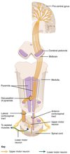
Briefly recap the UMN pathway
UMN cell bodies are arranged in a homuncular distribution in the primary motor cortex, at the posterior limit of the frontal lobe.
The axons of the UMNs descend through the subcortical white matter (corona radiata and then internal capsule).
The axons descend in the midbrain as the cerebral peduncles and then in the anterior pons and medulla where they cross as the pyramidal decussation.
During their brainstem course, some of the UMN axons synapse in motor nuclei (cranial nerve nuclei III, IV, V, VI, VII, X, XI, XII). These axons are called the corticobulbar fibres because the motor nuclei in the brainstem are known as the ‘bulbar nuclei’.
The remaining axons form the corticospinal tracts and descend in the lateral white matter of the spinal cord.

Briefly recap the LMN pathway
At each spinal level, some of the corticospinal fibres enter the anterior horn of the grey matter and synapse with cell bodies of the LMN, the anterior horn cells or motor neurons.
When these cell bodies are damaged, the syndrome is referred to as an anterior horn cell disorder.
Each LMN sends out an axon in the ventral (motor) root which joins a dorsal (sensory) root at each spinal level in the intervertebral foramen to form a spinal segmental or mixed nerve. When this nerve is damaged, it is called a radiculopathy.
The spinal nerve in the cervical and lumbosacral regions joins other adjacent spinal nerves in a junctional network referred to as a plexus. If these structures are damaged, the clinical syndrome is a plexopathy.
From the plexi emerge peripheral nerves and, when these are damaged, the patient develops a neuropathy.
The axons in the peripheral nerves split into many fibres just before synapsing with muscle fibres. • • •

What are the signs of lower motor neurone disease?
Note: inspection, tone, power, reflexes and plantar reflexes
Inspection: wasting and fasciculation of muscles.
Tone: decreased (hypotonia) or normal.
Power: different patterns of weakness, depending on the cause (e.g. classically a proximal weakness in muscle disease, a distal weakness in peripheral neuropathy).
Reflexes: reduced or absent (hyporeflexia or areflexia).
Plantar reflexes: normal (downgoing/flexor) or mute (i.e. no movement).
What are the signs of upper motor neurone disease?
Note: inspection, tone, power, reflexes and plantar reflexes
Inspection: no fasciculation or significant wasting (there may however be some disuse atrophy or contractures).
Tone: increased (spasticity or rigidity) +/- ankle clonus.
Power: classically a “pyramidal” pattern of weakness (extensors weaker than flexors in arms, and vice versa in legs).
Reflexes: exaggerated or brisk (hyperreflexia).
Plantar reflexes: upgoing/extensor (Babinski positive).
What are the cerebellar signs?
DANISH:
- Dysdiadochokinesia
- Ataxia (gait and posture)
- Nystagmus
- Intention tremor
- Slurred, staccato speech
- Hypotonia/heel-shin test
What is pyramdial weakness?
Pyramdial weakness: loss of power is most marked in the extensor muscles in the arms and the flexors in the legs. This is characteristic of UMN lesions involving the pyramidal tract within the brain or spinal cord.
What is proximal weakness?
Proximal weakness, affecting the shoulders, hips, trunk, neck and sometimes face. This is characteristic of muscle disease (myopathy) and also a common pattern in myasthenia gravis (neuromuscular junction).
What is distal weakness?
Distal weakness, affecting the hands and feet. This is typical of peripheral motor neuropathy, often affecting the lower limbs first and more severely.
What is global weakness?
Where is the lesion in pyramdial weakness?
Unilateral and bilateral UMN.
In an UMN lesion where is the site of pathology and what clinical syndrome does this result in?

In an LMN lesion where is the site of pathology and what clinical syndrome does this result in?

In a neuromucular junction where is the site of pathology and what clinical syndrome does this result in?

In a muscle where is the site of pathology and what clinical syndrome does this result in?


