Leg Structures Flashcards
(65 cards)

Pectineus M.
N: Femoral nerve, occasionally obturator nerve
M: Adduction and Flexion of thigh
A: Medial circumflex femoral artery, obturator artery
O/I: Pectineal Line of Pubis / Pectineal Line of Humerus

Adductor Longus M.
N: Obtrurator n., Anterior Division
M: Adduction and flexion of thigh
O/I: Body of the pubis/ Middle 1/3 of linea aspera
A: Deep femoral a.; femoral a.; medial circumflex femoral a.

Adductor Brevis M.
N: Obturator N.
M: Adduction and thigh flexion
I/O: Pubis body and Inferior Ramus / proximal linea aspera
A: Deep femoral a.; medial circumflex femoral artery.; obturator a.

Adductor Magnus M.
N: Adductor part = Obturator Nerve
Hamstring = Tibial Nerve
M:
Adductor part: flexes thigh, adducts
Hamstring part: extends thigh, adducts
O/I: Hamstring = Ischial Tuberosity / Gluteal Tuberosity, Lina Aspera, Medial super condylar Line
Adductor = Ischiopubic ramus / Adductor tubercle (Medial epicondyle)
A: Deep femoral artery

Gracilis M.
N: Obturator nerve (L2–L3)
M: Adducts thigh, flexes and medially rotates leg
O/I: Pubis and inferior ramus / Superior medial part of tibia (Pes Anserinus)
A: Profunda femoris artery, medial circumflex femoral artery

Iliopsas (Composed of Illiacus and Psoas Major) M.
N: Femoral Nerve (Illiacus) and L1 - L3 ventral rami (Psoas Major)
M: Chief Flexor of the Thigh
O/I: Lateral portion of T12 to L5 / Lesser Trochanter of Humerus
A: Lumbar branches of iliolumbar artery

Sartorius M.
N: Femoral Nerve
M: Abducts, laterally rotates, and flexes thigh; flexes knee joint (the tailor sit)
O/I: ASIS / Pes Anserinus
A: Femoral Artery

Rectus Femoris M.
N: Femoral N.
M: Extends leg at knee joint and flexes thigh
I/O: AIIS / Tibial Turbosity
A: Profunda femoris and lateral circumflex femoral arteries

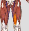
Vastus Medialis M.
N: Femoral Nerve
M: Extension of the knee
O/I: Shaft of Femur / AIIS
A: Femoral Artery

Vastus Lateralis M.
N: Femoral Nerve
M: Extension of the knee joint
O/I: Shaft of Femur / AIIS
A: Femoral Artery

Vastus Intermedius M.
N: Femoral Nerve
M: Extension of the Knee
O/I: Shaft of Femur / AIIS
A: Femoral Nerve

Articular Genu M.
N: Femoral Nerve
O/I: Lower anterior surface of femur / Articular capsule of knee joint
M: Retraction of supratellar bursa and elevation of the articular capsule of the knee during extention (Don’t need to know I think)
A: Femoral Artery

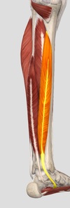
Tibialis Anterior M.
N: Deep fibular nerve
M: Dorsal flexion
O/I: Laterial tibeal condyle / superolateral 1/2 of tibia
A: Anterior tibial artery; anterior tibial recurrent artery; medial malleolar network

Obturator Externus M.
N: Obturator Nerve
M: Abduction, external rotation, stabilization of pelvis
O/I: External margins of obturator foramen / trochanteric fossa of femur
A: Medial circumflex . femoral a.

Gluteus Maximus M.
N: Inferior gluteal nerve
M: Chief extensor and lateral rotation, slight extension via tensor fascia lata
O/I: Posterior Glueteal Line of ilium, sacrum, lateral margin of coccyx / Upper fibers: the illio tibial tract and tensor fascia lata, Lower: The gluteal tuberosity of the femur
A: Superior Inferior Glureal Artery

Gluteus Medius M.
N: Superior Gluteal Nerve
M: Thigh abduction and medial rotation, pelvic stabilization
I/O: Between the anterior and posterior gluteal line/ Greater trochanter of the femur
A: Superior Gluteal Artery (Superficial Branch)

Tensor Fascia Lata M.
N: Superior Gluteal N.
M: Abducts, medially rotates, and flexes thigh. Slight rotation of the knee when combined with gluteus maximus
O/I: Iliac Turbecle / Gerdy’s turbecle on the the lateral tibial condyle
A: Superior Gluteal A.

Iliotibial Tract
M: Laterally stabilizes knee, Assists in deacceleration of adduction of the thigh, pulls patella laterally, antagonist of vastus medialis, and synergist with flexing vastus lateralis.
CN: Can compensate for quadraceps parallysis and can be stretched to treat chondropatella malecia.

Obturator Internus M.
N: Nerve to Obturator Internus and Superior Gemellus
M: Lateral rotation, abduction, and extension of hip
O/I: Margin of the obturator foramen / Medial surface of the greater trochanter of the femur

Superior Gemellus M.
N: Nerve to Obturator internus and Superior Gemellus
M: Lateral rotation, abduction and extension of hip
O/I: Ischial spine / Medial surface of greater trochanter of femur

Inferior Gemellus M.
N: Nerve to Quadratus Femoris and Inferior Gemellus
M: Lateral rotation, abduction and extension of hip
O/I: Ischial turberosity / Greater trochanter of the femur

Quadratus Femoris M.
N: Nerve to Quadratus Femoris and Inferior Gemellus
M: Lateral rotation, abduction and extension of hip
I/O: Lateral border of the ischial tuberosity / Quadrate line (below the intertrochanteric crest) of femur

Semitendinosous M.
N: Tibial division of the sciatic nerve
M: Extend thigh, flex leg, medial rotation of the knee
O/I: Ischial tuberosity / Medial surface of the superior aspect of the tibia (Pes anserinus)

Semimembranosis M.
N: Tibial division of the sciatic nerve
M: Extend thigh / Flex leg
O/I: Ischial tuberosity / Posterior part of the Medial condyle of tibia


