Lecture 3 Radiographic anatomy part 1 Flashcards
(54 cards)
What is the labelled arrow?

Root canal
Labelled arrow?
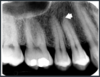
Root canal
Are these root canals visible in the last 2 mm?
Is the cementum discernible?

Frequently not visible in the last 2 mm.
Cementum is not discernible since it has same density of dentine.
What is this?
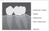
Cervical burnout.
- Overexposure of lateral portion of roots between enamel and alveolar crest.
What is this?

Cervical burnout
Labelled arrow?

Lamina dura
Name the labelled arrows

Lamina dura
- Visualization depends on the orientation of the beam and shape of tooth/root
Name this supporting structure

Alveolar crest
Name this supporting structure

Alveolar crest
Name this supporting structure

Periodontal ligament space
Name this supporting structure

Periodontal ligament space
Name this supporting structure

Periodontal ligament space - may appear double depending on the anatomy of the root
Describe what’s happening here

Periodontal ligament space may appear double depending on the anatomy of the root.
Name this supporting structure

Trabecular or cancellous bone
Name this supporting structure

Cancellous bone - small trabecular (marrow) space as pointed by arrow.
Name this supporting structure

Cancellous bone - aroow is pointing to large trabecular (marrow) spaces
Name the arrow pointed structure

Intermaxillary suture - also called the median suture
Name this structure pointed by arrows

Intermaxillary suture - also called the median suture
Name the arrow pointed structure and describe

Anterior nasal spine - opaque, irregular and V-shaped
Name this structure pointed by arrows

Anterior floor of the nasal cavity
Name the arrow pointed structure

Anterior floro of the nasal cavity
Name the labelled black arrow structure and when is it frequently seen?

Nasal septum - frequently seen covered by mucosa (white arrow)
Name the white arrow structure

Inferior concha - also called inferior turbinate
Name the white arrow labelled structure

Incisive canal - also called nasopalatine foramen


