Lecture 2 - Flow Cytometry Flashcards
(29 cards)
What is flow cytometry
Test that allows rapid measurement of several parameters of a single cell as it moves through a beam of light
Detectors of a flow cytometer
Forward scatter (detects cell size) Side scatter (detects cell granularity) Fluorescence detector (detects colour of emitted light)
What ferries cells into laser beam?
Fluidics system
What does each dot on a flow cytometry dot plot represent?
A single cell
Populations commonly seen in a blood sample
Three populations: Lymphocytes Monocytes Granulocytes

blood 2

Uses of fluorochromes 1) 2) 3) 4)
1) Direct conjugates 2) Indirect conjugates (secondary labelling) 3) DNA labelling 4) Cytoplasmic dyes
How is fluorescence used in flow cytometry? 1) 2) 3)
1) Attached fluorescent tag emits light when stimulated with a particular wavelength. 2) There is a wavelength which results in the greatest light emission (peak). 3) Detect EG: +/-30nm either side of peak for a particular fluorochrome
What is spectral overlap?
Where the emission spectra of two different fluorochromes overlap. Is detected by the wrong detector
How is spectral overlap compensated for?
Need to subtract FITC from PE

Uses of flow cytometry 1) 2) 3) 4) 5) 6) 7)
1) Cell surface molecule expression 2) Intracellular molecule expression 3) Cell function 4) DNA content cycle and analysis 5) Apoptosis 6) Antigen-specific cell function 7) Cell sorting
5 x 10^6 Splenocytes are stained with an:-CD4 FITC and an:-CD8 PE. 15% are CD4+ cells and 10% are CD8+ cells. How would a forward scatter vs side scatter dot plot look?

5 x 10^6 Splenocytes are stained with an:-CD4 FITC and an:-CD8 PE. 15% are CD4+ cells and 10% are CD8+ cells. How would a FITC vs empty channel dot plot look?

5 x 10^6 Splenocytes are stained with an:-CD4 FITC and an:-CD8 PE. 15% are CD4+ cells and 10% are CD8+ cells. How would a PE vs empty channel dot plot look?

5 x 10^6 Splenocytes are stained with an:-CD4 FITC and an:-CD8 PE. 15% are CD4+ cells and 10% are CD8+ cells. How would a FITC vs PE dot plot look?

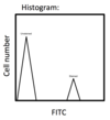
5 x 10^6 Splenocytes are stained with an:-CD4 FITC and an:-CD8 PE. 15% are CD4+ cells and 10% are CD8+ cells. How would a histogram comparing the relative amount of unstained to FITC stained cells look?
How can the number of cell divisions be measured using flow cytometry?
Carboxyfluorescin succinyl ester injected into cells. Splits evenly between daughter cells. With each generation, CFSE fluorescence decreases.
What is CFSE?
Carboxyfluorescing succinyl ester. Binds to cytoskeletal proteins, begins emitting light. Divides evenly between daughter cells.
How is CFSE detected?
Using FL-1 FITC detector
Why can’t FITC and CFSE be used together?
CFSE emits at a very similar wavelength to FITC
How would a CFSE vs empty channel dot plot look?

How would a CFSE vs empty channel histogram look?

How can specific T cell responses be measured with flow cytometry?
1) Tetramer staining 2) Cytokine staining
Tetramer staining 1) 2) 3)
1) Four peptide-loaded MHCI/MHCII/CD1d bound to streptavidin via biotin. 2) Fluorescent molecules directly conjugated to tetramer 3) This detects T cells expressing TCR specific to MHC/CD1d and antigen

