Lecture 10: Active Membrane Transport Flashcards
Glucose Transporters
Rate of erythrocyte Glucose Transporter GLUT1
- Glucose enters the erythrocyte by facilitated diffusion via a specific glucose transporter
- rate 50,000 time greater than uncatalyzed membrane diffusion
General structure of GLUT1
- 12 hydrophpbic segments
- each beleived to form a membrane spanning helix
Model of GLUT1
- side by side assemply of several helices produces a transmembrane corridor lined with hydrophilic residues that can hydrogen bond with a glucose as it movves through
Ser, Leu, Val, Thr, Asn, Phe, Ile
4 steps of GLUT1 trasnport
4 steps:
- binding
- transport
- dissociation
- recovery
Similarities of GLUT1 to enzymatic reaction
substrate is glucose outside of cell
product is glucose inside cell
enzyme is the trasnporter
plot of glucose uptake as a function of external glucose concentration
hyperbolic (like enzymes)
lineweaver burke
1/V0 = Km / (Vmax x [S]out) + 1/Vmax
GLUT2
- in epitherlial cells
- Na+ glucose symporter and glucose uniporter (GLUT2) opporate on opposite sides of epitherlial cells
- transporter systems located asymmetrically, each confined to one sid eof plasma membrane
GLUT4
- mainly in skeletal and heart muscle as well as adipose tissue
- can be distinguished from other glucose transporters by its response to insulin
- Activity increases when insulin signals a high blood glucose concentration, thus increasing the rate of glucose uptake into muscle and adipose tissue (15 fold or more)
Diagram of GLUT4
Comparison of GLUT transporters to Chloride Bicarbonate exchangers
GLUTs are uniport
Chloride bicarbonate are antiporters –> for each HCO3- ion that moves in one direction, one Cl- ion moves in the opposite
chloride bicarbonate exchangers
- for each HCO3- that moves in one direction, one Cl- ion moves in the opposite
- coupling is required for transport activity
- no net transfer of charge and exchange is electroneutral
- humanms have 3 AE isoforms, AE1 is predominant in erythrocytes
AE1 transporter
- in erythrocytes
- important CO2 trasnport and buffering blood pH
CO2 in blood
- CO2 that is produced by cellular respiration enters the blood and erythrocytes (RBCs) where it is quickly converted to bicarbonate ions and protons in a reaction catalyzed by carbonic anhydrase
- carbonic anhydrase catalyzes the formation of carbonic acid from carbon dioxide in water
Importance of carbonic anhydrase
- protons produced by the carbonic anhydrase reaction induce the Bohr shift, which makes the hemoglobin more likely to release the oxygen
- PCO2 in blood drops when CO2 is converted to bicarbonate, maintaining a strong partial pressure gradient favoring the entry of CO2 into red blood cells
* carbonic anhydrase activity makes CO2 uptake from tissues more efficient
Bohr Shift and oxygen release by hemoglobin
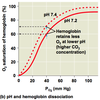
MEchanism of CO2 transport
- H+ produced by dissociation of carbonic acid is taken up by hemoglobin (hemoglobin also acts as a buffer, minimizing changes in pH)
- in the alveoli, a partial pressure gradient favors the diffusion of CO2 from plasma RBCs to the atmosphere
- hemoglobin releases protons, which combine wwith bicarbonate to form CO2, which then diffuses into the alveoli and is exhaled
- hemoglobin picks up O2 in inhalation and the cycle begins again
Important aspects of active transport
- results in accumulation of a solute above the equilibrium point
- thermodynamically unfavorable (endergonic)
- takes place only when coupled to exergonic process
- ie: coupled trasnport, ATP hydrolysis
Types of active trasnport
primary: uses ATP
Secondary: uses coupled trasnport
Primary active transport
- solute accumulation is coupled directly to an exergonic reaction, such as the hydrolysis of ATP
- P-type, V-type, F-type ATPases –> act like uniport or antiport pumps
- ABC (ATP binding cassette) trasnporters –> acts as uniporter
Secondary Active transport
- energy comes from an ion concentration gradient that is established by primary active trasnport
- sodium-solute or proton-solute contrasnporters, which are symporters or antiporters
Energy dependent pumps
- two principal conformational states
- each open to one side of the membrane
- to pump a molecule (such as an ion) in a single direction across a membrane, the invested energy (eg. free energy of ATP hydrolysis) must be coupled to the interconversion between these conformational states
Diagram of energy dependent pumps
P Type ATPases
cation trasnporters that are reversibly phosphorylated by ATP as part of the transport cycle
–> phosphorylation forces are a conformational change that is central to movement of the cation
–> they are sensitive to inhibition by vandate and can act as uniporters (eg Ca2+ ATPases) or antiporters (eg Na+K+ ATPases)
Na+K+ ATPases
Kinetics for active transport of Na+ and K+
Cardiotonic steroids
- inhibit Na+ K+ ATPases
Maintaining resting potential
- every cell has a voltage (difference in electrical charge) across its plasma membrane called membrane potential
- resting potential is the membrane potential of a cell such as a neuron no sending signals (-60mv)
- changes in membrane act as signals, transmitting and processing information
- in a mammalian neuron at resting potential, the concentration of K+ is highest inside cell, Na+ abnd Cl- are highest outside
Importance of sodium potassium pumps
- maintain resting potential
- use energy of ATP to maintain K+ and Na+ gradients across the plasma membrane
- concentration gradients represent chemical potential energy
- opening of ion channels in the plasma membrane converts chemical potential to electrical potential
V-Type ATPase
- proton trasnporting ATPases responsible for acidifying intracellular (and in some cases extracellular) compartments (eg: vacuoles, lysosomes, endosomes, and the golgi complex) in many eukaryotic organisms
–> have similar complex structure, with an integral (transmembrane) domain (V0) that serves as a proton channel and a peripheral domain (V1) that contains the ATP binding site and has ATPase activity
structure of VType ATPase
have similar complex structure, with an integral (transmembrane) domain (V0) that serves as a proton channel and a peripheral domain (V1) that contains the ATP binding site and has ATPase activity
Osteoclasts
tear down bone tissue
osteoblasts
build it back up
Osteoclasts and vacuolar ATPases
- in osteoclasts, vacoular ATPases are present on the portion of the plasma membrane that attaches to the bone (ruffled border)
- ATPases secrete acid (protons) into the space between the bone surface and the cell, lowering the pH of this extracellular space to about 4
- respults in dissolving the Ca2+ phosphate matrix of the bone
Bone remodeling by osteoclasts (diagram)
F-type ATPases
(ATP synthases) - structurally related to V-type ATPases with a trasnmembrane F0 domain and a peripheral F1 domain
- in vivo, a proton gradient generated by electron trasnport processes (involving pumps powered by substrate oxidation or light) is used to supple the energy to drive ATP synthesis (reaction is reversible though)
Diagram of F Type ATPases
Coupling ofr Electron trasnport and ATP synthesis in cellular systems
F-type ATPase catalytic reaction cycle
- F1 has 3 nonequivalent adenine nucleotide-binding sites, one for each pair of a and b subunits
–> b STP conformtaion binds ATP tightly
–> b ADP = loose binding conformation
–> b empty = very loose binding conformation
- proton motive force causes rotation of the central shaft which comes into contact with each ab subunit pair in succession
- produces a cooperative conformational change and hence interconversion
catalytic reaction cycle of F type ATPase
ABC transporters
- 2 nucelotide binding domains (NBDs) and 2 membrane-spanning domains (with usually six trasnmembrane helices)
–> the two NBDs do not interact strongly in the absence of nucleotide (ATP)
- substrate binding between membrane spenning domains induces conformational changes that increases the affinity of the NBDs to ATP and hence their ability to associate
- this association induces the substrate release to the opposite side of the cell
- ATP hydrolysis and ADP+Pi release resets trasnporter
Catalytic cycle of ABC transporters
Open and closed forms of ATP driven trasnporters
P-loop
ATP binding cassette
Bacterial and Eukaryotic ABC trasnporters
P class pumps
- plasma membrane of plants, fungi, bacteria (H+ pumps)
- plasma membrane of higher eukaryotes (Na+, K+ pump)
- apical plasma membrane of mammalian stomach (H+/K+ pump)
- sarcoplasmic reticulum membrane in muscle cells (ca2+ pumps)
V alcas proton pumps
- vacuolar membranes in plants, yeast, other fungi
- endosomal and lysosomal membranes in animal cells
- plasma membrane of osteoclasts and some kidney tubule cells
F-class proton pumps
- bacterial plasma membrane
- inner mitochondrial membrane
- thylakoid membrane of chloroplasts
ABC superfamily
- bacterial plasma membranes (amino acid, sugar, and peptide transporters)
- mammalian plasma membranes (transporters of phospholipids, small lipophilic drugs, cholesterol, other small molecules)
Secondary transporters
- ion gradients formed by primary trasnport of Na+ or H+ can provide the driving force for cotransport of other solutes
- ions, sugars or amino acids via secondary trasnporters (aka cotransporters)
- cotrasnporters can be symporters and antiporters
Lactose permease
- lactose H+ symporter
- pumps lactose inside along with H+ in too
- proton pump pumps H+ out

Lactose permease transport mechanism
R144
E216
E269

Lactose H+ symporter

Gastric H+ K+ ATPase
- parietal cells fo the gastric mucosa (lining of the stomach) have an internal pH of 7.4
- H+, K+ ATPases pump protons fromt he cells into the stomach to maintain a pH differences across a single plasma membrane of 6.6
- largest known transmembrane gradient in eukaryotic cells
gastric h+ K+ ATPase mechanism
Net: H+ out, CL- out
H+ out of mucous, K+ into mucous
K+ and Cl- out

chloride bicarbonate exchanger and carbonic anhydrase in gastric H+K+ ATPase mechnaism
- support net transport of H+ and CL- into the stomach