Lecture 1: Cellular Responses to Stress and Toxic Insults: Adaption, Injury, and Disease Flashcards

Based on Robbins Outlines

1
Q

FLeakage of intracellular enzymes (i.e., lactate dehydrogenase) is a marker of?

A

Irreversible cell injury

2
Q

What kind of cellular adaptation is a result of increased demand and stimulation?

A

Hyperplasia or hypertrophy

3
Q

What kind of cellular adaptation is a result of chronic irritation (physical or chemical)?

Is it irreversible or reversible?

A

Metaplasia

Reversible

4
Q

What cellular adaptation is most common in the uterus during pregnancy; stimulated by?

Endometrium from estrogen?

A
  • Hypertrophy during pregnancy; stimulated by estrogenic hormones
  • Hyperplasia (pathologic) of endometrium from estogen, but still remains as a thin lining to the muscular wall and does not contribute as much to the change in size!
5
Q

What cellular adaptation is most common in muscle disuse?

Barrett esophagus?

A
  • Muscle disuse = atrophy
  • Barrett esophagus = goblet cell Metaplasia (squamous to columnar type)
6
Q

Metabolic alterations to a cell that are due to chronic injury result in what?

A

Intracellular accumulations, calcification

7
Q

The most common stimulus for hypertrophy of skeletal muscle is __________

A

Increased work load

8
Q

The most common stimulus of hypertrophy for cardiac muscle is ________.

Usually resulting from what?

A
  • Increased hemodynamic load
  • HTN or faulty valves
9
Q

What is the key characteristic of hypertrophy?

A

Increased protein synthesis

10
Q

During the increased workload on the heart growth factors are stimulated and 2 biochemical pathways activated, which is thought to be the most important in physiologic muscle hypertrophy and which in pathologic hypertrophy?

A
  • PI3K/AKT in physiologic (i.e., exercise induced) hypertrophy
  • Signaling downstream GPCR’s in pathologic hypertrophy
11
Q

Cumulative sub lethal injury over long life span induce what cellular response?

A

Cellular aging

12
Q

Which TF’s are activated in the molecular pathogenesis of cardiac hypertrophy and work to increase the synthesis of muscle proteins responsible for hypertrophy?

A

GATA4, NFAT, and MEF2

*NFAT, GATA4, and MEF2 inhibitors are in phase 1 or 2 clinical trials; hoping to prevent cardiac failure from excessive hypertrophy

13
Q

What 2 adaptions does cardiac hypertrophy elicit that are associated with fetal heart functions?

What is the purpose of these adaptions?

A

1) Switch of contractile proteins from adult myosin heavy chain α isoform to the fetal/neonatal β isoform
- The β isoform has a slower, more energetically economical contraction
2) Increased expression of ANP (atrial natriuretic factor), which is down-regulated after birth
- Kidneys secrete Na , water follows, reduces blood volume

14
Q

What type of necrosis is associated with tuberculous infection?

What type specifically?

A
  • Caseous necrosis
  • Myobacterium tuberculosis
15
Q

*Caseous necrosis is described as what?

How does it look on microscopic examination?

A
  • Yellow-white and “cheese-like”
  • Structureless collection of fragmented or lysed cells and amorphous granular debris enclosed within a distinctive inflammatory border (Granuloma)
16
Q

The female breast is an example of what 2 cellular growth adaptions?

A
  • Hormonal hyperplasia: proliferation of glandular epithelium at puberty
  • Hypertrophy: during pregnancy
17
Q

What are 2 examples of compensatory hyperplasia (liver and bone marrow)?

A
  • Donating a lobe of liver for transplantation, remaining cells proliferate so that organ grows back to original size
  • After acute bleed or hemolysis, there is great expanse of red cell progenitors due to EPO
18
Q

What are the causative agents involved in pathologic hyperplasia of the endometrium and in benign prostatic hyperplasia; can these abnormalities be fixed?

A
  • Endometrial hyperplasia = estrogen
  • BPH = androgens
  • Can be treated by simply fixing the hormonal imbalances, allowing them to revert back to normal
19
Q

How is hyperplasia related to cancer?

A
  • Hyperplasia itself is NOT cancer
  • Pathologic hyperplasia constitutes a “fertile soil” in which cancerous proliferations may eventually arise
20
Q

What is the characteristic signs of atrophy?

A

Autophagy and decreased protein synthesis

21
Q

In cancer, what is responsible for the suppression of appetite and depletion of lipid stores that culminates in muscle atrophy?

A

TNF

22
Q

The degradation of cellular proteins seen in atrophy occurs mainly through which pathway?

A
  • Ubiquitin-proteasome pathway
  • Nutrient deficiency and disuse may activate ubiquitin ligases, which attach the small peptide ubiquitin to cellular proteins and target these proteins for degradation in proteosomes
23
Q

What is one of the hallmarks of atrophy and how can this be visualized?

A
  • Autophagy: starved cell eats its own components in an attempt to reduce nutrient demand to match supply
  • Some cell debris may resist digestion and persist in cytoplasm as residual bodies
  • Example of residual bodies is lipofuscin granules, which can impart a brown discoloration to the tissue
24
Q

What is the most common epithelial metaplasia and what are some examples?

A
  • Columnar to squamous (squamous metaplasia)
  • Respiratory tract in response to chronic irritation; in smoker the normal ciliated columnar of the trachea and bronchi are replaced by stratified squamous cells
  • Stones in the excretory ducts of salivary glands, pancreas, or bile ducts, may also lead to squamous metaplasia
25
Q

Vitamin A deficiency induces what type of metaplasia, where?

A

Induces squamous metaplasia in respiratory epithelium

26
Q

What is the purpose of squamous metaplasia (gained and lost)?

Can progress to?

A
  • Stratified squamous epithelium is better able to hold up against irritation/insult
  • Loses its specialized capacity (such as mucus secretion and ciliary action)
  • Influences that predispose to metaplasia, if persistent, can initiate malignant transformation in metaplastic epithelium
27
Q

Barrett’s esophagus is an example of what kind of metaplasia?

A

Squamous to columnar (columnar metaplasia)

*Metaplasias are named for what it turns into

28
Q

Myositis ossificans is what kind of cellular adaption?

A
  • Connective tissue metaplasia (mesenchymal variety)
  • Creation of bone, cartilage, or adipose tissue in tissues that do not normally contain these elements (i.e., muscle)
29
Q

What is the mechanism of metaplasia?

A
  • Does NOT result from a change in the phenotype of an alreadu differentiated cell type
  • Is the result of a re-programming of stem cells due to signals from cytokines, GFs, and the ECM
30
Q

What are the hallmarks of reversible cell injury?

A
  • Reduced oxidative phosphorylation and lower ATP
  • Cellular swelling from changes in ion concentrations and water influx
31
Q

What is the mechanism for necrosis and the hallmark?

Necrosis is caused by and is always?

A
  • Lysosomal enzymes enter cytoplasm and digest the cell\
  • Cellular contents leak thru damaged PM into EC space, where they elicit inflammation = hallmark
  • Caused by toxins, infections, ischemia, and trauma. Is ALWAYs pathologic
32
Q

Apoptosis is characterized by?

A
  • Nuclear dissolution, fragmentation of the cell without complete loss of membrane integrity, and the rapid removal of cell debris = NO inflammation
  • Can be pathologic AND/OR physiologic
33
Q

Oxygen deprivation leads to cell injury how?

A

Reduces the cell’s ability to do aerobic oxidative respiration and oxidative phosphorylation

34
Q

What are the 4 ultrastructural changes of reversible cell injury?

A

1) Plasma membrane alterations, blebbing, loss of microvilli
2) Mitochondrial changes, swelling and appearance of small amorphous densities
3) Dilation of the ER, w/ detachment of the ribosomes
4) Nuclear alterations, w/ diaggregation of granular and fibrillar elements

35
Q

If there is ischemia to a cell, what happens in the mitochondria?

A

Decreased ox-phos and decreased ATP production

36
Q

What are the 3 consequences of decreased ATP production in the mitochondria?

A

1) Decreased Na pump
2) Increased anaerobic glycolysis
3) Detachment of ribosomes (decreased protein synthesis)

37
Q

What 2 features of reversible cell injury can be recognized under the light microscope?

Cause of each of these features?

A
  • Cellular swelling: failure of energy-dependent ion pumps in plasma membrane
  • Fatty change: occurs in hypoxic injury and various forms of toxic or metabolic injury. Apperance of lipid vacuoles in the cytoplasm
38
Q

The morphological changes seen in reversible cell death are associated with what 5 biochemical and structural changes?

A

1) Decreased generation of ATP
2) Loss of membrane integrity
3) Defects in protein synthesis
4) Cytoskeletal damage
5) DNA damage

39
Q

What is it called on microscopic examination, small clear vacuoles may be seen within the cytoplasm, representing distended and pinched-off segments of the ER?

The may show what kind of staining?

A

- Hydropic change or vacuolar degeneration

  • Increased eosinophillic staining
40
Q

Are the appearance of intracytoplasmic myelin figures associated with reversible or irreversible cell injury?

A
  • NOT associated with reversible cell injury
  • More likely associated with irreversible cell injury, and a characteristc of necrosis
41
Q

What are 4 reversible morphological changes seen in the cell with injury?

A

1) Cellular swelling
2) Blebbing of the plasma membrane
3) Detachment of ribosomes from the ER
4) Clumping of nuclear chromatin

42
Q

What happens to maintenance of the membrane in necrosis, where are the enzymes that digest necrotic cells derived from?

A
  • Necrotic cells cannot maintain membrane –> contents leak out
  • Enzymes from lysosomes of the dying cells themselves and from lysosomes of recruited leukocytes called in as part of inflammatory reaction
43
Q

Necrotic cells show increased _________ with H/E stains due to loss of _______ (stains blue from hematoxylin) and ____________ (stains red from eosin)

A

Necrotic cells show increased eosinophilia with H/E stains due to loss of RNA (stains blue from hematoxylin) and denatured protein (stains red from eosin)

44
Q

Nuclear changes in necrosis happen in one of 3 patterns all due to nonspecific breakdown of DNA, what are they in order?

A

1) Karyolysis: basophilia of chromatin fade from loss of DNA due to enzymatic degradation by endonucleases
2) Pyknosis: nuclear shrinkage and inreased basophilia from the chromatin condensing into solid basophilic mass
3) Karyorrhexis: pyknotic nucleus undergoes fragmentation and eventually dissapears

45
Q

What are the 3 major causes of ATP depletion?

A

1) Reduced supply of oxygen and nutrients
2) Mitochondrial damage
3) Actions of toxins (Cyanide)

46
Q

Characteristics of coagulative necrosis?

Localized area of coagulative necrosis is called what?

Caused by and affects what organs?

A
  • Tissue that remains firms, cell shape and organ structure preserved
  • Nucleus dissapears
  • Typically caused by an obstruction of blood vessel leading to tissue in ALL organs EXCEPT the brain
  • Localized area of coagulative necrosis = infarct
47
Q

Liquefactive necrosis is characterized by and typically a result of what?

Where is this type of necrosis seen?

A
  • Digestion of the dead cells that turns the dead tissue into a liquid viscous mass
  • Bacterial and fungal infections due to stimulation or large #s of leukocytes to an area and liberation of enzymes from these cells
  • Frequently appears creamy yellow and is called pus
  • Hypoxic death of CNS causes liquefactive necrosis
48
Q

What is gangrenous necrosis?

What is wet gangrene?

A
  • Not a specific pattern of cell death, but commonly used in practice
  • Essentially coagulative necrosis of an extremity, often seen in limbs of uncontrolled diabetic patients
  • Wet gangrene is when a bacterial infection is present as well and there will also be some liquefactive necrosis
49
Q

Fat necrosis is commonly seen where and what reaction causes its appearance?

A
  • Focal areas of fat destruction from release of pancreatic lipase into the substance of the pancreas and peritoneal cavity = acute pancreatitis
  • Released enzymes split TAG esters within fat cells and these then combine with Ca2+ to produce chalky-white appearance from fat saponification
50
Q

Fibrinoid necrosis is a special form usually seen in?

Appear how in H/E stains?

A
  • Seen in immune reactions involving blood vessels (i.e., Malignant HTN and Vasculitis)
  • Complexes of antigens and antibodies are deposited in the wall of arteries – Type III hypersensitivity
  • These deposits + fibrin leaked from vessels result in a bright-pink and amorphous apperance in H/E stains, called “fibrinoid” (fibrin-like)
51
Q

What happens if necrotic cells are not taken back up by leukocytes?

A
  • They provide a nidus for the deposition of calcium salts and other minerals and become calcified
  • Known as dystrophic calcification
52
Q

In cell deprived of oxygen or glucose, proteins may become misfolded and accumulation of misfolded proteins in the ER triggers what?

A

Cellular reaction called the unfolded protein response (that may culminate in cell injury or even death)

53
Q

Mitochodrial damage leads to the formation of what pore, which causes what problems?

What is a component of this pore and possible drug target?

A
  • Mitochondrial permeability transition pore
  • Opening of this conductance channel leads to loss of membrane potential, failure of OxPhos, progressive depletion of ATP –> necrosis
  • Structural component is cyclophilin D, targeted by cyclosporine (immunosuppressive)
  • Cyclosporine reduces injury by preventing opening of the pore - targeted therapy for cellular injury
54
Q

Which proteins are sequestered between the outer and inner membranes of the mitochondria and may leak out into the cytosol with increased permeability caused by damage to the mitochondria?

A

Cytochrome c and proteins that indirectly activate apoptosis inducing enzymes called caspases

55
Q

Increased intracellular Ca2+ levels lead to cell injury in what 3 ways?

A

1) Accumulation of Ca2+ in mito. results in opening of mitochondrial permeability transition pore = failure of ATP generation
2) Activates enzymes like phospholipase**s (membrane damage), proteases (break down membrane and cytoskeletal proteins), endonucleases (DNA and chromatin fragmentation), and ATPases (speeds up** ATP depletion)
3) Result in induction of apoptosis, direct activation of caspases and by increasing membrane permeability

56
Q

When are ROS produced in a cell and how are they dealt with?

A
  • A normal product of the cell during mitochondrial respiration and energy generation
  • Typically degraded and removed by cellular defense systems
  • Cell is able to maintain a steady state in which free radicals are present transiently at low concentrations but DO NOT causes damage
57
Q

Which free radicals are generated from the reduction-oxidation that occur during normal metabolic processes?

What is the redox reaction occuring to generate these by-products?

A
  • O2-<b>.</b>, one electron
  • H2O2
  • <strong>.</strong>OH, three electrons

* Molecular O2 is reduced by transfer of four electrons to H2 to make two water molecules

58
Q

How does the absorption of radiant energy generate free radicals?

A
  • UV light, and X-rays can hydrolyze water into <strong>.</strong>OH and hydrogen (H) free radicals
59
Q

How are leukocytes involved in the formation of free radicals?

A
  • Leukocytes use NADPH oxidase during inflammation
  • Pivotal in the oxidative burst conversion of molecular O2 to superoxide anion O2
60
Q

Which transition metals are involved in the production of free radicals?

Which specifically with the Fenton reaction and describe this reaction?

A
  • Iron and copper donate or accept free electrons during intracellular reactions and catalyze free radical formation
  • Iron does the Fenton reaction (H2O2 + Fe2+–> Fe3++ OH* + OH-)
  • Most intracellular free iron found in Fe3+ (ferric) state, must be reduced to Fe2+ (ferrous) to participate in Fenton reaction
  • Production of the Fe3+ form, which is the most abundant form, produces a hydroxyl free radical (OH*), which is the most damaging!
61
Q

How are hydroxyl free radicals (OH*) inactivated?

A

Conversion to H2O by glutathione peroxidase

62
Q

What are some examples of antioxidants?

A

Vitamins E, A, ascorbic acid (C), and glutathione

63
Q

What are 3 enzymes located near the sites of generation of the oxidations that scavenge free radicals and break down ROS?

A
  1. Catalase: present in peroxisomes, decomposes H2O2 (catalase rxn when pouring hydrogen peroxide on wound with bacteria present)
  2. Superoxide dismutase (SOD): convert O2-<strong>.</strong> to H2O2
  3. Glutathione peroxidase: breaks down OH- or H2O2
64
Q

What is the importance of the intracellular ratio between oxidized glutathione (GSSG) to reduced glutathione (GSH)?

A

Reflection of the oxidative state of the cell and is an important indicator of the cell’s ability to detoxify ROS

65
Q

What is lipid peroxidation?

A
  • Free radicals attack the double bonds found in the lipid membrane of cells, this interaction yields peroxides
  • Autocatalytic chain reaction ensues (called propogation) that can result in extensive membrane damage
66
Q

How do free radicals effect proteins?

Enhances the action of what enzyme?

A
  • Oxidative modification of proteins, through oxidation of AA side chains, formation of covalent protein-protein cross-links, and disruption of backbones
  • Damages the active sites of enzymes and disrupts structural conformations
  • Enhances proteosomal degradation of unfolded or misfolded proteins, raising havoc in the cell.
67
Q

How can free radicals effect the DNA?

A

Cause single or double strand breaks in DNA that leads to cell aging and cancer

68
Q

The major actions of superoxide anion (O2-*) stem from its ability to do what?

A

Stimulate the production of degradative enzymes rather tha direct damage of macromolecules

69
Q

What are the 4 mechanisms of membrane damage?

A

1) ROS cause injury to the cell membranes (lipid peroxidation)
2) Decreased phospholipid synthesis: defective mito. function or hypoxia = decreased ATP needed for biosynthesis
3) Increased phospholipid breakdown: increased levels of Ca2+ activating phospholipases = increased lipid breakdown products (FFA’s, acyl carnitine, lysophospholipids) which have detergent effect on membrane
4) Cytoskeleton abnormalities: activation of proteases by increased intracellular Ca2+

70
Q

Two phenomena consistently characterize irreversible changes in cell injury, what are they?

A

1) Inability to reverse mitochondrial dysfunction (lack of OxPhos and ATP generation)
2) Profound disturbances in membrane function

71
Q

What provides a means for the detection of tissue-specific cellular injury and necrosis using blood serum samples?

A

Leakage of intracellular proteins through the damaged cell membrane and ultimately into the circulation

72
Q

What are the biomarkers from the cardiac muscle, liver (and specifically bile duct epithelium), and hepatocytes which can be used clinically to assess damage/irreversible cell injury occuring in these tissues?

A

Cardiac = cardiac specific isoform of Creatine Kinase and Troponin

Liver/bile duct epithelium = Alkaline phosphatase

Hepatocytes = Transanimases

73
Q

Accumulation of damage DNA and misfolded proteins triggers what kind of cell death?

A

Apoptosis

74
Q

What kind of cellular adaptation is a result of decreased nutrients and and decreased stimulation?

A

Atrophy

75
Q

What are the the GFs of mechanical sensors in hypertrophy?

Vasoactive agents?

A

TGF-B, IGF-1, FGF = GFs

A-adrenergic agonists, Endothelin-1, Angiontensin II = Vasoactive

76
Q

What are the 3 basic steps in the molecular pathogenesis of cardiac hypertrophy?

A

1) Actions of mechanical sensors
2) Signals that activate a signal transduction pathways
3) Signaling pathways activating TFs

77
Q

What are the 6 different causes of atrophy?

A

1) Dec workload
2) Loss of innervation
3) Diminished blood supply
4) Inadequate nutrition / malnutrition
5) Loss of endocrine stimulation
6) Pressure (tissue compression)

78
Q

Some of the cell debris within autophagic vacuoles may resist digestion and persist in the cytoplasm as what?

Example of one?

A

Residual bodies

Lipofuscin granule (brown color)

79
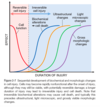
Q

What is the order of change in a cell during irreversible cell injury? (Fig. 2-7)

A

Biomechanical alterations leading to cell death

Ultrastructural

Light microscopic

Gross morphologic

BULGe

80
Q

If there is duct obstruction that may occur in the pancreas, parotid, or kidney what may this lead to?

A

Pathologic apoptosis

81
Q

How is hydrogen peroxide converted to oxygen (2 ways based on location)?

A

1) Catalase (in Peroxisomes)
2) Glutathione peroxidase (cytosol, mitochondria)

82
Q

Differentiate ischemia vs. hypoxia w/o ischemia

Which is a bigger problem?

A
  • Ischemia prevents the blood from bringng more substrates and taking away degradation products to the cell for it to conduct more glycolysis = aerobic and anaerobic metabolism is compromised

- Hypoxia = glycolysis (anaerobic) may continue

  • Ischemia tends to cause more rapid and severe cell and tissue injury than does hypoxia in the absence of ischemia
83
Q

Large, flocculent, amorphous densities develop in the mitochondrial matrix, what are these an indication of in myocardium and how quickly can they be seen?

A
  • Indication of irreversible injury
  • Seen as early as 30-40 mins. after ischemia
84
Q

Mammalian cells have developed protective responses to deal with hypoxic stress, the best defined is what TF and what does it do?

A
  • Hypoxia-inducible factor 1
  • Promotes new blood vessel formation (angiogenesis), stimulates cell survival pathways, and enhances glycolysis
85
Q

What is the best known strategy for treating ischemia (and traumatic) brain and spinal cord injury?

A
  • Inducing hypothermia (CBT < 92 F)
  • Reduces metabolic demands of stressed cells, decreases cell swelling, suppresses formation of free radicals, and inhibits the host inflammatory response
86
Q

What are the 4 mechanisms that contribute to ischemia-reperfusion injuries?

A

1) Oxidative stress: reoxygenation may increase ROS’s and reactive nitrogen species; also a decrease in activity of antioxidants
2) Intracellular Ca2+ overload: exacerbated during reperfusion (more Ca2+ brought by blood) = favors opening of mitochondrial permeability transition pore w/ resultant decrease in ATP (#1 contributor to cell death!)
3) Inflammation: neutrophils brought to site of inflammation by reperfusion and exacerbate initial insult
4) Complement system activation: some IgM antibodies have propensity to deposit in ischemic tissues, and when blood flow resumes, complement proteins bind the Ab’s and cause more cell injury and inflammation

87
Q

What is the most frequent reason for terminating the therapeutic use or development of a drug?

A

Toxic liver injury

*Many drugs are metabolized in liver

88
Q

How does mercury (mercuric chloride) poisoning contribute to direct toxicity of cells?

Which cells sustain the greatest amount of damage?

A
  • Binds sulfhydryl groups of the cell membrane proteins
  • Increased membrane permeability and inhibition of ion transport
  • Greatest damage to cell of GI tract and kidney (cells that use, absorb, secrete or concentrate the chemical)
89
Q

How does Cyanide contribute to direct toxicity of cells?

A

Binds cytochrome oxidase and inhibits OxPhos in the mitochondria of ALL cells

90
Q

Which drugs therapeutic drugs induce cell damage by direct cytotoxic effects?

A

Many antineoplastic chemotherapy agents and antibiotics

91
Q

CCl4 which was once widely used in the dry cleaning industry can contribute indirectly to cell toxicity how?

Acetaminophen?

A
  • Converted by cytochrome P-450 to highly reactive *CCl3
  • Causes lipid peroxidation and damages other cell structures
  • Acetaminophen is converted to a toxic product during detoxification in the liver
92
Q

Endometrial cell breakdown during the menstrual cycle, ovarian follicular atresia in menopause, regression of the lactating breast after weaning, and prostatic atrophy after castration are all examples of what?

A
  • Physiological apoptosis
  • Involution of hormone sensitive dependent organs with hormone withdrawl
93
Q

Regression of lymphocytes that fail to express useful antigen receptors, elimination of self-reactive lymphocytes are examples of what?

A

Physiologic apoptosis

94
Q

What are 4 examples of pathologic apoptosis?

A

1) DNA damage: if repair mechanisms are not enough, cell triggers intrinsic mechanisms of apoptosis
2) Accumulation of misfolded proteins: could be from mutations in the genes encoding these proteins, or other extrinsic factors
3) Virus induced cell death: as in adenovirus and HIV infections
4) Pathologic atrophy in parenchymal organs after duct obstruction (pancreas, parotid gland, and kidney)

95
Q

What is the most characteristic feature of apoptosis seen with electron microscope?

Describe it

A
  • Chromatin condensation
  • Chromatin aggregates peripherally, under the nuclear membrane, into dense masses, may fragment
96
Q

What happens to the size of the cell in necrosis vs. apoptosis?

A

Necrosis the cell swells

Apoptosis the cell shrinks

97
Q

Which pathway is the major mechanism of apoptosis in all mammalian cells?

A

Mitochondrial pathway

98
Q

What are the anti-apoptotic proteins?

What domains do they contain?

What do they prevent?

A
  • BCL2, BCL-XL, MCL1
  • BH1-4 (four BH domains)
  • Cytochrome c leakage
99
Q

What are the pro-apoptotic proteins?

What domains do they have?

What do they promote?

A
  • BAX and BAK
  • BH1-4
  • Promote mitochondrial outer membrane permeability, allows leakage of Cytochrome C
100
Q

What are the major sensors of apoptosis?

Domains?

Function?

A
  • BAD, BIM, BID, Puma, Noxa
  • BH3 ONLY
  • Sensors of cellular stress and damage, regulate the balance between the anti-apoptotic and pro-apoptotic proteins
101
Q

What stimulates the production of anti-apoptotic proteins, such as BCL2, thus preventing the leakage of death-inducing proteins from the outer mitochondrial membrane?

A

Growth factors and other survival signals

*When cells are deprived of these signals, the BH3-only proteins “sense” such damage and are activated

102
Q

Once the BH3-only proteins sense damage what do they do?

A
  • Activate BAX and BAK, which insert into mito. membrane and allow proteins to leak out
  • BH3-only proteins bind to and block the function of BCL2 and BCL-XL (anti-apoptotic proteins)
103
Q

What happens once cytochrome c is released into the cytosol?

A
  • Binds APAF-1 forming apoptosome
  • Binds caspase 9 which activates caspase 3
104
Q

During apoptosis which mitochondrial enzymes enter the cytoplasm and bind/neutralize proteins that function as physiologic inhibitors of apoptosis (called IAPs)?

A

Smac and Diablo

105
Q

What are the death receptors of the extrinsic pathway of apoptosis?

A
  • Fas (CD95)
  • TNFR1
106
Q

What is the adaptor protein that begins the pathway of apoptosis once Fas binds FasL?

A

FADD

107
Q

Once the adaptor protein of the extrinsic pathway is activated what happens leading up to apoptosis (series of activations)?

A

Caspase 8 and 10 are activated, which will activate executioner caspases 3 and 6

108
Q

What protein is capable of inhibiting the death receptor pathway of apoptosis?

How?

What contains this protein?

A
  • FLIP
  • Binds pro-caspase-8, but cannot cleave and activate because it lacks protease domain
  • Viruses and normal cells use this inhibitor to protect themselves from Fas-mediated apoptosis
109
Q

How are the extrinsic and intrinsic pathways of apoptosis interconnected in tissues like hepatocytes and pancreatic β cells?

A
  • Caspase-8 from Fas signaling cleaves and activates the BH3-only protein BID, which then feeds into the mitochondrial pathway
  • Activation of both pathways delivers a fatal blow to the cells
110
Q

What are the proteins in the execution phase of apoptosis (both pathways)?

A
  • Caspase 3 and 6 are the executioner caspases
  • Cleave an inhibitor of cytoplasmic DNase, chops up DNA
  • Degrade the nuclear matrix
111
Q

What are the most prominent (“eat me”) signals for apoptotic cells ready to be engulfed by phagocytes?

A
  • Phosphatidlyserine (ligand for macrophage receptor)
  • Thrombospondin (adhesive glycoprotein recognized by phagocytes)
  • C1q (antibody of the complement system, recognized by phagocytes)
112
Q

DNA damage leading to the induction of apoptosis is relient on what tumor-suppressor gene, which has what function?

A
  • TP53
  • p53 arrests the cell cycle at G1 phase for repair
  • If damage too great, p53 triggers apoptosis
113
Q

Li-Fraumeni syndrome is a result of DNA damage to what gene?

A

TP53

114
Q

When the unfolded protein response is triggered due to accumulation in the ER the cell has options with how to deal with the problem, what are they?

A
  • Increase production of chaperones, enhance proteosomal degradation of abnormal proteins, and slow protein translation, in an attempt to reduce the load of misfolded proteins
  • If unable to cope with the load, cell activates caspases and induces apoptosis
115
Q

Intracellular accumulation of abnormally folded proteins, is now recognized as a feature of which diseases?

A
  • Neurodegenerative, including:
  • Alzheimers
  • Parkinsons
  • Huntington
  • Possibly T2DM
116
Q

Tay-Sachs disease is due to what defective protein?

A

Hexoaminidase Beta subunit

117
Q

Creutzfeldt-Jacob disease is due to what defective protein?

A

Prions (abnormal folding of PrPsc)

118
Q

What is the most common genetic abnormality found in human cancer?

A

Mutation of TP53

119
Q

What is the distinguishing feature of necroptosis?

Triggered by mostly what? What else?

A
  • No activation of Caspases (“caspase-independent” programmed cell death)
  • TNFR1 and by viruses
  • If caspase-8 is not acitvated via the normal extrinsic pathway, RIP1 and RIP3 may be able to induce cell death through a necrotic like pathway.
120
Q

Ligation of TNFR1 recruits what in necroptosis?

A

RIP1 and RIP3

121
Q

After recruitment of RIP1 and RIP3 by TNFR1 in necroptosis the downstream pathway is still not fully understood, but what are the known terminal events?

A
  • Permeabilization of lysosomal membranes
  • Generation of ROS
  • Damage to mitochondria
  • Reduced ATP levels
122
Q

Necroptosis occurs in what normal physiological process?

Associated with cell death in what diseases?

A
  • Formation of mammalian bone growth plate
  • Cell death in steatohepatitis, acute pancreatitis, reperfusion injury, neurodegenerative diseases (Parkinsons)
123
Q

Necroptosis also acts as a backup mechanism when?

A
  • Host defense against certain viruses that encode caspase inhibitors (i.e., Cytomegalovirus)
  • If caspase-8 is inhibited, cell death may still be able to occur, but will be necrotic-like
124
Q

What is the function of the inflammasome (activates what)?

Causes release of?

Involved in what kind of cell death?

A
  • Activate caspase-1 (AKA interleukin-1β converting enzyme), which cleaves a pre-cursor form of IL-1 leading to the:
  • Release of IL-1 (mediator of leukocyte recruitment and fever)
  • Pyroptosis (has “pyro” in the name, think fever!)
125
Q

Caspase-1 works with what other caspase to induce cell death?

Describe the morphology of pyroptosis?

A
  • Caspase-11
  • Cell swelling, loss of plasma membrane integrity, and release of inflammatory mediators
  • Not apoptosis, but useful for killing microbes that gain entry into the cytosol and promotes the release of inflammasome-generated IL-1
126
Q

What is the major form of autophagy involving the sequestration and transportation of portions of the cytosol in a double-membrane bound autophagic vacuole (autophagosome)?

A

Marcophagy (AKA autophagy)

127
Q

What cell processes involves inward invagination of lysosomal membranes for delivery?

A

Microautpohagy

128
Q

What marker is used to identify cells in which Autophagy is occurring?

What is the function of this marker in autophagy?

A
  • LC3
  • Contributes to the “selectivity” in autophagy by targeting protein aggregates and effete organelles
129
Q

Activation of autophagy proceeds through several phases what are they?

A

Initiation –> Nucleation and Elongation of isolation membrane –> Maturation of Autophagosome –> Fusion w/ Lysosome –> Degradation

130
Q

Deletion of what gene leads to increased susceptibility to tuberculosis?

A

Atg5

131
Q

Autophagy plays a role in what main diseases?

A

1) Cancer
2) Alzheimer’s (formation of autophagosomes accelerated)
3) Huntington’s (mutant huntingtin impairs autophagy)
4) IBD and Crohn’s (SNPs in autophagy related genes)
5) Infections diseases (mycobacteria, shigella, and HSV-1)

132
Q

What are the major functions of autophagy?

A
  • Save cell during starvation (cell will cannibalize itself)
  • Clean up debris (accumulations from aging, stress, etc)
  • Turn over cellular organelles
  • Recycle critcal nutrients
133
Q

Autophagy can trigger cell death if it is inadequate to cope with the stress imposed on the cell, which pathway of cell death does it use?

What autophagic process often precedes or accompanies cell death?

A
  • Pathway is distinct from apoptosis or necrosis, but the mechanism is unknown
  • Nevertheless, autophagic vacuolization often precedes or accompanies cell death
134
Q

Intracellular accumulation of cholesterol within macrophages is also characteristic of acquired and hereditary hyperlipidemic states is called what?

Found where?

A
  • Xanthomas (clusters of foamy cells)
  • Subepithelial CT of the skin and tendons
135
Q

The focal accumulations of cholesterol-laden macrophages in LP of the gallbladder is referred to what?

A

Cholesterolosis

136
Q

What is the name of a lysosomal storage disease caused by mutations affecting enzyme involved in cholesterol trafficking, resulting in cholesterol accumulation in organs?

A

Niemann-Pick type C disease

137
Q

Large, homogenous eosinophilic inclusions produced from a distended ER are called what?

A

Russell bodies

138
Q

In developed nations, the most common causes of significant fatty change in the liver (fatty liver) are what?

A
  • Alcohol abuse
  • Nonalcoholic fatty liver disease, which is often associated with diabetes and obesity
139
Q

Extracellular cholesterol esters may crystallize in the shape of?

A

Long needles, producing distinct clefts in tissue sections

140
Q

In atherosclerotic plaques, smooth muscle cells and macrophages within the intimal layer of the aorta and large arteries are filled with?

A

Lipid vacuoles that are mostly composed of cholesterol and cholesterol esters

141
Q

Intracellular accumulation of proteins usually appear as what (morphology)?

A

Rounded, eosinophilic droplets, vacuoles, or aggregates in the cytoplasm

142
Q

In α1-antitrypsin deficiency may cause what in the hepatocytes?

What in the lungs?

A
  • Storage of non-functional protein in hepatocytes causing apoptosis
  • Absence of enzymatic activity in lungs causes destruction of elastic tissue —-> emphysema
143
Q

This subclass of intermediate filaments resists forces applied to it in epithelial cells?

Muscle cells?

CT cells?

A

Keratin filaments

Desmin

Vimentin

144
Q

What are the microscopic features of coagulative necrosis in the heart?

A
  • Wavy fibers
  • Widened spaces bw dead fibers containing edema fluid and scattered neutrophil
145
Q

What is the most common exogenous pigment?

Accumulations of this tissue in the lung is called what?

A
  • Carbon (coal dust)
  • Anthracosis (blackend lung)
146
Q

What is an insoluble pigment produced from wear-and-tear?

A

Lipofuscin (AKA lipochrome or wear-and-tear pigment)

147
Q

What is the only endogenous brown-black pigment?

What is the only other that could be considered in this category and is a result of a rare disase?

A
  • Melanin
  • Homogenistic acid, which occurs in pt’s with alkaptonuria, a rare metabolic disease, where the pigment deposits in skin, CT, and cartilage and is called ochronosis
148
Q

What is of a yellow-brown color and is a tell-tale sign of free radical injury and lipid peroxidation?

A

Lipofuscin (AKA lipochrome or wear-and-tear pigment)

*Physiologically seen with aging

149
Q

What is of a golden yellow-to-brown, granular or crystalline pigment?

Major storage form of what?

A
  • Hemosiderin
  • Iron
150
Q

When there is local or systemic excess of iron, what will form hemosiderin granules?

A
  • Ferritin
  • Hemosiderin pigment represents ferritein micelles
151
Q

What are the major causes of hemosiderosis (deposited in organs and tissues)?

A
  • Increased absorption of dietary iron due to inborn error of meabolism, hemochromatosis
  • Hemolytic anemia, premature lysis of RBC’s leads to release of increased iron
  • Repeated blood transfusions, transfused red cells constitute an exogenous load of iron
152
Q

What is the abnormal deposition of calcium salts, together with iron and magnesium called?

A

Pathologic calcification

153
Q

What kind of calcification occurs locally in dying tissues with normal serum levels of calcium?

Frequently occurs where?

A
  • Dystrophic
  • Aging or damaged heart valves
  • Encountered in areas of necrosis
154
Q

Deposition of calcium salts in otherwise normal tissues is what?

Results from what?

A
  • Metastatic calcification
  • Hypercalcemia secondary to disturbance in calcium metabolism/homeostasis
155
Q

What may be formed in the focus of calcification?

A

Heterotopic bone

156
Q

Papillary cancers (thyroid) are apt to develop what?

Describe them

A
  • Psammoma bodies
  • Grains of sand, lamellated configurations, swirly looking things
157
Q

Where does metastatic calcification most often occur?

A

Gastric mucosa, kidneys, lungs, systemic arteries, pulmonary vein

158
Q

Patients with Werner syndrome show what?

Defective gene is what?

A
  • Premature aging
  • DNA helicase
159
Q

What does the CDKN2A locus encode for?

Controls what phase progression of the cell cycle?

A
  • p16 (INK4a): protects cells from uncontrolled mitogenic signals and pushes cells along senescence pathway
  • G1 to S

*Involved in controlling replicative senscence

160
Q

Alcoholic hyaline is an eosinophilic cytoplasmic inclusion in liver cells that is characteristic of alcoholic liver disease and is composed predominantly of?

A

Keratin intermediate filaments

161
Q

What is the best example of localized hemosiderosis?

Briefly explain this process

A
  • The common bruise (hemorrhage in tissues)
  • Extravasated red cells at injury site are phagocytosed over several days by macrophages, which break down the hemogloblin and recover the iron
  • After removal of iron, the heme moiety is converted first to biliverdin (green) and then to bilirubin (red)
  • In parallel, the iron released from heme is incorporated into ferritin and eventually hemosiderin
162
Q

What are the serum calcium levels like in dystrophic calcification and the effect o of hypercalcemia?

A
  • Serum calcium levels are normal
  • Hypercalcemia may accentuate dystrophic calcification, but does NOT cause it
163
Q

Dystrophic calcification found in areas of what kind of necrosis?

A
  • Coagulative, caseous, or liquefactive type
  • Also in foci of enzymatic necrosis of fat – fat saponification
164
Q

Bloom syndrome and ataxia-telangiectasia are caused by a mutation in protein that does what?

A

Repairs double strand breaks

165
Q

What is the telomerase like in germ, stem, and somatic cells and why is this important?

A
  • Expressed in germ cells, and in low levels in stem cells
  • Not expressed in somatic cells
  • As somatic cells age their telomeres become shorter and they eventually exit the cell cycle
166
Q

What is replicative senescence?

A

All normal cells have a limited capacity for replication, and after a fixed number of divisions cells become arrested in a terminally nondividing state

167
Q

How does telomerase work and how does it function in cancer cells?

A
  • A specialized RNA-protein complex that uses its own RNA as a template for adding nucleotides to the ends of chromosomes
  • Not expressed in somatic cells, but is reactivated in cancer cells and allows them to proliferate indefinitely
168
Q

Administration of rapamycin inhibits which pathway, which has what affect in middle aged mice?

A
  • Inhibits the mTOR (mammalian target of rapamycin) pathway
  • Increases the lifespan of middle aged mice
  • Rapamycin promotes autophagy
169
Q

Caloric restriction is thought to increase longevity by what 2 ways?

A

1) Reducing signaling intensity of IGF-1g
- Leads to lower rates of cell growth and metabolism and possibly reduced cellular damage
2) Increasing sirtuins
- Particularly sirtuin-6 contributes to metabolic adaptions of caloric restriction and promotes genomic integrity by activating DNA repair enzmes through deacylation

170
Q

How are Sirtuins thought to promote longevity and which one is particularly important?

May be target for the treatment of which disease?

A
  • Promotes expression of proteins that inhibit metabolic activity, reduce apoptosis, stimulate protein folding, and inhibit the harmful effects of ROS
  • Particularly Sirtuin-6: activates DNA repair enzymes through acylation
  • Also increases insulin sensitivity and glucose metabolism, and may be target for tx of diabetes
171
Q

IGF-1 has what function and activates what downstream targets?

How is this related to Rapamycin?

A
  • Informs the cell about the availability of glucose and promotes cell growth and replication – Anabolic state
  • Activates 2 kinases: AKT and AKT’s downstream target mTOR
  • Rapamycin inhibits mTOR pathway and increases life span of middle aged mice
  • Longevity is increased when caloric restriction INHIBITS this pathway
172
Q

What are Sirtuins (what type of protein)?

A

Family of NAD-dependent protein deacetylases

173
Q

Which GFs and vasoactive agents along with the mechanical sensors of the heart work to activate the signal transduction pathways involved in cardiac hypertrophy?

A

GFs: TGF-B, insulin-like GF-1, FGF

Vasoactive agents: α-adrenergic agonists, endothelin-1, angiotesin II

174
Q

In response to an acute bleed or premature lysis of red cells, the bone marrow can undergo what cellular adaption?

Involve which GF?

A
  • Hyperplasia
  • Erythropoietin
175
Q

Papillomavirus (HPV) can cause what characteristic cellular adaption?

A
  • Hyperplasia
  • Skin warts and mucosal lesions
176
Q

What occurs in Sarcoidosis?

Can lead to?

A
  • Macrophages activate a Vitamin D precursor
  • Can lead to hypercalcemia