Lecture 1 - Anatomy Flashcards
describe the anatomy of the upper airway and the role of the nose, pharynx, larynx, and epiglottis
How does nose filter, humidify and warm the air? – there are hairs (called bursae), it is not smooth, there are projections in the nose that increases SA, and the mucus traps the particles and filters it
Cilia move/beat (5-10 mm) and are throughout entire resp. system
Smokers destroy cillia – cause irritation of airways, more susceptible to infection and no cilia to clear (double whammy)
Nasopharynx = back of nose, larynx = 4-6 vertebrae
Soft palate is to close off nasal cavity allowing us ot propel food down (not up through nose) – allows us to increase force of air through the mouth ie when bowing out candles
Epiglottis = prevents food from going into trachea – increases pressure/force – for example it closes and then when it opens the force of the cough is generated, also force with valsava maneuver
Larynx = involved in talking and coughing – 9 complex cartilages with muscles

describe how the larynx functions
Vocal chords aka glottis
Rima glottidis = opening of vocal cords at rest is diamond shape – 2 circled muscles adduct and close the vocal cords
With forced expiration, vocal cord opening enlarges (due to these 2 muscles)
- Speech = control of these muscles that allow us to talk
- Larynx also helps from food going down from upper airway

describe the anatomy of the R and L mainstem broncha
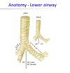
The right and left mainstem bronchus are not the same size nor is the angle
Right mainstem bronchus = shorter, wider, 25 deg angle
Left mainstem bronchus = narrower, longer, 40-60 deg angle
When you aspirate something will go into right mainstem bronch because it is more vertical!
Breathing tube improperly inserted may only be going into right mainstem bronchus (bc it is wider and angle) – therefore cant hear any air going to L lung (tube may slip too far down even though its taped causing this to occur)

describe the anatomy of the lung lobes
Right = 3 lobes, left = 2 lobes
Each lobe has different segments – important bc you will treat different segments in cadioresp
Upper L lobe has lingula (segment similar to what we find in the R lung as the middle lobe)
When positioning patients in optimal position lingula (left upper lobe) similar to middle lobe
Medial segment in R lower lung that is not in the L
- Gravity will help drainage specific to each segment based on positioning

describe the zones of the airway from trachea to aveoli
No gas exchange in conductive zone (no alveoli), some in transitional (some alveoli), and most exchange in resp zone (alveoli)
- Cartilage in first 16 generations
- Lung maintained open in lower lungs due to network of elastic tissue and pleura attaching lung to chest wall via fibres
- How large is the dead space? About 150 mL (space where no gas exchange occurs)

what is the acinus
Acinus = respiratory bronchiole, alveolar ducts, and alveoli
Alveolar sac = ~ 17 alveoli per sac
We have 300 million alveoli total in our lungs
All alveoli covered with capillaries – where gas exchange occurs (via diffusion)
Flow greater in upper airways and lowest in terminal bronchiole level – this is where dust will settle if inhaled

describe the histology of the airways
1) More columnar type epithelium
2) Lose cartilage, cuboidal epithelial cells, mucus glands disappear, have primarily clara cells producing the mucus
3) cilia epithelial cells disappear

describe the cell types in the lungs
Type 2 cells produce surfactant (which reduces surface tension, therefore prevents collapse)
Capillaries = endothelial cells!
Interstitial space = important bc with heart failure, back pressure from heart goes to the capillaries of the lung, this pushes fluid out of capillaries into the lungs – if a little, it can go into interstitial space = “Interstitial pulmonary edema” – if it gets to be more, start getting fluid in the alveoli which is then “pulmonary edema”
There are pores of kohn (openings/connections btw 2 alveoli)
Communication pathways btw terminal bronchioles and adjacent alveoli = canals of lambert – good because used as a collateral form of ventilation (air can flow around blockage through these openings), bad because if you have an infection it can propagate faster this way throughout the lung

describe the gas exchange process in the lungs
Capillary = 10 microns (size of a RBC) – very thin and small – gas exchange happens here
Therefore anything that increases membrane thickness of the alveoli can cause problems for gas exchange
Things that cause thickening = fibrosis, but also edema/ infection or lung abscess can also cause gas exchange problems

describe the anatomy of the boy thorax - what part is thickest?
Thickest bone is he manubrium, body of sternum is relatively thin (drs can cut through it)
Top of manubrium = sternal notch and first rib (under the clavicle)
Sternal angle/angle of louis – underneath is the bifercation/chrina
Xiphoid process = cartilage in young and solidifies over time
Behind of sternum = mediastinum, and heart
12th rib is a bit more posterior, 11th you can feel at your side

describe the anatomy of the thoracic cavity (side view)
This is the mediastinum looking from the side
There are many vessels
Heart in the inferior
Aorta and other vessels are in superior
Under heart is diaphragm (the main muscle of breathing) – heart sits on diaphragm
Anterior mediastinum = space btw sternum and heart
Middle mediastinum = heart
*Posterior mediastinum = lung tissue is there on L and R mediastinum

describe the anatomy of the thoracic cavity (transectional view)
- Note the pleura
- Parietal and visceral pleura – in btw there is pleural fluid
- Is someonw needs open heart surgery – open anteriorly without interfering with lungs (pleura are intact – lungs unaffected) – by piercing sternum

describe the anatomy of the pleura
Right and left lung have independent pleura
Therefore if someone needs to have part of lung removed, can cut lung (only that one lung will collapse and not the other side) – can do surgery while other lung is fine – same with stab wounds
- Think of lung being pushed into a fluid-filled balloon (balloon = pleura and fluid = pleural fluid) = each one independent!

describe the thoracic wall anatomy/ribs (how they move/how wide AP/laterally)
Ribs connect on transverse processes of vertebrae
The angle of the trandverse process determines the orientation of the ribs
The AP diameter is less than the transverse diameter by 2:1
In children they have a more rounded thorax – changes with age
Angle of transverse process determines the movement of the rib
Below each rib is a little groove where artery vein and nerve pass through – important bc if dr needs a lung biopsy or chest tube, will put needle above the rib, not below
There is an angle to the rib – this is commonly where fractures occur (just anterior to the angle)
- First rib does not normally fracture but if it does, damage to brachial plexus may also occur
- Lower rib fractures may disrupt diaphragm and diaphragmatic hernia may occur

describe the 2 ways in which the ribs move with respiration
For upper ribs articulation parallel to frontal plane – therefore upper ribs moving and increasing AP diameter of chest when breathing known as pump handle movement
For lower ribs when ribs move, increases lateral chest wall diameter = bucket handle movement
Therefore chest moves differently with breathing for upper vs lower

what are the respiratory muscles? (in their respective categories)
1) Diaphragm = main inspiration muscle
2) IAM – called accessory bc if we are setting at rest, inspiration is active (primary insp muscles – not IAM), expiration is passive – when doing exercise recruit more muscles to try and increase that flow (exp muscles)
- Note breathing is both automatic and voluntary
- For healthy people at rest we will not see our expiratory or IAM activating
- For COPD however, at rest they might have exp muscles working (have troubles getting air out) – can be measured with EMG – if condition is very severe you have hyperinflation and air trapping in lung and therefore may have IAM recruited at rest
- When diaphragm is flattened (due to hyperinflation of lung), not as efficient as normal dome shape – may see IAM recruited and they have dyspnea (shortness of breath)

for the diaphragm: which side is higher, which cavities does it separate, what percent of VC does it contribute to, and what percent of VT does it contribute to
- On left stomach and R liver underneath diaphragm (therefore R higher than left due to liver)
- Diaphragm differs for TV contribution when lying down bc when standing abd contents away from diaphragm, but when lying down they are pushing against dprm and therefore lengthening it
- Muscle can generate more force in lengthened position – in supine position diaphragm is lengthened it is able to contract and contribute more to tidal volume

describe the innervation and sections of the diaphragm
- pt with SCI level C4 therefore would have problems with diaphragm – partially innervated and not as efficient

what is the zone of apposition?
- Function of diaphragm depends on how much of a dome shape you have
- SCI of middle thoracic will not have abd muscles (involved with inspiration and posture) – if no tone in abs, abdomen will protrude, abdominal contents no longer held in place, and therefore not as much pressure can be generated

describe the anatomy of the trachea and landmarks
- Horseshoe shaped cartilages on trachea (open on the back and connected by muscle at the back) – this positioning protects the trachea from trauma in the front, also the esophagus is in the back, and also the cartilage prevents the trachea from collapsing on itself
- Between cartilage = elastic tissue, fibrous tissue, and muscle
- Trachea extends down to the 4th/5th thoracic vertebrae where it divides (called the bifercation)
- Landmark for bifercation (aka carina) = the angle of louis aka sternal angle (or posteriorly 4/5th T vertibrae)
- Trachea is 10cm long, 2.5 cm wide


