Large animal Abdomen and Viscera Flashcards
What is the most superficial abdominal muscle deep to the cutaneous trunci?
External Abdominal Oblique

Abdominal Coat or Yellow Coat
Tunica Flava

The tunica flava is deep fascia to the _______ abdomen which provides support for the muscular wall and the heavy abdominal contents.
ventral

The tunica flava is a deep fascia that consists of dense ____________.
elastic fibers

Ventrally, the tunica flava is interconnected with the ____________ of the external abdominal oblique.
aponeurosis

What is this arrow pointing to?

Lateral Fold
What is the remnant subsequent to dissection, in the lateral fold.
Cutaneous Trunci

What is outlined here?

Muscolotendinous Junction
What is the junction of external abdominal oblique with its aponeurosis?
Musculotendinous Junction

What forms the “heavy line” in horses with emphysema?
Muscolotendinous Junction

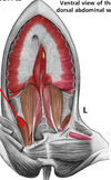
This is an Equine Superficial view of the ventral abdominal wall.
What are these arrows pointing to?

Cutaneous Trunci
This is an Equine Superficial view of the ventral abdominal wall.
What is this arrow pointing to?

Lateral Fold
This is an Equine Superficial view of the ventral abdominal wall.
What is this arrow pointing to?

Tunica Flava and aponeurosis of the external abdominal oblique.
This is an Equine Superficial view of the ventral abdominal wall.
What is this arrow pointing to?

The medial crus of the external abdominal oblique, superficial inguinal ring.
This is an Equine Superficial view of the ventral abdominal wall.
What is this arrow pointing to?

Lateral crus of the external abdominal oblique, superficial inguinal ring.
This is an Equine Superficial view of the ventral abdominal wall.
What is this arrow pointing to?

Right superficial inguinal ring
This is an Equine Superficial view of the ventral abdominal wall.
What is this arrow pointing to?

Prepubic Tendon
This is an Equine Superficial view of the ventral abdominal wall.
What is this arrow pointing to?

Inguinal Arch or Ligament
This is the Equine Abdominal Wall (with external abdominal oblique removed)
What is this arrow pointing to?

Rectus Abdominis
This is the Equine Abdominal Wall (with external abdominal oblique removed)
What is this arrow pointing to?

Rectus Sheath
This is the Equine Abdominal Wall (with external abdominal oblique removed)
What is this arrow pointing to?

Internal Abdominal Oblique
This is the Equine Abdominal Wall (with external abdominal oblique removed)
What is this arrow pointing to?

Cremaster Muscle
This is the Equine Abdominal Wall (with external abdominal oblique removed)
What is this arrow pointing to?

Aponeurosis, internal abdominal oblique
This is the Equine Abdominal Wall (with external abdominal oblique removed)
What is this arrow pointing to?

Inguinal Canal