Lab Practicals Micro Flashcards

1
Q

Catalase

A

An enzyme produced by bacteria that respire using oxygen, and protects them from the toxic by products of oxygen metabolism. Catalase positive bacteria include strict aerobes as well as facultative anaerobes. It helps us differentiate between some gram positive bacteria.

2
Q

Oxidase test

A

Used to determine if a bacterium produces certain cytochrome c oxidases. Only present in aerobic organizms. It is used to differentiate between some gram negative bacteria.

3
Q

Microsporum canis

A

FUNGUS. Mycetoma (chronic subcutaneous infection). Most common cause of ringworm.

Zoophilic dermatophyte (animal- infection of the skin)- cats, dogs, horses, monkeys

Colonies are flat, spreading, white or yellow, dense cottony surface

Clinical significance: cause of ringworm in humans and skin lesions in animals- particularly dogs and horses.

4
Q

What does oil immersion do?

A

Enables you to get very close to the specimen and allows you to get better magnification- you need transmitted light- dial up the light. Used with fixed specimens (staining and heating).

5
Q

How can you ID yeast vs. bacteria?

A

Size and replicate by budding

6
Q

What does gram stain do?

A

Allows you do ID gram neg or gram negative. And allows you to determine what type of antibiotic (more specifically)– though broad spectrum could be used on either. Also helps ID shape and allows you to begin to determine the type of organism.

7
Q

What is acid-fast (or Ziehl-Neelsen) stain?

A

Used to ID mycobacteria (TB, Leprosy, Johne’s disease)– will hold the stain even when they’ve been decolorized with a strong solution.

8
Q

What is Giemsa stain?

A

Aka Diff quick- blood smears- blood borne bacteria. Often stain differentially.

9
Q

Trichophyton mentagrophyte var. mentagrophytes

A

Fungus. Zoophilic dermatophyte.

Mice, Guinea pigs, roos, cats, horses, sheep, rabbits

Animal bedding and food, floors, and soil.

Frow fairly fast- after 4-5 days

Small white, fluffy, button- colony spreads rapidly; can be diagnosed after 2-3 weeks. (Upper side cream to light under side dark brown)

Clinical significance: produces inflammatory skin lesions, nodular and tumourous lesions called kerions, most commonly seen in dogs.

10
Q

How do you ID thin or small organisms?

A

Silver stain, dark field microscopy (look just at refracted light), electron microscopy (electrons have such a small wavelength)

11
Q

What is one of the most sensitive ways to detect an organism?

A

Culture.

12
Q

What is Sheep Blood Agar?

A

SBA is a nonselective medium and therefore allows the growth of a wide variety of organisms. It is used in the primary isolation of organisms from clinical specimens. (Pseudomonas aeruginosa- blue/green pigment and sweet odour, Streptococcus agalactiae- small colonies that are often beta haemolytic- i.e. complete lysis of sheep blood cells in the vicinity of the colonies, and Staphylococcus aureus- produce smaller colones compared to P. aeruginosa)

Peptides, salt, glucose, and sheep blood cells- nutrition that pathogenic bacteria like. Grow most common bacterial pathogens for vet medicine.

13
Q

What is cell free media?

A

Cell free media- most bacteria will grow (has intert red cells)- can be broth (liquid) or solidified with agar.

Agar often preferred: you can see what they look like after they’ve grown. You can also spread out the bacteria in your sample so you can see the colony derived from a single bacterial cell.

* many different additives depending on nutritional req. of the organism

* Grow at different temperatures– usually we do 37C. However, this is under most animal’s body temperature. So it’s a habit from medical microbiology. Birds are even at 40C. There are some pathogens that will grow only at lower temps- ones that grow on the skin.

14
Q

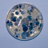
Penicillium species

A

Fungus.

Reservoir: air, house dust, plants; man and animal

Growth- quick, can be differentiated in just a few days

Powdery or velvety surface

Many colour tones- esp. green and blue green with FINE EXTENSIONS

Enlarged Lymph Nodes.
15
Q

How do you grow a microaerophilic organisms?

A

Increase partial pressure of CO2 and decreased the partial pressure of O2- stick a candle in a jar- light the candle and put the culture in with it. Not anaerobic, but has more CO2 than O2.

16
Q

Obligately intracellular parasites

A

ALL viruses and some bacteria- culture animal cells and grow them inside cells. Primary cultures freshly from an animal, established lines (easier to maintain in a lab- live in liquid nitrogen). Grown in complex media, often containing serum.

17
Q

Aspergillus species

A

Fungus.

Reservoir: almost everywhere on every conceivable type of substrate; especially soil and decaying organic debris.

Growth- fast growing, recognizable within a few days.

Growth form- velvety to flaky surface due to marked sporulation

Color- white- yellow (A. flavus), grey- green (A. fumigatus), black (A. niger)

Clinical significance: A. fumigatus is the most common cause of all forms of invasive and non-invasive aspergillosis (nasal discharge- can be aggressive and cause turbinate destruction)

18
Q
A

Trichophyton mentagrophytes var. mentagrophytes

19
Q

Rhizopus sp.

A

High prevalence in sub tropical and tropical regions

Reservoir: isolated from many substrates including a wide variety of soils, decaying vegetation, foodstuffs, and animal and bird dung.

Growth- fast growing, colonies cover an agar surface with dense cottony growth, first white becoming grey or yellowish brown with sporulation.

Clinical significance: R. oryzae is the most common causative agent of cygomycosis (infection due to a member of the zygomycetes- aprophytic fungi)– accounting for some 60% of reported culture positive cases. Often used in the fermented foods and alcoholic beverages in Asia.

20
Q

Epidermophyton floccosum

A

Antrhopophilic dermatophyte (prefers humans)

Commonsal, common shower or gym facilities

Growth- slow growing- suede-like surface, raised and folded in the center. Can develop white tufst of myslium.

Clinical significance: often causes tinea pedis (athlete’s foot), tinea cruris, tinea corporis (groin), and onychomycosis (fungla infection of the nail)- not known to invade hair invivo and no specific growth req. reported. Tinea= contagious skin fungal infection.

21
Q

What is cytopathic effect (CPE)?

A

Viruses multiply in cells and in most cases cause CPE- i.e. the cell looks sick: inclusion bodies

22
Q

Viral culture

A

Uninfected equine foetal kidney (EFK) culture and one that was infected with EHV1. Virus “plaque” formed by virus growth leading to lysis of the infected cells. Note the different morphology of the cells surrounding the plaque (infected) compared to cells away from it (uninfected).

* When EFK cells are infected with EHV1- the virus multiplies in the first cell it infects then spreads to neighboring ones forming a “focus” of infection. These foci slowly spread or enlarge until the entire cell culture is wiped out.

23
Q

Nutrient agar

A

Like sheep blood agar it is a nonselective medium. It is less nutritious than SBA and therefore does not support the growth of the same variety of organisms. S. agalactiae is not supported on NA. Easier to view P. aeruginosa.

24
Q

MacConkey’s Agar (MAC)

A

Commonly used to isolate and enumerate coliforms (gram negative organisms that inhabit the GI tract of mammals) and intestinal bacterial pathogens from a variety of sources including water, food, dairy products, and clinical specimen. Clinical specimen are typically placed on MAC and SBA in the initial steps of isolation.

It is an example of a SELECTIVE and DIFFERENTIAL medium.

The selective ingredient: bile salts. The aim is to inhibit organisms that do not tolerate the prescence of bile (GRAM NEGATIVE inhabit GI tract of mammals!).

Organisms that can grow on MAC are differentiated by their effect on neutral red, a pH indicator, which acts as an indicator for lactose fermentation. Pink (acid) colonies indicate that the organism is able to ferment lactose.

e.g. E. coli- lactose fermentation- yes!

Salmonella typhimurium- no lactose fermentation

25
Q

XLD agar

A

Used for isolation and presumptive ID of Salmonella sp. Most isolates of Salmonella ferment xylose and decarboxylate lysine, producing the alkaline conditons that are required for H2S production. On XLD, Salmonella sp. colonies are black, indicative of H2S production. The black colour is precipitate of iron sulphide formed when H2S and soluble iron salts in the medium react.

26
Q

Mannitol Salt Agar (MSA)

A

MSA is another example of a selective differential medium. The high salt concentration selects organisms that tolerate these conditions (commonly experienced on the skin. The organisms are differentiated by their ability to use mannitol and this is detected by a pH indicator. S. aureus colonies appear opaque and are surrounded by a yellow halo.

27
Q

ORSAB

A

Used to directly screen for MRSA. Based on MSA agar but the antibiotic, oxacillin is included. It inhibits the growth of S. aureus colonies that are susceptible to it. Aniline blue is the pH indicator used in ORSAB. So MRSA appears blue.

28
Q

Antimicrobial susceptibility plate

A

Tests for the resistance and susceptibility of the isolate to a range of antimicrobial agents. Growth within an annular radius of less than 6 mm indicates the isolate is resistant.

29
Q

Cell culture

A

* obligately intracellular parasites, only grow in animal cells (viruses), Cultured cells: Primary cell culture, established lines, grown in complex media, often containing serum

30
Q

Organ cultures

A

If you cannot grow a virus or bacteria in a cultured cell. Foetal tissue is best because it has the longest lifespan. Tracheal organ cultures will last for a couple of weeks- might be enough to get a virus up and replicating. Problem- tissue from adult animals- chance that the adult carries a virus- some contamination.

31
Q

Why are chick embryos still used often?

A

Influenza grows best- you can get the best replication which is good for making vaccines. Aim for Specific Pathogen Free or germ free. Will be some germs there but not too many.

32
Q

Animal inoculation

A

* Natural or artificial host (usually mice)

( Specific pathogen free- of the pathogen you are looking for

Gnobiotic- no viral, non bacterial population)

33
Q

What are you trying to detect with a toxin-antitoxin test, Labelled antibody test, ELISA, neutralization or inhibition assay, haemagglutination, or latex agglutination?

A

Detection of microbial proteins or carbohydrates

34
Q

What is a toxin-antitoxin test looking for? What is it?

A

Detecting toxin. Cow with botulism. Gut contents/ serum- filter it to sterilize it. Put it in a mouse- see if it reproduces botulism in mouse. If it does, then mix serum from animal with a specific antibody directed against the toxin and put it into the mouse and if it protects the mouse- then you know that is the toxin responsible for the disease.

35
Q

What is a labelled antibody test?

A

Apply label to animal tissue- will bind to spots where protein occurs- generally where protein is replicating (fluorescence).

36
Q

Haemagglutination

A

Some viruses bind to RBCs carbohydrates on the surface. If you take a filtered suspension of faeces from a dog with potentially parvo- mix it with RBCs from pigs. Then parvo will clump the RBCs (agglutinate- cross link RBCs).

37
Q

What is latex agglutination?

A

Ab against protein or carbohydrate stuck to latex beads- see if they cross link. If that happens then you know the specific pathogen is present.

38
Q

Immunoflourescence

A

Caused by a gram positive clostridial organism. Using fluorescently labelled Ab against the cell surface proteins of the bacterium. Touch a slide to affected piece of muscle- dry it out- apply Ab with label- and if there are cells there- the Ab will bind and fluoresce under a microscope. Will distinguish from other organisms in an animal that has recently died for example.

39
Q

Detecting Nucleic Acids

A

Mostly commonly PCR (hybridization, genome itself, whole genome sequencing). PCR is sensitive but it is very specific- you need to know what you are looking for. Can compare if diarrhoea in two foals has the same cause. Extract from faeces- stain silver. Can get epi info and specific diagnosis. Not applicable to too many viruses need double stranded RNA.

40
Q

Real Time PCR vs. Conventional and Melting Temperature Determination

A

* Conventional PCR- run rxn through 30 cycles- take sample and run it in a gel. Look for characteristic DNA band of a specific size- if present, we say it is positive. Very qualatitive. Need to be careful- this band is lighter than there is more agent here than there, for example.

* Real time PCR- include fluorescently labelled dye that binds to the DNA product as it gets made. Measure the amount of that dye in the DNA products. Exponential curve- 40 cycles- measuring amount of fluorescence. When exp. curve crosses threshold (arbitrarily) you can see how many copies of target RNA was sitting in the original sample. POWERFUL TEST BECAUSE you can tell if this animal has more than that animal. You can start to make conclusions of stages of disease. (often only use it as positive or negative, however. AKA quantative PCR)

** another thing you can do is analyze the melting curve- if you have the melting temperature- it is affected by the position and number of Gs and Cs in the sequence. If you have the dye sitting in the middle- the dye gets released- by plotting the curve- you can distinguish DIFFERENT STRAINS!!! You can distinguish a SINGLE DIFFERENT NUCLEIC STRAIN.

ARE THERE TWO OUTBREAKS LINKED??

41
Q

Detection of Specific Immune Responses

When can you use a single test?

A

* Acute and convalescent test (2 weeks later)

* you are looking for the Ab response- if you see an increase- then you can be pretty certain than that animal was infected with that agent at the time you saw it

** You can use a single test if you can detect IgM specifically (produced at the start of the immune response). Only if you think the animal has never been exposed before NOT ENDEMIC.

** OR a foetal test or newborne who has not had its Mother’s colostrum yet.

* picture agglutination test- whole blood agglutination- stained bacteria…. whole blood with Ab against that agent.

42
Q

How do you detect Antibody concentrations?

A

Agglutination, ELISAs, neutralizations, western blots, immunoprecipitation, complement fixation (detect IgG bound to an Antigen), immunofluorescence, haemagglutination inhibition (agent which clumps RBCs- then use an agent to block the agglutination to see if it is there)

43
Q

What fluid can you use of an animal’s if you want to find out if an Ab is present?

A

Blood or cerebral spinal fluid

44
Q

What is cell mediated response?

A

TB test- if you’ve been previously exposed- you’ll get an influx of T cells and macrophages– you’ll get redenning and a lump and therefore a positive reaction.

* In vivo (tests on a living organism)

* Lymphocyte stimulation test- IN VITRO- mix a bit of the Antigen with the whole blood (sample from the animal)- look to see if it stimulates lymphocytes to replicate and also to produce cytokines- interfeuron gamma or interleukin 2 (BOVINE TB- developed at Melbourne!!)– take the fluid off the top and test for interfeuron gamma… advantage- you don’t have to visit the animal twice!

CMI can last 20 years for example

45
Q

What is the most important thing for us as vets regarding pure infections and not disease?

A

Production loss!

46
Q

Normal Flora importance for vets

A

* Important for health in an animal

* Can cause disease if disrupted/ opportunistic infection

* Protectic role

* Important because it can confuse a diagnosis (how well did you take the sample!)

* Mouth and nasopharynx, stomach, duodenum, jejunum, ileum, large intestine, trachea, bronchi, lungs, vulva, prepuce, vagina, skin, mammary gland

47
Q

Problems with Koch’s postulates

A

Leprosy bacteria cannot be grown!

48
Q

If you detect an organism or don’t detect an organism, what do you have to consider?

A

If I detect an organism: is it from the animal?

Is it the one causing disease?

Is it the only one causing the disease?

If I don’t detect an organism:

Is it because ithere wasn’t ones there?

Is it because it didn’t survive the trip?

Did I look for the right one?

49
Q

What is an important thing the laboratory needs to know about the animal you took the sample from? And two other things?

A

History! Method of sampling and transport. Site of sampling. For example, with a dead animal- sterilize the lesion first before you take the sample.

50
Q

Class I- IV containment facilities

A

We work in a class II containment facility, for example- the pathogens we work with cannot be aerolosilized in large quantities. The air out of the lab gets filtered.

* Class III and IV containment: negative pressure- even if a break in glove- air will suck it in the cabinet and it will pass through a filter. Still a risk- stab for example. Not optimal as hard to manipulate things. OR positive pressure suit- air being puffed in. So even if a hole in the suit- no agents can get in. Protect him but also to stop him from becoming a vector. Needed for exotic agents and where the person can be a vector. Or if the agent poses a particular risk.

51
Q

Sterilisation v. Disinfection

A

Absolute- the material is either sterile or it is not. Disinfection is removal of potential infectivity. Not removing all the agents- you are decreasing the risk- not completely eliminating.

* Environment- disinfect

52
Q

Antisepsis

A

* Animal’s skin-you cannot remove even the potential infectivity- you can only reduce the load, not eliminating- swabbing with iodine for example

53
Q

Talk through the different levels of removing the risk when cleaning

A

Sterilization, Disinfection, and Antisepsis

** always incorporate cleaning!! “removing faeces from the surface”

54
Q

Bacteriostatic vs. Bactericidal

A

Bacteriostatic- stop the agents from growing don’t kill them. Useful for treating an animal where there is an immune response where you can clean up those stopped organisms afterwards. Useful for chemotherapy but not useful disinfection (bacteriocidal). Or useful in cases of streotococcal and clostridial gangrene because they inhibit the production of the toxins that cause much of the morbidity.

55
Q

Different basis of action for disinfectants

A

* Damage the genome- cause a break in DNA or RNA

* Damage the cell envelop

* Damage crucial metabolic proteins- critical for ongoing agent (e.g. break in the cell surface)

* Kinetics

56
Q

Physical agents

A

* heat

  • pasteurisation (removes 90% of bacteria that are there, wipes out nearly all of the pathogens, some of the spoilage organisms that make the milk curdle- but they don’t cause disease… good at eliminating TB, lysteria, etc.) (FLASH PASTEURIZATION- heat it up more for a shorter period of time)
  • moist heat (MOST EFFECTIVE)- boiling & autoclaving (steam)– pressures above atmospheric pressure- 120C– the best way!! And FASTEST (destroys proteins, cell envelopes, kills even the most resistant)
  • dry heat (much higher temps 160C– need to do this for an hour or two- much more destructive than wet heat and more time consuming)

* Radiation

  • UV light (any shadowing, won’t reach those areas- good for flat surfaces– and CANNOT GET THROUGH GLASS- OK for OR environments- won’t completely sterilize materials)
  • Ionizing radiation (need cobalt 60 source- uncommon- used to sterilize needles, syringes, scapel blades– useful for fragile materials (heat labile materials))

* Filtration (good for liquids– cannot filter out soluble molecules like the endotoxin– left over after gram negative bacteria are killed– need to have reduced the load/ eliminated bacteria from the liquid before you inject it into an animal’s body otherwise they can be sent into endotoxic shock)

57
Q

Chemical Agents

A

Effective disinfecants. The only exception: Ethylene Oxide- 18-20 hours- completely enclosed space.

More selective– none are broadly effective.

Need to know:

* Activity spectrum (what will it kill?)

* inhibition vs. inactivation

* rate of action

* Compatability problems (with soaps for example)

* effect of organic material (blood, pus, faeces– residual after removal)– think of bleach! Or iodine what it does to material.

* Side effects to patient, staff, client

* Price and stability