Janis Notes Flashcards

1
Q

Define Maffucci’s syndrome.

A

Enchondromatosis associated with multiple cutaneous hemangiomas.

2
Q

Indications for Mohs micrographic surgery (5).

A
  1. Recurrent
  2. Cosmetically sensitive (periorbital, periauricular, perinasal)
  3. Morpheaform and sclerosing or aggressive features
  4. Poorly delineated margins in scar tissue
  5. Other tumor types: SCC with perineural involvement, dermatofibrosarcoma protuberans, microcystic adnexal carcinoma.
3
Q

Define Nevus of Ota.

A

Nevus of Ota: Onset at birth or less than 1 year and around puberty. Found more commonly in blacks and Asians. Blue-brown, unilatreal, periocular macula. Size varies from few cm to covering half the face. Areas follow distribution of V1 and V2.

4
Q

Describe Tessier Cleft Number 5.

A

Tessier Cleft Number 5.

One liner: Cleft begins lateral to the canine and courses lateral to infraorbital foramen terminating in the lateral aspect of lower eyelid and orbital floor.

Soft tissue:

  • Begins medial to oral commisure and courses along the cheek lateral to the nasal ala
  • Terminates in the lateral half of the lower eyelid

Skeletal involvement:

  • Begins lateral to the canine.
  • Courses lateral to infraorbital forarmen
  • Terminates in the lateral aspect of the orbital floor and rim.
  • Lateral orbital wall possibly thickened and greater wing of sphenoid abnormal.
5
Q

Risk of malignant transformation with congenitcal nevocytic nevi.

A
  • Small (<1.5 cm^2): 1-5% lifetime risk (rarely before puberty).
  • Medium (1.5 - 20 cm^2): uncertain (rarely before puberty).
  • Giant (> 20 cm^2): 5-10% result in melanoma with 50% arising between the age of 3-5 years old.
6
Q

Risk factors (11) for recurrence of BCC and SCC.

A
  • Location/size: >20 mm on trunk/extremities; >10mm cheek, forehead, scalp, neck; >6mm central face, genitalia, hands, feet.
  • Poorly defined borders.
  • Recurrent lesion.
  • Immunopsuppresion
  • Previous radiotherapy.
  • Pathology: morpheaform, sclerosing, micronodular, mixed infiltrative, adenoid, desmoplastic
  • Perineural involvement
  • Lymphovascular invasion
  • Radpidly growing
  • Depth: >2mm, Clark IV and V
  • Poorly differentiated
7
Q

Timing of normal suture fusion.

A
  • Metopic: 6-8 months.
  • Sagittal: 22 years.
  • Coronal: 24 years.
  • Lambdoidal: 26 years.
8
Q

Risk factors for tetanus (5).

A
  1. Time since injury (>6 hours)
  2. Depth of injury (>1 cm)
  3. Mechanism of injury (crush, burn, GSW, puncture)
  4. Presence of devitalized tissue
  5. Contamination (grass, soil, saliva, retained foreign body)
9
Q

Treatment of phenol burns.

A

Irrigate and treat with polyethylene glycol.

10
Q

Define Parkes Weber syndrome.

A
  • Similar to Klippel-Trénaunay syndrome (patchy port-win stain on an extremity overlying a deeper venous and lymphatic malformation with associated skeletal hypertrophy).
  • Distinguished by the prescence of an arteriovenous fistula.
11
Q

Steps to PIP contracture release after Dupuytren’s disease excision and continued stiffness.

A
  1. Chein rein ligament release.
  2. Volar capsulomtomy.
  3. Collateral ligament release.
12
Q

Name two otoplasty techniques for addressing lower third prominence.

A
  • Modified fishtail excision of lobule (Wood-Smith)
  • Inferior conchal mastoid sutures
13
Q

Horizontal (5) and vertical buttresses (4) of the face.

A

HORIZONTAL BUTRESSES

  1. Frontal bar
  2. Inferior orbital rim / upper transverse maxillary
  3. Maxillary alveolar and hard palate / lower transverse maxillary
  4. Mandibular alveolar / upper transverse mandibular
  5. Inferior border of mandible / lower transverse mandibular

VERTICAL BUTTRESSES

  1. Zygomaticomaxillary / lateral maxillary / lateral buttress
  2. Nasomaxillary / medial maxillary / medial buttress
  3. Pterygomaxillary / posterior maxillary / deep buttress
  4. Vertical mandible / posterior verticle
14
Q

Describe ‘Stahl’s ear’.

A

The presence of the third and/or horizontal superior crus with a pointed upper helix. Results in upper and middle third prominence.

15
Q

Describe the intracranial, cranial, orbit, midface, and extremity findings in Apert syndrome.

A
  • Intracranial: elevated ICP.
  • Cranial: bicoronal synostoses with turribrachycephaly, enlarged anterior fontanel, bitemporal widening, occipital flattening.
  • Orbits: exorbitism, downslanting palpebral fissures, and mild hypertelorism.
  • Midface: hypoplasia, parrot beak deformity, high arch or cleft palate, anterior open bite, class III malocclusion.
  • Extremity: Complex syndactyly of hands and feet.
16
Q

Treatment of hydroflouric acid burns.

A

Calcium gluconate.

17
Q

Surgical management of trigonocephaly (metopic synostosis).

A

Overcorrection of frontal dysmorphology and bitemporal contriction with bifrontal craniotomy, frontal reshaping and expansion of frontoorbital bar and frontal bone flaps with interpositional bone graft, wedge osteotomies, bone grafting.

18
Q

Alkali burns cause what time of injury.

A

Liquefactive necrosis.

19
Q

Where is Stenson’s duct located intraorally?

A

Opposite the second maxillary molar.

20
Q

Describe the cranial, extremity, and other findings of Muenke syndrome.

A
  • Cranial: uni or bicoronal synostosis.
  • Midface: typically no hypoplasia.
  • Extremities: Thimble like hypoplasia.
  • Other: sensorineural hearing loss.
21
Q

Describe Tessier Cleft Number 30.

A

Tessier Cleft Number 30

One liner: Mandibular cleft originating between the central incisors and extending inferiorly, yielding notching of the lower lip and a bifid tongue.

Soft tissue:

  • Notch in lower lip
  • Bifid anterior tongue with attachment to mandible by dense fibrous band.

Skeletal involvement:

  • Cleft between central incisors extending into mandibular synthesis
  • Hyoid bone may be absent
  • Thyroid cartilage incompletely formed
22
Q

Surgical management of lambdoid synostosis.

A

Biparietal-occipital craniotomies, occipital switch, and contouring.

23
Q

Formula to estimate caloric needs in burns.

A

Curreri formula.

Caloric needs = [25 kcal/kg/day] + [40 kcal/%TBSA/day]

Caloric needs per day = [25 kcal]*[weight in kg] + [40 kcal]*[%TBSA]

24
Q

Compared with nasal ideals for whites, describe the characteristic differences of black and middle eastern noses?

A

Blacks

  • Wide, low nasal dorsum
  • Descreased nasal length and tip projection
  • Poor nasal tip definition
  • Acute columellar-labial angle
  • Alar flaring

Middle Eastern

  • Wide nasal bones
  • Thick, sebaceous skin
  • Ill-defined bulbous tip
  • High dorsum and over projecting radix
  • Acute columellar-labial angle
  • Slight alar flaring
25
Q

Histological zones of distraction osteogenesis (5).

A
  1. Central zone: cellular proliferation.
  2. Transitional zone of vasculogenesis
  3. Paracentral zone: Parallel orientation of collagen fibers with osteoid production.
  4. Transitional mineralization front: Primary mineralization found with bone spicule formation.
  5. Mature bone zone: Progressive calcification of primary mineralization front with formation of cortical and cancellous elements.
26
Q

Natural history and malignant degeneration of Nevus sebaceus of Jadassohn.

A
  • Present at birth on scalp or face.
  • Well-circumscribed, hairless, yellowish plagque that becomes verrucous and nodular at puberty.
  • 10-15% malignant degeneration to BCC.
27
Q

Cells of the epidermis and their function.

A
  • Keratinocyte: Major cell type. Protective barrier.
  • Melanocyte: Neural crest origin. Pigmentation. UV protection.
  • Merkel cells: Neural crest origin. Constant pressure and touch. Static two-point discrimination.
  • Langerhands cells: Mesenchymal origin (from precursor cells of bone marrow). Antigen presenting cells.
28
Q

Ratio of Type I:III collagen in mature skin, hypertrophic scars, and keloid scars.

A
  • Mature skin = 4:1
  • Hypertrophic = 2:1
  • Keloid = 3:1
29
Q

Options for surgical access to the orbital floor.

A
  1. Subciliary (‘lower bleph’)
  2. Subtarsal (‘mid lid’)
  3. Infraorbital (‘inferior orbital rim’)
  4. Tranconjunctival
30
Q

TIming of normal fontanelle closure.

A
  • Posterior fontanelle: 3-6 months.
  • Anterior fontanelle: 9-12 months.
31
Q

Options for recipient veins in breast reconstruction.

A
  1. Thoracoacromial.
  2. Lateral thoracic.
  3. Axillary.
  4. Retrograde flow in IM vein.
  5. Cephalic turned down.
  6. External jugular turned down.
32
Q

Describe Tessier Cleft Number 8.

A

Tessier Cleft Number 8

One liner: Largely isolated to the orbit with the cleft affecting the frontozygomatic suture with a hypoplastic or absent zygoma with coloboma of the lateral commisure.

Soft tissue:

  • Largely isolated to the orbit.
  • Coloboma of the lateral commissure with abscence of the lateral canthus.

Skeletal involvement:

  • Involves frontozygomatic suture.
  • Zygoma hypoplastic or absent with lateral orbital wall missing.
  • Continuit of orbital and temporal fossa.

Other:

  • Almost always concurrent with another rare cleft
  • Associated with Goldenhar’s syndrome
33
Q

Optimal centrifuge for fat grafting.

A

3 minutes at 3000 rpm.

34
Q

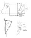
Describe the correct placement of the ear (height, width, angulation)?

A
35
Q

Define Sturge-Weber syndrome.

A
  • Large facial port-wine stain with V1 and commonly V2 trigeminal nerve distribution.
  • Associated with leptomeningeal venous malformations and frequent mental retardation.
36
Q

Define Riley-Smith syndrome.

A

Pseudopapilledema, microcephaly, vascular malformations.

37
Q

Prognostic signs of hemangiomas (7).

A
  1. Size of hemangioma or sex of patient has no influence on the speed or completeness of resolution.
  2. Site of hemangioma has minimal effect on final result.
  3. Multiple hemangiomas may resolve at different rates.
  4. Presence of subcutaneous tumor elements has no effect on final outcome.
  5. Early dramatic growth is not a prognostic sign of resolution.
  6. Time of involution is indicative of outcome.
  7. Prescence of ulceration has no prognostic significance except for scar consequences.
38
Q

Advantages and disadvantages of alloplastic breast reconstruction.

A

Advantages:

  1. Lower initial costs compared with autologous reconstruction.
  2. Technical ease.
  3. No donor site morbidity.
  4. Shorter operative time and recovery time compared with autologous.

Disadvantages:

  1. Early cost advantage disappears over time as revisions surgeries are required.
  2. Potential capsular contracture.
  3. Potential implant rupture / failure.
  4. Potential infection requiring removal.
  5. Increased probability for future surgery.
  6. Difficult to acheive symmetry with unilateral reconstruction.
39
Q

List the basic goals of otoplasty (10)?

A
  1. Correction of upper-third protrusion.
  2. Visibility of the helix beyond the anithelix when viewed from the front.
  3. Smooth and regular helix.
  4. No marked distortion or decrease in the depth of the postauricular sulcus.
  5. Correct placement of the ear; helix-to-mastoid distance falls in the normal range of 10-12mm at the top, 16-18mm in the middle, and 20-22 in the lower third.
  6. Bilateral symmetry to within 3mm at any given point.
  7. Smooth, rounded, and well-define antihelical fold.
  8. Concoscaphal angle of 90 degree.
  9. Conchal reduction or reduction of the conchomastoidal angle.
  10. Helical rim that projects laterally farther than the lobule.
40
Q

Describe the Mustarde otoplasty technique.

A
41
Q

Name the most common anatomical deformities yielding a prominent ear (3).

A
  1. Poorly defined antihelical fold.
  2. Conchoscaphal angle >90 degrees.
  3. Conchal excess.
42
Q

Describe Tessier Cleft Number 4.

A

Tessier Cleft Number 4

One liner: Cleft begins between lateral incisor and canine teeth extending superiorly lateral to the piriform and medial to the infraorbital foramen into the lower eyelid.

Soft tissue:

  • Begins lateral to Cupid’s bow between commisure of mouth and philtral crest -> passes onto cheek lateral to nasal alar -> curves into lower eyelid to terminate medial to the punctum
  • Lower canniculus disrupted along with most of the inferior supporting structures of the eye.
  • Colobomas.
  • Medial canthal ligament and lacrimal aparatus usually intact.

Skeletal involvement:

  • Begins between lateral incisor and canine teeth
  • Extends onto anterior surface of maxilla, lateral to piriform aperature, medial to infraorbital foramen
  • Medial and inferior portions of orbital disrupted
  • Nose displaced superiorly (especially in bilateral cases)
43
Q

List the contents of the superior orbital fissure and explain the presentation and pathophysiology of superior orbital fissue syndrome (SOFS). Differentiate with orbital apex syndrome.

A

Contents of superior orbital fissure:

The order of the nerves passing through the superior orbital fissure from superior to inferior (Lazy French Tarts Sit Nakedly In Anticipation):

  • L: lacrimal nerve (branch of CN V1)
  • F: frontal nerve (branch of CN V1)
  • T: trochlear nerve (CN IV)
  • S: superior division of the oculomotor nerve (CN III)
  • N: nasociliary nerve (branch of CN V1)
  • I: inferior division of the oculomotor nerve (CN III)
  • A: abducens nerve (CN VI)

Presentation of superior orbital fissue syndrome (SOFS):

  • Ophthalmoplegia: due to compression or damage to oculomotor, trochlear and abducens nerves
  • Ptosis: due to loss of oculomotor motor supply to the levator palpebrae superioris and loss of sympathetic input (third order postganglionic) to Muller’s muscle
  • Proptosis: due to decreased tension in the extraocular muscles with loss of innervation
  • Fixed dilated pupil: due to loss of parasympathetic supply to the pupil by the oculomotor nerve
  • Lacrimal hyposecretion and eyelid or forehead anaesthesia: due to damage to branches of the ophthalmic division of the trigeminal nerve
  • Loss of corneal reflex: due to loss of afferent input from the ophthalmic division of the trigeminal nerve.

Orbital apex syndrome presents the same but also has loss of vision because of involvement of the optic nerve at the orbital apex.

44
Q

Three predictors of mortality in burns (Baux score).

A
  1. Age
  2. % TBSA
  3. Inhalational injury
45
Q

Two mechanisms of skull growth.

A
  • Sutural growth: perpendicular to sutures.
  • Appositional growth: Bone resoprtion on the inner surgace and deposition on the outer surface of the skull.
46
Q

Options for burn wound coverage.

A

Temporary

  • Allograft
  • Xenograft
  • Biobrane (nylon fabric coated with porcine dermal collagen with silicone membrane)
  • TransCyte (biobrane and cultured human neonate fibroblasts)

Permanent

  • FTSG / STSG
  • Integra (bovine collagen, shark chondroitin-6-sulfate, silicone layer)
  • AlloDerm
  • Cultured epithelial autograft
47
Q

Keen approach to the zygomatic arch.

A

1-2cm incision made laterally in the buccal sulcus. Subperiosteal elevation allows the eleator to be placed behind the arch.

48
Q

Define PHACE syndrome.

A
  • Posterior fossa malformations
  • Hemangiomas (large facial hemangiomas)
  • Arterial anomalies
  • Coarctation of the aorta and other cardiac anomalies
  • Eye abnormalities
49
Q

Treatment algorithm of ZMC fractures based on energy.

A

Low-energy injuries:

  • ZM exposed with with upper buccal sulcus incision
  • ZF and ZS exposed with upper bleph incision
  • ZS articulation best to assess reduction
  • Stabilize ZF and then ZM; if needed ZS fixation can be performed as well
  • Lower lid incision not required unless floor exploration or reconstruction desired

High-energy injuries:

  • Require wide exposure with coronal approach to visualize temporal articulation
  • Will likely require exploration and fixation of the floor
50
Q

Prevalance of concurrent injuries in panfacial fractures (top 5).

A
  1. Intracranial injury / hemmorrhage (18%).
  2. Abdominal organ injury (16%).
  3. Pneumothorax (13%).
  4. Pulmonary contusion (13%).
  5. Cervical spine fracture (13%).
51
Q

Features consistent with metopic synostosis (4).

A

Collectively referred to as trigonocephaly (‘keel shaped, triangular’).

  1. Keel-shaped forehead
  2. Bitemporal narrowing
  3. Hypotelorism
  4. Bilateral supraorbital retrusion
52
Q

Describe Tessier Cleft Number 1.

A

Tessier Cleft Number 1.

One liner: Cleft begins between the lateral and central incisor moving superiorly through the margin of cupid bow and up between the nasal bones and the frontal process of the maxilla.

*Can continue as cleft number 13*

Soft tissue:

  • Pattern similar to cleft lip and palate.
  • Cleft through lateral margin of Cupid’s bow progresses into nose, causing notching of the dome and soft triangle but may travel medially to a malpositioned medial canthus.

Skeletal involvement:

  • Cleft between central and lateral incisor.
  • Cleft separates nasal floor from piriform and the nasal bone from the frontal process of the maxilla.
53
Q

Predisposing conditions / lesions for melanoma (7).

A
  1. Familial Atypical Multiple-Mole Melanoma (FAMMM Syndrome)
    • >100 melanocytic nevi measuring 6-15mm
    • one or more measuring > 8mm
    • 10% risk of melanoma
  2. Dysplastic nevus (6-10% risk of melanoma).
  3. Congenital nevus (6% lifetime risk of melanoma).
  4. Typical moles (risk if >50)
  5. Melanoma in situ
  6. Xeroderma pigmentosum
  7. Lentigo maligna (Hutchinson freckle)
54
Q

Describe the embrology of the face and its prominences.

A
  • Face develops from five prominences:
    • Frontonasal prominence (1) which is composed of bilateral medial and lateral nasal prominences.
    • Paired maxillary prominences (2)
    • Paired mandibular promincens (2)
  • Frontonasal prominence:
    • Pulled ventrally and caudally to form the forehead, nasal dorsum, and medial and lateral nasal prominences.
    • Outpouchings of the lateral nasal walls form the sinsuses (maxillary at 3 months gestations, ethmoid at 5 months gestation, sphenoid 5 months postnatally, and frontal 2-6 years postnatally).
    • Medial nasal prominence forms the primary palate, midmaxilla, midlip, philtrum, central nose, and septum.
    • Lateral nasal prominence forms the nasal alae.
  • Maxillary prominences:
    • Migrate medially to form the secondary palate, lateral maxilla, and lateral lip.
    • Junction with the lateral nasal prominences forms the nasolacrimal groove and nasolacrimal duct system.
  • Mandibular prominences:
    • Form the mandible, lower lip, and lower face.
55
Q

Features consistent with sagittal synostosis (3).

A

Collectively referred to as scaphocephaly or dolichocephaly (“boat shaped”)

  1. Elongated anteroposterior head.
  2. Frontal and occipital prominence.
  3. Biparietal narrowness
56
Q

Define Nevus of Ito.

A

Nevus of Ito: Usually appears at birth. Typically found in Asians and blacks. Large, blue-brown macula located on the posterior shoulder, areas innervated by the posterior supraclavicular and lateral cutaneous brachial nerves.

57
Q

Decribe Tessier Cleft Number 12.

A

Tessier Cleft Number 12

One-liner: Cleft lies medial to the medial canthus extending upward and coming out lateral to the cribriform plate, yielding hypertelorism.

Soft tissue:

  • Cleft lies medial the medial canthus, which is displaced laterally.
  • Colobomas extend to the root of the eyebrow.
  • Paramedian frontal hairline projects downward.

Skeletal involvement:

  • Orbital hypertelorism and telecanthus from increased dimension of ethmoid air cells.
  • Frontal and sphenoid sinuses pneumatized.
  • Lies lateral to cribriform plate, which remains normal.
  • Anterior and middle cranial fossa enlarged.
58
Q

Timing and indications for surgery (3).

A

Surgery typically delayed until school age when child may beging to have psychological consequences.

Indications for urgent surgery in proliferative phase:

  1. Visual obstruction
    • During first year of life, visual obstruction for 1 week can result in deprivation amblyopia or anisometropia.
  2. Nasolaryngeal obstruction
    • Hemangiomas in “beard distribution’ proliferating in subglottic airway = potentially life threatening. Requires aggressive treatment with beta-blockers, surgery, and potentially LASER.
  3. Auditory canal obstruction
    • Can result in conductive hearing loss.
59
Q

Describe the ossification of the cranium, including the neurocranium and the viscerocranium.

A

Neurocranium is the portion of the skull encasing and protecting the brain. It develops from both intramembranous ossification and endochondral ossification.

  • Membranous neurocranium
    • Forms via intramemranous ossification of neural crest origin.
    • Includes the paired frontal, squamosal, and parietal bones, and upper occipital bones.
  • Cartilaginous neurocranium (ie basicranium)
    • ​Forms via endochondral ossification of mesodermal origin.
    • Includes sphenoid and ethmoid bones, mastoid and petrous temporal bone, and the base of the occipital bone.

Viscerocranium is the bones of the facial skeleton and forms primarily via intramembranous ossification of pharyngeal arch I, except for Meckel’s cartilage (which forms the malleus and the mandibular condyles)

60
Q

Describe Tessier Cleft Number 2.

A

Tessier Cleft Number 2

One liner: Cleft originates at lateral incisor and margin of cupid bow and extends superiorly through the alar rim.

*Can extend as cleft number 12*

Soft tissue:

  • Originates at the lateral margin of cupids bow and extends into the lateral alar rim lateral to dome.
  • Ala typically hypoplastic.
  • Medial canthus displace but lacrimal duct not involved

Skeletal involvement:

  • Cleft through lateral incisor extends into piriform aperature.
61
Q

Define Klippel-Trénaunay syndrome.

A

Pathy port-win stain on an extremity overlying a deeper venous and lymphatic malformation with associated skeletal hypertrophy.

62
Q

Features (7) of Nevoid basal cell syndrome (Gorlin’s syndrome).

A
  1. Multiple basal cells
  2. Odontogenic keratocysts
  3. Palmar and plantar pits
  4. Calcification of falx cerebri
  5. Bifid ribs
  6. Hypertelorism
  7. Broad nasal root
63
Q

Describe the intracranial, cranial, orbit, and midface features of Crouzon syndrome.

A
  • Intracranial: hydrocephalus, ICP elevation, Chiari malformation.
  • Cranial: bicoronal most common; usually brachycephalic, but scaphocephally, trigonocephaly, cloverleaf deformity, and normocephalic described.
  • Orbit: Exorbitism.
  • Midface: hypoplasia with anterior open bite, class III occlusion, narrow and high arched palate.
64
Q

Ratios to assess on a lateral view facial analysis (3).

A
65
Q

Indications for distraction osteogenesis.

A
  1. Mandibular lengthening.
    • Hemifacial microsomia, micrognathia
  2. Le Fort I adavanacement
    • For large anterior movements > 1cm
    • Particularly helpful for significant class III occlusion in clefts secondary to maxllary hypoplasia.
  3. Le Fort III / monobloc
    • Syndromic craniosynostosis with severe midface hypoplasia and exorbitism.
  4. Posterior cranial vault reconstruction.
    • Can be used in some craniosynostosis.
66
Q

Mutations associated with craniosynostosis (4).

A
  1. FGFR2
  2. FGFR3
  3. TWIST1
  4. EFNB1
67
Q

Describe Tessier Cleft Number 10.

A

Tessier Cleft Number 10.

One liner: Cleft lateral to the supraorbital foramen, yielding encephaloceles and hypertelorism from inferolateral orbital rotation.

Soft tissue:

  • Begins at middle third of upper eyelid and eyebrow.
  • Coloboma in mid-upper lid and irregular retraced brow.

Skeletal involvement:

  • Cleft at middle orbital rim just lateral to supraorbital foramen.
  • Encephalocele common
  • Hypertelorism from inferolateral rotation of orbit
  • Anterior cranial base distortion
68
Q

Describe the Furnas otoplasty technique.

A
69
Q

Topical antimicrobial for burn wounds along with their antimicrobial coverage, notable clinical properties and side effects (4).

A
  • Silver sulfadiazine (silvadene)
    • Gram positive and gram negative coverage.
    • Transient leucopenia.
    • Penetrates eschar poorly.
  • Mafenide acetate (sulfamylon)
    • Gram positive.
    • Potent carbonic anyhydrase inhibitor.
    • Hyperchloremic metabolic acidosis.
    • Compensatory hyperventilation.
    • Penetrates deeply.
  • Silver nitrate (0.5% solution)
    • Staphylococcus species and gram negative (pseudomonas).
    • Hyponatremia and hypokalemia.
  • Sodium hypochlorite
    • Broad spectrum.
    • at 0.025% is bactericidal without inhibiting fibroblasts or keratinocytes.
70
Q

Treatment options for SCC.

A
  • Medical
    • Topical 5-FU for premalignant lesions
    • Chemotherapy for adjuvant therapy
  • Radiation (adjuvant or primary treatment; 90% cure)
  • Destructive
    • Curettage and electrodessication
    • Photodynamic therapy
  • Surgery
    • WLE (95% cure; 4-6 mm margins)
    • Mohs (95% cure)
71
Q

Describe the features of Treacher Collins Syndrome.

A

Bilateral Tessier cleft 6, 7, and 8.

  • Coloboma and retraction of the lower lid
  • Upper lid redundancy in lateral half gives impression of ptosis
  • Lateral canthus displaced inferiorly
  • Absence of zygomatic arch
  • Hypoplasia of temporalis
  • Ear malformations
  • Hairline abnormalities
  • Abscence of lateral inferior orital rim
  • Hypoplasia of malar ones and mandible
72
Q

Ratios to assess in the frontal view facial analysis (8).

A
73
Q

Cords in Dupuytren’s disease.

A

1. Pretendinous cord: in the palm.

2. Spiral cord: composed of pretendinous, spiral band, lateral digital sheet, and Graysons (can displace NVB centrally and superficially).

3. Central cord: continuation of pretendinous.

4. Lateral cord: runs from the natatory to the lateral digital sheet.

5. Natatory cord: natatory band.

6. Retrovascular cord.

74
Q

Risk factors for Dupuytren’s diathesis.

A
  1. Male gender.
  2. Earlier disease onset.
  3. Family history.
  4. Bilateral involvement.
  5. Ectopic involvement (Peyronies, Ledderhose, Garrods).
75
Q

Landmarks for Gillies approach.

A

Incision made 2cm anterior and superior to the helix.

Dissection carried down to and through the deep temporal fascia (overlying temporalis muscle). Elevator placed beneath the deep temporal fascia but superficial to temporalis allowing access to the medial aspect of the zygoma while protecting the facial nerve.

76
Q

Describe Tessier Cleft Number 7.

A

Tessier Cleft Number 7

One liner: Cleft begins at the oral commissure and extends outward affect the ramus/condyle/coronoid of the mandible and include the ear.

Soft tissue:

  • Begins at oral commissure and varies from mild broadening to complete fissure.
  • Extends toward the ear, ceasing at the anterior border of the masseter.
  • Ear involvement varies from a skin tag to microtic ear.
  • Paralysis of CN V and CN VII common.

Skeletal:

  • Cleft passes through pterygomaxillary junction.
  • Posterior maxilla and ramus (condyle and coronoid) are hypoplastic.
  • Zygomatic body is also severely hypoplastic and displaced.
  • Cranial base asymmetrical.
  • Open bite or cross bite seen.

Other:

  • Often accompanies crnaiofacial microsomia.
77
Q

Management algorithm for anterior table fracture of frontal sinus.

A

Depends on degree of displacement and involvement of nasofrontal duct.

78
Q

When does the the size of the ear mature (width, length)?

A
  • By the third year of life the ear has reached 85% of adult size.
  • Ear WIDTH reaches mature size by 6-7 years of age (girls-boys)
  • Ear LENGTH reaches mature size by 12-13 years of age (girls-boys).
79
Q

Risk of metastasis from primary SCC (trunk vs. face/extremity).

A
  • Trunk: 2-5%
  • Face / extremity: 10-20%
80
Q

Differential diagnosis for congenital nevocytic nevus (13).

A
  1. Café au lait spot
  2. Nevus spilus
  3. Epidermal nevus
  4. Acquired nevus
  5. Dysplastic nevus
  6. Blue nevus
  7. Becker’s nevus
  8. Halo nevus
  9. Mongolian spot
  10. Nevus of Ota
  11. Nevus of Ito
  12. Spitz nevus
  13. Nevus sebaceus
81
Q

Describe the intracranial, cranial, orbit, and other features of Saethre-Chotzen syndrome.

A
  • Intracranial: mental status usually normal.
  • Cranial: bi or uni-coronal synostosis.
  • Orbits: Ptosis.
  • Ears: Prominent crus helicis extending through the conchal bowl.
  • Midface: deviated nasal septum, narrow palate.
  • Extremities: Patrial syndactyly with short stature.
82
Q

Collagenase clostridium histolyticum injection protocol.

A
  1. Inject 0.58mg into a given cord (each vial contains 0.9mg).
  2. Up to TWO cords or TWO joints in the same hand can be treated each visit.
  3. A cord can be treated up to THREE times at 4 week intervals.
83
Q

Draw the anatomy of the ear.

A
84
Q

Define Bannayan-Zonana syndrome.

A

Microcephaly, multiple lipomas, multiple vascular malformations.

85
Q

Describe Goldenhar’s Syndrome.

A
  • Prominent frontal bossing.
  • Low hairline.
  • Mandibular hypoplasia.
  • Low set ears
  • Colobomas of the UPPER eyelid
  • Epibulbar dermoids
  • Bilateral anterior accessory auricular appendages
  • Vertebral abnormalities
86
Q

List the nervous, arterial, bony, and muscular elements of the pharyngeal arches.

A
87
Q

Three pathophysiological stages of Dupuytren’s Disease?

A

1. Nodule formation: type III collagen, disorganized, high cellular density, myofribroblasts.

2. Cord formation without contracture: decreasing cellularity, more organization to collagen.

3. Mature cords with finger contracture: organized collagen, hypocellularity.

88
Q

Types of SCC (4).

A
  1. Verrucous
  2. Ulcerative
  3. Marjolin
  4. Subungual
89
Q

Advantages and disadvantages of IMMEDIATE breast reconstruction.

A

Advantages:

  1. Superior cosmetic result previous of preservation of skin envelope and anatomic landmarks.
  2. Psychological benefit from immediate return of body image.
  3. Single-stage procedure with lower overall socioeconomic cost.
  4. Best for stage I/II breast cancer not expected to receive post-operative radiotherapy.

Disadvantages:

  1. Difficult to assess mastectomy flap viability.
  2. Risk of post-operative radiotherapy could jeopradize reconstruction.
  3. Increased risk of post-operative complications.
  4. Post-operative complications can delay adjuvant thereapy.
90
Q

Reasons for failure of Dupuytren’s release.

A
  1. Inadequate resection of fascia.
  2. Failure to address joint contracture.
  3. Failure to address central slip attenuation.
  4. Poor hand therapy compliance.
  5. Contraction of digital vessels over time.
91
Q

Define Spitz nevus and outline management.

A

Spitz nevus: Usually appears at birth. Also called juvenile melanoma. REd or pigmented smooth, dome-shaped papule. Telangiectasia is a frequent finding. Average diameter is 8mm. Most commonly found on head and neck. Recommend excision for both therapeutic and diagnostic reasons.

92
Q

Indications for transfer to a burn center (10).

A
  1. Partial thickness burns greater than 10% total body surface area (TBSA).
  2. Burns that involve the face, hands, feet, genitalia, perineum, or major joints.
  3. Third degree burns in any age group.
  4. Electrical burns, including lightning injury.
  5. Chemical burns.
  6. Inhalation injury.
  7. Burn injury in patients with preexisting medical disorders that could complicate management, prolong recovery, or affect mortality.
  8. Any patient with burns and concomitant trauma (such as fractures) in which the burn injury poses the greatest risk of morbidity or mortality.
  9. Burned children in hospitals without qualified personnel or equipment for the care of children.
  10. Burn injury in patients who will require special social, emotional, or rehabilitative intervention.
93
Q

Predictors of recurrence with SCC (4).

A
  1. Degree of differentiation (well differentiated = 7%, moderately differentiated = 23%, poorly differentiated = 28%)
  2. Depth of tumour invasion
  3. Perineural invasion
  4. Desmoplastic SCC
94
Q

Treatment options for Dupuytren’s disease.

A
  1. Collagenase (Clostridium histolyticum)
  2. Needle fasciotomy.
  3. Open fasciotomy.
  4. Limited fasciectomy.
  5. Radial fasciectomy.
  6. Dermatofasciectomy.
95
Q

Melanoma types (8).

A
  1. Superficial spreading
  2. Nodular
  3. Lentigo maligna
  4. Acral-lentiginous
  5. Desmoplasic
  6. Amelanotic
  7. Noncutaneous
  8. Ocular
96
Q

What is traumatic carotid-cavernous sinus fistula.

A

Traumatic carotid-cavernous sinus fistula arises when a fracture results in a laceration or tear in the arterial wall allowing blood to shunt fron the internal carotid artery to the cavernous sinus.

Signs:

  • Proptosis
  • Ocular bruit
  • Injection and chemosis (edema of the conjunctiva)
  • Ophthalmoplegia of CNs II, IV, VI
  • Dilated ophthalmic vein on CT of orbit

Management: Surgical ligation of carotid artery or interventional placement of coild to obliterate fistula.

97
Q

Describe Tessier Cleft Number 0.

A

Tessier Cleft Number 0

One liner: Like cleft number 14, can yield absent or excess midline skeletal and soft tissue involvement. Cleft begins between the upper incisors, extending through premaxilla, secondary palate (cleft), and nasal bones (potential absence OR potential bifid nose).

* Can continue as cleft number 14*

Deficient midline structures:

  • Soft tissue deficiency: Midline facial cleft includes upper lip/nose, leading to hypotelorism (holoprosencephaly).
  • Skeletal deficiency: Premaxilla is absent, secondary palate cleft, partial / total absence of nasal bones.

Excess midline structures:

  • Soft tissue excess: Duplicated frenulum, bifid nose, middorsal furrow. Hypertelorism.
  • Skeletal involvement: Diastema of the upper incisors, keel shaped maxillary alveolus, nasal maxillary process, nasal bones, septum broadened.
98
Q

Differentiate synostotic and deformational plagiocephaly.

A

Deformational plagiocephaly is often mistaken for either unilateral lambdoidal or coronal plagiocephaly.

In deformational plagiocephaly, the following changes are expected:

  1. Head assumes parallelogram configuration.
  2. Ipsilateral occipital flattening and frontal bossing.
  3. Ear displaced anteriorly.
99
Q

Describe Tessier Cleft Number 6.

A

Tessier Cleft Number 6

One liner: Cleft extends from the oral commissure along the junction of the maxilla and zygoma into the lower eyelid and inferior orbital fissure.

Soft tissue:

  • Cleft is a vertical furrow extending from the oral commissure to the lateral lower eyelid.
  • Lateral palpebral fissure is pulled downward and lateral canthus follows, causing antimongoloid slant.
  • Creates appearance of ectropion and colobomas and of the lower eyelid.

Skeletal involvement:

  • Essentially a cleft at the zygomaxillary junction.
  • Choanal atresia is common (blocking of the posterior aspect of the nasal passages).
  • Cleft connected to the inferior orbital fissure.
  • Zygoma is hypoplastic.
  • Anterior cranial fossa narrowed.
100
Q

Risk factors for melanoma (7).

A
  1. UV exposure
  2. Age
  3. Prior melanoma
  4. Family history
  5. Fitzpatrick skin type (I, II)
  6. Sex (male > female)
  7. Race (white > black)
101
Q

Describe the embryology of the palate.

A
  • Primary palate:
    • Develops at 5-6 weeks.
    • Medial and lateral palatine processes of the medial nasal prominences come together to form the primary palate.
    • Clefts of primary palate result from failure of fusion of he medial and lateral palatine processes.
  • Secondary palate:
    • Develops weeks 9-12 weeks.
    • Lateral palatine shelves of the maxillary prominences form the secondary palate.
    • Palatine shelves are initially vertical but migrate horizontally as the tongue drops with mandibular growth.
    • Right palate drops first, explaining the higher incidence of left sided clefts.
    • Clefts of the secondary palate result from the failure of fusion of the lateral palatine process with the nasal septum.
102
Q

Histological effects of aging within the EPIDERMIS and DERMIS.

A

EPIDERMIS

  • Compact laminated stratum corneum.
  • Flattened dermal-epidermal junction.
  • Fewer layers of keratinocytes.
  • Melanocyte density decreases.
  • Langerhans cells decrease.

DERMIS

  • Dermal atrophy.
  • Fribroblasts enlarge and decrease mitotic activity.
  • Pacini’s and Meissner’s corpuscles decrease in density.
  • Decreased elastic fibers.
  • Increased ground substance.
  • Increased Type III collagen.
103
Q

Indications for tracheotomy (4).

A
  1. Acute airway obstruction and failed endotracheal intubation.
  2. Expected prolonged mechanical ventilation.
  3. Multiple facial fractures associated with basilar skull fractures.
  4. Destruction of nasal anatomy associated with facial fractures.
104
Q

Principles of operative management of NOE fractures.

A
  • Treatment directed at reconstituting the intercanthal relationship, nasal projection, and internal orbital structures.
  • Broad exposure with coronal flap.
  • Bony fragments reduced and fixated with miniplates.
  • Cranial bone grafts may be required to reestablish nasal projection.
  • Canthal tendons reconstructed with transnasal wiring.
  • Soft tissues of nasoorbital valley redraped with bolsters.
105
Q

Describe the lacrimal apparatus.

A

The lacrimal system is made up of the superior and inferior canaliculi that coalesce into the common canaluculus. This empties into the lacrimal sac, which drains into the nose (inferior meatus) through the lacrimal duct.

106
Q

Describe Tessier Cleft Number 9.

A

Tessier Cleft Number 9

One liner: Cleft involves the lateral third of the upper eyelid and brow with a hypoplastic greater wing of the sphenoid; can have encephaloceles.

Soft tissue:

  • Hallmarks are lateral third of the upper eyelid and brow abnormalities
  • Micropthalmia.
  • CN VII palsy of the forehead and upper eyelid.

Skeletal involvement:

  • Hypoplastic greater wing of sphenoid causes lateral displacement of orbital rim.
  • Anterior cranial fossa reduced.
  • Can have encephaloceles.
107
Q

Surgical management of anterior plagiocephaly (unicoronal synostosis) or brachycephaly (bicoronal synostosis).

A

Bifrontal craniotomy, fronto-orbital advanacement, repositioning of the frontal bar, recontouring frontal bone, barrel staves.

108
Q

Name the sutures of the cranium and what implication synostosis has on growth.

A
109
Q

Features of lambdoid synostosis (5).

A

Collectively referred to as posterior plagiocephaly or occipital plagiocephaly.

  1. Ipsilateral occipital flattening
  2. Ipsilateral mastoid bulge
  3. Ipsilateral downward cant of the posterior skull base
  4. Ipsilateral ear inferior
  5. Contralateral large middle cranial fossa
110
Q

Types of BCC.

A
  1. Nodular.
  2. Micronodular.
  3. Superficial spreading.
  4. Infiltrative.
  5. Pigmented.
  6. Morpheaform (sclerosing or fibrosing).
111
Q

Surgical management options for nonsyndromic scaphocephaly (sagittal synostosis).

A
  1. Extended strip craniectomy (open vs. endoscopic) with helmet.
  2. Spring assisted cranioplasty.
  3. Open vault
    • Anterior and posterior in 2 stages (total cault in 1 stage rarely done).
    • Posterior reconstruction only: patients shown to normalize their forehead shape if an isolated posterior-middle vault expansion peformed.
112
Q

Acids cause what type of injury.

A

Coagulative necrosis.

113
Q

Name otoplasty techniques for addressing upper third prominence (3)?

A
  1. Mustarde (scaphaconchal sutures)
  2. Furnas (conchal mastoid sutures)
  3. Converse-Wood-Smith (cartilage breaking)
114
Q

What are the elements of an aesthetic nasal assessment made on the AP (6), lateral, and basal view?

A

AP

  1. Facial symmetry and nasal deviation: line drawn through the midglabella to the menton should bisect the nasal bridge, nasal tip, and cupids bow.
  2. Nasal length: Nasal length should be ~ 2/3 of the middle third of the face and/or roughly the distance from the stomion to the menton.
  3. Width of the nose and degree of alar flare: Alar base should be the same as the intercanthal distance and the width of an eye. Normal alar flaring is 2mm wider than alar base (caucasian females).
  4. Dorsal aesthetic lines: Lines should extend from the medial supraciliary ridges to the tip-defining points on the dorsum.
  5. Nasal tip evaluation: Tip-defining points bilaterally, supra-tip break superiorly, and columellar-lobular angle inferiorly should form 2 equilateral triangles.
  6. Columella evaluation: Columella should hang just inferior to alar rims, giving a gentle gull wing.

LATERAL VIEW

  1. Projection: Tip projection (from the alar base to the nasal tip) should be the same as the alar base width. Put differentialy, tip projection should be 2/3 of nasal length. Also, 50-60% of this projection should line anterior to a line drawn vertically from the most projecting portion of the upper lip.
    • If >60% is anterior to the vertical line, tip is overprojected and should be reduced.
    • If <50% is anteriot to the vertical line, tip is underprojected and should be augmented.
  2. Dorsum: Connect a line from the nasofrontal angle to the tip. In women the dorsum should lie 2mm behind this line with a slight supratip break. In men, the dorsum should lie slightly more anterior.
  3. Tip Rotation: Nasolabial angle is formed by a line through the most anterior and posterior elements of the nostril and a perpindicular line through the natural horizontal facial plane. In women 95-100 is preferred. In men, 90-95 is preferred.
  4. Tip: Columellar-lobule angle is the angle between the columella and the infra-tip lobule. It is typically 30-45 degrees.

BASAL VIEW

  • Nostrils: Should be tear drop shape.
  • Columella to lobule ratio: 2:1 ratio of the columella to lobule on basal view.
115
Q

Indications for SLNB in melanoma.

A

Both the ASCO-SSO and the National Comprehensive Cancer Network (NCCN) guidelines state that for patients with T1b (<0.8 mm with ulceration or 0.8-1.0 mm with or without ulceration) melanomas, SLNB may be considered and discussed with the patient.

116
Q

Treatment options for BCC.

A
  • Medical
    • Imiquimod 5% or 5-flurouracil (can be used for low risk superficial BCC and SCC in situ)
    • Vismodegib (adjuvant therapy when surgical and radiation options exhausted)
  • Radiation (92% cure)
  • Destructive
    • Curettage and electrodessication (96-100% cure for tumours <2mm)
    • Cryotherapy
    • LASER phototherapy (CO2)
    • Photodynamic therapy
  • Surgical excision
    • WLE (4mm for small primary BCC on face, 10mm for high risk larger tumors on trunk / extremities)
    • Mohs
117
Q

Qualities of a youthfull neck (5).

A
  • Distint inferior mandibular border.
  • Subhyoid depression.
  • Visible thyroid cartilage bulge.
  • Visible anterior border of sternocleidomastoid
  • Cervicomental angle of 105-120 degrees.
118
Q

Define Osler-Weber-Rendu disease (hereditary hemorrhagic telangiectasia).

A

Multiple malformed ecstatic vessels in the sin, mucous mebranes, viscera.

119
Q

Describe Tessier Cleft Number 3.

A

Tessier Cleft Number 3

One liner: Cleft begins between lateral incisor and canine and extends superiroly to create communication between oral, nasal, and orbital cavities with disruption of lacrimal system.

*Can continue as cleft number 10 or 11*

Soft tissue:

  • Originates from lateral margin of Cupid’s bow and extends across ala through the frontal process of maxilla.
  • Extends through the nasolacrimal duct and into lacrimal groove.
  • Alar base usually superioly displaced and the nose foreshortened.
  • Lacrimal system usually blocked with anomalous drainage into cheek.
  • Colobomas of the lower eyelid.
  • Globe positioned inferiorly and laterally.

Skeletal involvement:

  • Between lateral incisor and canine.
  • Direct communication between oral, nasal, and orbital cavities.
120
Q

Three zones of transplanted fat.

A

1. Surviving area: adipocytes survived.

2. Regeneration area: adipocytes died and adipose-derived stromal cells survived to replace dead adipocytes.

3. Necrotic area: adipocytes and adipose-dervied stromal cells died.

121
Q

Management approach for life threatening hemorrhage from internal maxillary artery with associated factial fractures.

A
  1. Secure airway.
  2. Posterior nasal and oropharyngeal packing.
  3. Immediate reduction of fractures.
  4. Selective embolization for stable patients or ligation of external carotid for hemodynamically unstable patients.
122
Q

Function of the face (5).

A
  1. Protect central nervous system.
  2. House specialized sensory organs (olfaction, gustation, vision, balance, hearing, touch).
  3. Initiate digestions.
  4. Facilitate respiration.
  5. Express emotion.
123
Q

Describe the intracranial, cranial, orbit, midface, and extremity findings in Pfeiffer syndrome.

A
  • Intracranial: hydrocephalus, potential chiari malformaiton. Usually normal mental status.
  • Cranial: bicoronal synostoses with turribrachycephaly.
  • Orbits: shallow exorbitism, downslanting palpebral fissures, strabismus.
  • Midface: hypoplasia, parrot beak, class III malocclusion, anterior open bite.
  • Extremity: Broad thumbs and halluces, mild cutaneous syndactyly.
124
Q

Which patients should get a tetanus toxoid vaccination and which patients also require tetanus immune globulin.

A

NON-TETANUS PRONE WOUND

  1. In a non-tetanus prone wound, a patient requires a tetanus booster (0.5 mL) if:
    • They have an unknown or incomplete tetanus vaccination series.
    • They received their booster >10 years ago.
  2. In a non-tetanus prone wound, a patient does not need tetanus immune globulin regardless of their tetanus vaccination status.

TETANUS PRONE WOUND

  1. In a tetanus prone wound, a patient requires a tetanus booster if:
    • They have an unknown or incomplete tetanus vaccination series.
    • They received their booster >5 years ago.
  2. In a tetanus prone wound, a patient requires tetanus immune globulin (250 units) if:
    • They have an unknown or incomplete tetanus vaccination series.
    • They received their booster >10 years ago.
125
Q

Risk factors for SCC (8).

A
  1. Fitzpatrick skin type (type I and II)
  2. Sun
  3. Carcinogen (pesticides, arsenic, hydrocarbons)
  4. Viral infection (HPV, herpes simplex)
  5. Radiation
  6. Immunopsupression
  7. Chronic wound
  8. Premalignant lesions (actinic keratosis, bowen’s, leuoplakia, keratoacanthoma)
126
Q

What are the main considerations in the timing of otoplasty?

A
  1. Ear development: ear is nearly fully developed at 6-7, so it is reasonable to proceed with surgery at this time.
  2. Child maturity and preferences: Decision should ideally be driven by child’s desire to fix the ear deformity.
127
Q

Contraindications to breast conservation therapy.

A
  1. Previous radiation
  2. Multifocial disease
  3. Serious connective tissue disorder
  4. Pregnancy
  5. Large tumours (>5cm)
  6. Inflammatory breast cancer
  7. Large cancer in a small breast
128
Q

Fascial structures affected in Dupuytren’s.

A
  1. Spiral bands.
  2. Lateral digital sheets.
  3. Natatory ligaments.
  4. Pretendinous bands.
  5. Grayson’s ligaments (NOT Cleland’s ligaments)
129
Q

Zones of injury in burns.

A
  1. Zone of coagulation (necrosis): Non-viable necrotic tissue in the center of the wound.
  2. Zone of stasis (edema): Surrounds zone of coagulation, initially viable.
  3. Zone of hyperemia (inflammation): Outermost zone, viable.
130
Q

Layers of epidermis and clinical significance.

A
  • Stratum corneum: Composed of non-viable keratinocytes. Responsible for thickness of glaborous skin.
  • Stratum lucidum: Composed of non-viable keratinocytes.
  • Stratum granulosum: Composed of marginally viable keratinocytes. Thickens most with tissue expansion.
  • Stratum spinosum: Composed of viable keratinocytes.
  • Stratum basal: Mitotically active keratinocytes, melanocytes, tactile cells, nonpigmented granular dendrocytes. Origin of various skin cancers.
131
Q

Define neuromelanosis and neurocutaneous melanosis.

A

Neuromelanosis is a melanocytic proliferation (benign or malignant; nodular or diffuse) within the leptomeninges and brain parenchyma.

Neurocutaneous melanosis is neuromelanosis associated with congential nevocytic nevi (CNN).

132
Q

Describe the embroyology of the ear?

A
  • Anterior hillock is derived from the first branchial arch (mandibular), which contributes with upper third of the ear:
    • tragus
    • root of helix
    • superior helix
  • Posterior hillock is derived from the second branchial arch (hyoid), which contributes the lower two thirds of the ear:
    • antihelix
    • antitragus
    • lobule
133
Q

Treatment of phosphorus burns.

A

Stain with 0.5% copper sulfate and surgically remove.

134
Q
A
135
Q

Describe Tessier Cleft Number 14.

A

Tessier Cleft Number 14

One liner: Midline cleft often with significant CNS abnormalities (including holoproencephaly), midline encephalocele and cyclopia - consequently, limited life expectancy often.

*Can be paired with a Tessier Cleft Number 0*

Soft tissue abundance:

  • Hypertelorism
  • Midline encephalocele
  • Long midline frontal hairline

Soft tissue agenesis:

  • Holoproencephaly (forebrain does not develop into two hemispheres), hypotelorism, microcephaly, severe CNS abnormalities
  • Cyclopia
  • Malformation of forebrain to proportional degree of facial deformity

Skeletal involvement:

  • Medial frontal defect with encephalocele
  • Midline structure bifidity: crista galli, perpendicular plate of ethmoid.
  • Cribriform plate displaced caudally
  • Harlequin deformity of orbits from upslanting anterior cranial fossa

Other:

  • Accompanying central nervous system abnormalities.
  • Life expectancy limited
  • Like a cleft 0, can create agenesis or overabundance of tissue
136
Q

Define Kasabach-Merritt syndrome.

A

Profound thrombocytopenia with kaposiform hemangioendothelioma.

137
Q

Critical factors (3) and the management algorithm for combined anterior and posterior frontal sinus fractures.

A

Three important factors:

  1. Degree of displacement of the posterior table.
  2. Presence or absences of CSF leake.
  3. Infolvement of the nasofrontal duct.

Management options:

  1. ORIF anterior table alone:
    • Reserved for isolated anterior table fractures without nasofrontal duct involvement and without CSF leakage.
  2. Sinus obliteration:
    • Indicated for fractures of anterior table involving nasofrontal ducts.
    • Used when posterior table displacement minimal and no CSF leakage.
  3. Cranialization:
    • Indicated for fractures of anterior table with involvement of CSF leakage, significant displacement or comminution of posterior table.
    • Posterior table removed, remaining mucosa removed, nasofrontal ducts obliterated, nasal cavity isolated from cranial cavity with pericranial flap along floor of anterior cranial fossa. Anterior table recontructed.
138
Q

Desribe the innervation of the ear?

A
  • Auricular temporal nerve (branch of trigeminal) provides sensation to tragus and crus helicis.
  • Great auricular nerve (branch of cervical plexus) divides into anterior and posterior devisions and supplies the rest of the ear.
  • Lesser occipital nerve has some contribution.
139
Q

Breast cancer risk factors.

A

Strong risk factors (RR>4.0)

  1. Female.
  2. Age (>65)
  3. Biopsy confirmed atypical hyperplasia
  4. Personal history of breast cancer
  5. BRCA1/BRCA2
  6. Mammographically dense breasts

Moderate (RR 2.1-4)

  1. Two first-degree relatives with breast cancer
  2. High dose radiation to chest
  3. High endogenous estrogen or testoterone levels
  4. High bone density (postmenopausal)

Low (RR 1.1-2.0)

  1. EtOH
  2. Ashkenazi Jewish heritage
  3. Early menarche (<12)
  4. Late menopause (>55)
  5. Never breast fed child
  6. Nulliparity
  7. Late age at first full term pregnancy (>30)
  8. Hormone replacement therapy
  9. Personal history of endometrial, ovarian, or colon cancer
  10. Obesity
  11. Height (tall)
  12. High socieconomic status
  13. Jewish heri
140
Q

Options for recipient arteries in breast reconstruction.

A
  1. Internal mammary.
  2. Thoracodorsal.
  3. Lateral thoracic.
  4. Axillary.
  5. Retrograde IM artery.
  6. Thoracoacromial.
141
Q

Treatment options for infantile hemangiomas.

A
  1. Conservative management.
  2. Beta-blockers (propranolol oral, timolol topical).
  3. Steroids (oral or intralesional).
  4. LASER (pulsed dye and Nd:YAG).
  5. Surgery.
142
Q

Describe the development of the frontal sinus.

A
  • Pneumatization begins at age 2.
  • Reaches adult size by age 12.
  • Not identifiable radiographically until age 8.
143
Q

Describe the embryology of the external ear.

A
  • Arch I form three anterior hillocks, which form essentially the superior half of the ear:
    • Tragus
    • Root of helix
    • Superior helix
  • Arch II form three posterior hillocks, which form essentially the inferior half of the ear:
    • Antitragus
    • Antihelix
    • Lobule
144
Q

Differentiate intramembranous ossification and endochondral ossification.

A
  • Intramembranous ossification: Cartilaginous precursor resorb; mesenchymal cells directly differentiate into osteoblasts without a cartilaginous intermediate.
  • Endochondral ossification: A cartilaginous template is directly and gradually replaced with a bony matrix.
145
Q

Dermal appendages and their function.

A
  • Hair follicle: hair growth.
  • Sebaceous gland: sebum.
  • Eccrine sweat gland: thermoregulation.
  • Apocrine sweat gland: sweat.
  • Naked nerve fiber: pain, temperature, chemoreceptor.
  • Meissner’s corpuscle: light touch, dynamic two-point discrimination.
  • Pacinian corpuscle: vibration, deep pressure.
  • Bulb of Krause: temperature (cold).
  • Ruffini ending: sustained pressure, temperature (hot)
146
Q

Features of bilateral coronal synostosis (3).

A

Collectively referred to as brachycephaly.

  1. Bilateral retruded forehead and supraorbital rims.
  2. Increased biparietal diameter.
  3. Elevation of height of skull but short in AP direction.
147
Q

Describe Tessier Cleft Number 11.

A

Tessier Cleft Number 11.

One-liner: Cleft extending upward from the medial third of the eyelid accompanied by hypertelorism and encephaloceles.

Soft tissue:

  • Medial third of upper eyelid with coloboma.
  • Disruption extends to the hairline.

Skeletal:

  • Extensive pneumatization of ethmoid cells produces hypertelorism / enecephalocele.
  • Cranial base is normal.
148
Q

Diagnostic tests for CSF (2).

A
  1. Beta-transferrin test.
  2. Ring test.
149
Q
A
150
Q

Describe the blood supply of the ear (2)?

A
  1. Posterior auricular artery
  2. Superficial temporary artery
151
Q

Strategies to prevent intra-arterial injections when fat grafting.

A
  1. Do not use sharp cannulas or needles; instead use larger, blunt cannulas.
  2. Use epinephrine; a vasocontricted vessel is much harder to cannulate than a dilated one.
  3. Inject small volumes around danger areas.
  4. Use small syringes; controlled injected volume is much easier with a 1cc Luer-lock syringe han a 10-20cc syringe.
  5. Do not use mechanical devices for injecting that can generated high pressures.
152
Q

Perforator flap options for breast reconstruction.

A
  1. Deep inferior epigastric artery perforator (DIEP)
  2. Periumbilical perforator (PUP)
  3. Superior gluteal artery perforator (SGAP)
  4. Inferior gluteal artery perforator (IGAP)
  5. Posterior thigh
  6. Anterolateral thigh
  7. Thoracodorsal artery perforator (TDAP)
  8. Transverse upper gracilis myocutaneous (TUG)
  9. Superficial inferior epigastric artery (SIEA)
  10. Rubens (based on cutaneous perforators of the deep circumflex iliac artery and vein)
153
Q

Describe Tessier Cleft Number 13.

A

Tessier Cleft Number 13

One liner: Paramedian cleft with disruption of cribriform plate, hypertelorism, and frontal encephalocele.

Soft tissue:

  • Paramedian frontal encephalocele locaed between nasal bone and frontl process of maxilla
  • V-shaped frontal hairline projection

Skeletal:

  • Hypertelorism from widening of cribriform plate and ethmoid cells
  • Cribriform plate displaced inferiorly by frontal encephalocele
  • Orbital dystopia
154
Q

In distraction osteogenesis, what is the typical time period form osteomy to distraction.

A

In neonates range is form 0-72 hours.

Typically initiated after 5-7 days in most patients.

155
Q

Advantages and disadvantages of DELAYED reconstruction.

A

Advantages:

  1. No delay in adjuvant thereapy as a result of reconstructive complications.
  2. Allows careful monitoring for advanced malignancy prior to reconstruction.
  3. Decreased risk of complications.
  4. Skin damage to mastectomy flap can be addressed at time of reconstruction.
  5. Improved body image and increase vitality.

Disadvantages:

  1. Loss of breast skin envelope and natural landmarks.
  2. Recipient vessel dissection more tedious because of scarred / irradiated axilla or chest wall.
  3. Flap size requirement usually greater than with immediate reconstruction.
  4. Psychological morbidity of living with mastectomy defect.
156
Q

Surgical margins for melanoma.

A
  • In situ: 0.5cm
  • <1mm: 1cm margin
  • >1mm: 2cm margin
157
Q

Basal cell carcinoma (BCC) risk factors.

A
  • Fitzpatrick skin type
  • Sun exposure
  • Age
  • Immunosuppression
  • Carcinogenic exposure (UV, ionizing radiation, arsenic, hydrocarbons)
  • Genetic mutuations / syndrome (Albinism, Nevoid basal cell syndrome / Gorlin’s syndrome, Xeroderma pigmentosum)
158
Q

Phases of infantile hemangiomas.

A
  1. Proliferative phase (0-12 months)
    • 80% of maximum size at 3-5 months.
  2. Involuting phase (12 months to 7-10 years)
    • Tumor shrinks, color fades, lesion flattens.
  3. Involuted phase (complete at > 10 years)
    • 50% of cases will have residual bulk, color, or skin redundancy.
159
Q

Features consistent with unilateral coronal synostosis (7).

A

Collectively referred to as anterior plagiocephaly.

  1. Ipsilateral frontal flattening, retruded forehead and supraorbital rim.
  2. Ipsilateral raised eyebrow.
  3. Ipsilateral widened palpebral fissure (increased diameter of orbital alters origin of levetor muscle and leads to widened palpebral fissure).
  4. Ipsilateral deviated nasal root.
  5. Ipsilateral ear anterior and superior.
  6. Contralateral frontoparietal bossing.
  7. Anteriorly displaced glenoid fossa causes chin to point to the contralateral side.
160
Q

Describe the facial canons of devine proportion (11).

A
161
Q

Relative frequency of CL/P for the left, right, and bilateral.

A

6:3:1

162
Q

What the main emobryological event that leads to an isolated cleft lip?

A

Failure of medial nasal process to contact maxillary process.

163
Q

During what weeks of gestation does the lip form?

A

Weeks 4-7.

164
Q

Describe the following as they relate to the surface anatomy of the life:

  • Philtral column
  • Philtral dimple
  • White roll
  • Vermillion
  • Red line
  • Cupid’s bow
  • Tubercle
A
  • Philtral column: Bilateral verticle buldge created by dermal inseration of the orbicularis rosi fibers.
  • Philtral dimple: Concavity between the phitral columns created by a relative paucity of muscle fibers.
  • White roll: Prominent ridge just above the cutaneous-vermillion border.
  • Vermillion: Red mucosal portion of the lip divided into dry (keratinized) and wet (nonkeratinized); widest at peaks of cupids bow.
  • Red line: Line separting wet and dry vermilion.
  • Cupid’s bow: Curvature of the central white roll; two lateral peaks are the inferior extension of the philral columns
  • Tubercle: Vermillion fullness at central inferior apex of cupid’s bow.
165
Q

Describe the anatomy of a unilateral cleft lip (5).

A
  1. Cleft lip results in projection and outward rotation of the premaxilla and retropositioning of the lateral maxillary segment.
  2. Orbicularis oris muscle in lateral lip element ends at the margin of the cleft and inserts into the alar wing.
  3. There is hypoplasia and disorientation of the pars marginalis of the orbicularis oris.
  4. Philrum is short.
  5. Vermillion width is decreased on the cleft side and increased laterally.
166
Q

Describe the anatomy of a bilateral cleft lip.

A
  1. Two deep clefts separate prolabium from paired lateral elements.
  2. Prolabium has no cupid’s bow, no philtrum or philtral columns, and no orbicularis.
  3. Lateral lip elements muscle fibers run parallel to cleft edges toward alar bases.
167
Q

Describe a Simonart’s band.

A

Simonart’s band: Residual skin bridge spanning upper portion of cleft lip.

168
Q

Features of cleft nasal deformity (10).

A
  1. Deviated nasal tip.
  2. Attenuated lower lateral cartilage (obtuse angle between medial/lateral crura as well as bucklng of lateral crura).
  3. Flattened alar/facial angle.
  4. Widened nostril floor.
  5. Nasal tip and nostrils asymmetrical.
  6. Hypertrophic inferior turbinate on cleft side.
  7. Alar base displaced latereally, posteriorly, and sometimes inferiorly.
  8. Deficient vestibular lining on cleft side.
  9. Columella shorter on cleft side.
  10. Caudal spetum deviate to noncleft side with posterio septum convex on cleft side.
169
Q

Risk of familial recurence in cleft lip, with or without cleft palate for:

  • 1 afffected parent.
  • 1 affected child
  • Affected parent and affected child
A
170
Q

Risk factors for cleft lip and palate.

A
  • Family history
  • Phenytoin.
  • Exposure to anticonvulsants.
  • Smoing in first trimester.
171
Q

Elements of microform cleft (forme fruste) (3).

A
  1. Small notch in vermillion.
  2. Band of fibrous tissue running from edge of red lip to nostril floor
  3. Deformity of the ala on the side of the notch.
172
Q

Describe the timeline for surgery for a patient with CL/P (cleft lip, palate, VPI, alveolar bone grafting, rhinoplasty, orthognathic surgery)?

A
173
Q

Methods of unilateral cleft lip repair (4).

A
  1. Rose-Thomspon
    • Straight line repair
    • May be useful in microform clefts and repair of vermillion notching.
  2. Triagnular flap repair
    • Uses single z-plasty as vermillion-cutaneous junction.
  3. Quadrangular flap repair
  4. Rotation-advancement
    • Millard
    • Rotates medial lip element downward and fills resulting defect with lateral lip.
    • Places scar along proposed philtral column.
    • Most common method of unilateral repair.
174
Q

Options for bilateral cleft lip repair.

A
  • Mulliken repair
  • Millard repair
  • Modified Manchester repair
175
Q

Describe the markings and steps for a Mulliken bilateral cleft lip repair.

A

Mulliken Repair for Bilateral Cleft Lip

  • Markings:
    • subnasale (sn), subalaris (sbal), labiale superisu (ls), crista philtri superioris (cphs), and crista philtri inferioris (cphi), red line.
    • Prolabium flap dimenions:
      • 2mm wide superiorly (cphs-cphs)
      • 4mm wide inferior (cphi-cphi)
      • 6-7mm in height (sn-ls)
      • Flanking flaps 2-3mm width on each flap to be deepithelialized
    • Noordhoff point marked on lateral labial elements (ensure 3mm of white roll medial to this)
    • Cutaneous advancement flaps drawn just above white roll up to sbal.
  • Operative steps:
    • Key landmarks tattooed and markings scored with scalpel
    • Local anesthetic infiltration.
    • Deepithelialization of flanking flaps.
    • Discard remaining prolabial skin and vermillion with preservation of prolabial mucosa. Excise prolabial submucosa.
    • Lateral labial flap are incised at the superior aspect and maxilla exposed.
    • Alar base released sharply from piriform rim.
    • Submuscular (supraperiosteal) dissection continued laterally to the level of the malar eminence using Tessier elevator to mobilize the cheek and decrease tension on labial repair.
    • Vermillion / mucosal flap is then divided from the cutaneous portion of the lateral advancement flap.
    • Orbicularis separated from overlying skin and oral mucosa.
    • Mucosal flap secured to the premaxillary periosteum at the anterior nasal spine and gingivoperiosteoplasty is performed.
    • Vestibular mucosa approximated and lateral labial mucosa is closed.
    • Orbicularis muscle is approximated in the midline, secured to anterior nasal spine, and closed.
    • Identify the cphi on each lateral labial flap and mark 3mm medial to cphi. These two points approximated and will later receive the ls of the philtral flap.
    • Prolabial c-flaps approximated to the alar base flaps.
    • Alar cinch stich placed to control alar bases (24-26 mm in infants).
    • Nasal correction (if any).
    • Lateral labial cutaneous advancements trimmed and closed.
176
Q
A
177
Q

Long term complications after cleft lip repair.

A
  • Short lip
  • Long lip
  • Whistling deformity: Central vermillion deformity that presents as notching or inadequate vermillion with exposure of central incisiors in repose.
  • Widened scar
  • Lip landmark abnormalities
178
Q

Syndromes with variable degrees of auricular malformations (5).

A
  • Branchial arch syndromes
  • Goldenhar’s syndrome
  • Treacher Collins Syndrome
  • Oculoauriculovertebral syndrome
  • Hemifacial microsomia
179
Q

What is the typical timing of autologous auricular reconstruction.

A
  • Brent technique
    • Wait until age 4-6 years to allow for ear maturity and to reach appropriate school age.
  • Nagata technique
    • Wait until age 10 OR when chest circumference at xyphoid is 60cm to allow for sufficient cartilage for recontruction.
180
Q

Diagnostic studies to be performed when considering autologous auricular reconstruction.

A
  • Complete audiometric testing to determine conductive vs. sensorineural defect.
  • High resolution CT to assess middle ear ossicles.
  • MRI to assess course of facial nerve which can be displaced and to assess for presence of cholesteatoma (4-7% of atretic ears)
181
Q

What are options for reonstruction in the context of microtia (4).

A
  • Autogenous costal cartilage.
    • Originally described by Tanzer, and revised by Nagata and Brent.
    • Positives: Best long-term recontruction.
    • Negatives: Donor site morbidity, chest wall deformity, multiple stages.
  • Silastic Framework
    • Positive: Excellent aesthetic appearnce, no donor site.
    • Negatives: extrusion, long-term failure, susceptible to trauma.
  • Porous polyethtylene implant
    • Positive: good short-term aesthetic results and extrusion rates.
    • Negatives: Limited long term data.
  • Posthestic / Osteointegrated Reconstruction
    • Positive: good alternative to more invasive options
    • Negatives: limited by anaplastologist
  • Tissue engineering
    • Still in development
182
Q

Describe the Brent Technique for microtia reconstruction.

A

Brent Technique for microtia reconstruction.

  • Timing: Begining at 4-6 years of age.
  • Stage I:
    • High-profile ear framework fabricated from contralateral costochondal rib cartilage of synchondrosis of 6th-8th rib. Placed in subcutaneous pocket at posterior/infeior border of ear vestige.
  • Stage II:
    • Lobule transposition several months after framework.
  • Stage III:
    • Projection established.
    • Incision along margin of the rim. Posterior capsule elevated. Projection established with wedge of cartilage placed subfascially.
    • Retroauricular skin advanced. STSG + bolster to cover the posterior defect.
  • Stage IV:
    • Tragus construction, conchal excavation, and symmetry adjustment.
    • Tragus fashioned from composite graft from contralateral conchal cault or anteriorly based conchal flap with cartilage support.
183
Q

Describe the Nagata technique for microtia recontruction.

A

Nagata technique for microtia recontruction.

  • Timing: Involves two stages starting at 10 years old.
  • Stage I:
    • Ipsilateral rib cartilage from 6th-9th ribs.
    • Perichondrium left in situ to minimize chest well deformity.
    • Framework recontructed with tragal component.
    • Placed subcutaneously through W-shaped flap.
    • Lobule transposed at this stage.
  • Stage II:
    • Framework elevated 6 months later.
    • Additional cartilage elevated from 5th rib to use as wedge.
    • Temporoparietal fascia flap elevated and tunneled subcutaneously to cover posterior cartilage grafts.
    • Advancement of retroauricular skin and coverage of defect with STSG.
184
Q

What are the optimal angles for fusion of the MCP, PIP, and DIP of the hand?

A
185
Q

Staging of lymphedema.

A

International Society of Lymphology staging of lymphedema.

Stage 0: Latent or subclinical. No edema evident, but lymph transport impaired. Can occur months or years before overt edema.

Stage I: Early accumulation of proteinaceous fluid with edema that resolves with limb elevation. Pitting edema can occur.

Stage II: Pitting edema may or may not be present. Tissue fibrosis develops. Does not resolve with limb elevation.

Stage III: Lymphostatic elephantiasis with absent pitting. Acanthosis, fat deposits, warty growth, and other trophic skin changes.

186
Q

Name and describe the three subtypes of PRIMARY lymphedema.

A

Congenital Lymphedema (Milroy’s Disease)

  • Occurs within first 2 years of life.
  • Commonly presents as bilateral lower extremity lymphedema but can afect upper extremity as well (3:1, LE:UE incidence).
  • Possible associations: lymphangiectasia (pathologic dilation of lymph vessels) and cholestasis

Familial Lymphedema Praecox (Meige’s Disease)

  • Presents during puberty.
  • Most common form of primary lymphedema.
  • Commonly presents unilateral affecting the foot and calf.
  • Possible associations: vertebral, cerebrovascular malformation, hearing loss, double row of eyelashes (distichiasis)

Lymphedema Tarda

  • Usually occurs after 35 years old.
  • Histologically lymphatics are tortuous, hyperplasics and absent competent valves.
187
Q

Describe the pathophysiology and treatment of secondary lymphedema from filariasis.

A
  • Filariasis (parasitic disease caused by an infection with roundworms) is caused by Wuchereria bancrofti. Whereby, the adult forms obstruct the lymphatic sytem.
  • Drug of choice for treatment is ivermectin.
  • Ciculating filarial antigens are pathognomonic and used to test efficacy of treatment.
188
Q

Complications of lymphedema (3).

A
  1. Erysipelas.
  2. Lymphangitis.
  3. Lymphangiosarcoma (Stewart-Treves Syndrome)
    • Rare malignant tumour associated with chronic lymphedema.
    • Presents with multiple blue/red subcutaneous nodules in the affected extremity.
189
Q

Define Stemmer’s sign.

A

Stemmer’s sign: inability to grasp the skin of the dorsum of the second digit of the feet.

190
Q

Describe the conservative management of lymphedema.

A

Conservative management of lymphedema is first line management in all cases, and is called Complete Decongestive Therapy (CDT). It is composed of a REDUCTIVE PHASE followed by a MAINTENANCE PHASE. CDT can yield 40-60% reduction in excess volume in those with pitting edema.

Reductive Phase (3-8 weeks)

  • Manual lymphatic drainage
  • Compression bandaging
  • Therapeutic exercise
  • Skin care
  • Education in self-management
  • Elastic compression garments

Maintenance Phase (ongoing)

  • Lifelong self-lymph drainage
  • Exercise
  • Skin care
  • Compressions garments
191
Q

What three prevenative measures have the best evidence in the treatment of lymphedema?

A
  1. Maintence of normal weight
  2. Avoidance of weith gain
  3. Participation in supervised exercise programs
192
Q

What are the resection and microsurgical options in the management of lymphedema? Please include a differentiation of the Charles, Modified Charles, and Miller procedure.

A
  1. Resection procedures
    • Charles procedure: circumferential excision of lymphedematous tissue down to the deep fascia with coverage with STSG.
    • Modified Charles procedure: circumferential excision of lymphedematous tissue down to the deep fascia, with temporary coverage with negative pressure dressing for 5-7 days prior to definitive coverage with STSG.
    • Miller procedure: longitudinal excision of medial tissue (skin and subcutaneous tissue) with a layered closure and placement of drains.
  2. Suction assisted-lipectomy
    • Circumferential liposuction of affected extremity
  3. Microsurgical procedures
    • Autologous lymph node transfer: Flap including myphnodes, artery, and vein is harvested from donor site and placed into lymphedematous tissue follow scar release.
    • Interpositional lymph vessel transplation: Healthy lymphatic vessels used as interpositional grafts to bypass non-functioning area of lymphatic system.
    • Lymphovenous bypass: Subdermal lymphatics anastomosed to neighboring venules.
193
Q

What is the name, foramen, innervation, and function of the cranial nerve?

A
194
Q

What are the boundaries of the oral cavity?

A

Oral aperature to the palatoglossal fold.

195
Q

What are is the name, function, and location of the ducts within the oral cavity?

A

Wharton’s ducts: Salivary ducts for the submandibular and sublingual salivary glands on the floor of the mouth.

Stensen’s ducts: Salivary ducts for the parotid gland located on the buccal mucosa across from and at the level of the maxillary second permanent molar.

196
Q

Describe primary dentition (pediatric), including number and name of teeth.

A
  • Total teeth: 20
  • 4 incisors, 2 canines, 4 molars per arch
  • No premolars
  • Numbering begins with the right maxillary second molar and counts maxillary teeth (A-J). Then begins with the left mandibular second molar and counts mandibular teeth (K-T).
197
Q

Describe secondary dentition (adult), including number and name of teeth.

A
  • Total teeth: 32
  • 4 incisors, 2 canines, 4 premolars, 6 molars per arch
  • Numbering begins with the third maxillary molar on the right and counts all the maxillary teeth (1-16). Then continues with left third mandibular molar adn counts all mandibular teeth (17-32).
198
Q

Define the following terms:

  • Occlusal:
  • Apical:
  • Incisal:
  • Medial:
  • Distal:
  • Buccal:
  • Lingual:
  • Palatal:
  • Overbite:
  • Overjet:
  • Proclined:
  • Retroclined:
  • Buccal version:
  • Lingual version:
  • Centric occlusion:
  • Centric relation:
  • Crossbite:
  • Open bite:
A
  • Occlusal: functional surface of the tooth.
  • Apical: toward the root tip.
  • Incisal: occlusal surface of anterior teeth.
  • Medial: toward midline.
  • Distal: away from midline.
  • Buccal: toward the cheek.
  • Lingual: toward the tongue (mandibular).
  • Palatal: toward the palate (maxillary).
  • Overbite: amount of vertical overlap of incisal edges.
  • Overjet: amount of horizontal overlap of incisal edges.
  • Proclined: anterior tooth angled toward the lip.
  • Retroclined: anterior tooth angled angulated toward the tongue.
  • Buccal version: posterior tooth angulated toward the cheek.
  • Lingual version: posterior tooth angulated toward the tongue.
  • Centric occlusion: occlusion of teeth in maximal intercuspation.
  • Centric relation: occlusion of teeth with condyle in its most anterosuperior position.
  • Crossbite: horizontal malrelationship of teeth; may be classified as anterior or posterior.
  • Open bite: occlusal surfaces not in contact when in centric occlusion.
199
Q

Describe normal occlusion and the classification of malocclusion.

A

Occlusion is defined by the relationship of the first maxillary and mandibular molars. Specifically, the normal relationship is that the mesiobuccal cusp of the first maxillary molar occludes in the buccal grove of the first mandibular molar.

Normal occlusion exists when the mesiobuccal cusp of the mandibular first molar occludes in the buccal groove of the first mandibular molar AND there is no malposed or malrotated teeth.

Angle classification of malocclusion

  • Class I: mesiobuccal cusp of the first maxillary molar occludes in the mesiobuccal groove of the mandibular first molar (normal), BUT teeth or malposed or malrotated.
    • NOTE: Class I is NOT normal occlusion.
  • Class II: The mandibular molar is distally positioned relative to the maxillary molar. Two divisions describe the relationship of the incisor.
    • Division I: Overjet with normal angulation of incisor.
    • Division II: Incisor retroclined to some degree, resulting in less overbite and increased overjet.
  • Class III: The mandibular molar is mesially positioned relative to the maxillary molar.
200
Q

Describe the location of the infraorbital nerve.

A

4-7mm below the inferior orbital rim along a line dropped from the medial edge of the limbus.

201
Q

Describe the location of the greater palatine nerve and nasopalatine nerve.

A

Foramen for the greater palatine nerve is located halfway between the teeth and the palatal midline at about the second molar.

Foramen for the nasopalatine nerve is located at the midline 5-7mm behind the maxillary incisors.

202
Q

Describe location of the inferior alveolar and mental nerve.

A
  • Inferior alveolar nerve* enters the mandibular formamen on the medial ramus 5-10mm above the mandibular occlusal plane and 15-20mm posterior to the anterior border of the ramus.
  • Mental nerve* exits through its foramen below the second premolar.
203
Q

Describe the soft tissue cephalometric landmarks.

A
204
Q

Describe the bony cephalometric landmarks.

A
205
Q

Describe presentation and management of maxillary excess.

A

Anteroposterior excess characteristically has class II malocclusion with protrusion of maxillary incisors and convex facial profile. Vertical excess can also include elongation of the lower third, narrow nasal base, excessive incisal amd gingival show, and lip incompetence.

Primary surgical correction involves LeFort I osteomtomy.

206
Q

Describe presentation and management of maxillary deficiency.

A

Features typically include: deficiency of infraorbital / paranasal regions inadequate upper tooth show, short lower third, deficient upper lip.

Primary surgical correction involves LeFort I osteotomy.

207
Q

Describe the presentation and management of mandibular excess (prognathism).

A

Typically demonstrates class III molar and canine relationship and reverse overjet of the incisors.

Facial features include a prominent lower third.

Primary correction involve maxillary advancement or mandibular setback. Mandibular setback can be accomplished by bilateral saggital split osteotomies (BSSO) or intraoral vertical ramus osteotomies (IVRO). IVRO requires MMF.

208
Q

Describe the presentation and management of mandibular deficiency (retrognathism).

A

Typically class II molar and canine relationship.

Facial features include: retruded position of chin, acute labiomental fold, abnormal lip posturing, and short thyromental distance.

Primary surgical treatment includes mandibular advancement with BSSO.

209
Q

Classification of mandible fractures.

A

Anatomic classification of mandible fractures.

  • Dentoalveolar: A fracture without disruption of the underlying osseous structures of the mandile and only involving the tooth-bearing area.
  • Condyle: Any fracture the affects the condylar process of the mandible, further classified into:
    • Intracapsular
    • Extracapsular
    • Neck
  • Coronoid: Any fracture that affects the coronoid process.
  • Ramus: Superior to the gonial angle to the sigmoid notch.
  • Angle: Region of the gonial angle extending to the region of the third molar.
  • Body: Any fracture between the mental foramen and the distal aspect of the second molar.
  • Parasymphysis: Any between the mental foramen (second premolar) and the distal aspect of the lateral incisor.
  • Symphysis: Fracture in the region of the incisors in a vertical or near-vertical orientation.
210
Q

Indications for teeth removal in the context of mandible fractures (5).

A
  1. Grossly mobile teeth showing evience of periapical pathology or advanced periodontal disease.
  2. Partially erupted third molars with associated dental pathology (cycsts or periocoronitis).
  3. Teeth prevent fracture reduction.
  4. Fractured roots.
  5. Exposed root apices.
211
Q

Define the following terms:

  • Rigid (absolute) stability
  • Functional stability
  • Load-sharing stability
  • Load-bearing stability
A
  • Rigid (absolute) stability: No movement across the fracture gap.
  • Functional stability: Movement is possible across the fracture gap but is balanced by external forces and remains within the limits that allows the fracture to progress to union.
  • Load-sharing stability: Functional stability is achieved by the fixation system in conjunction with stablizing forces provided by anatomic abutment of noncomminuted fracture segments.
  • Load-bearing stability: Functional stability, provided solely by the fixation system. Accomplished only by reconstruction plates.
212
Q

Management of condyle fractures.

  • Condyle fracture without malocclusion:
  • Condyle fracture with malocclusion:
A

Management of condyle fractures.

  • Condyle fracture without malocclusion:
    • Soft diet and close observation
    • If malocclusion develops (usually deviation to affected side with contralateral posterior open bite) can usually be treated with arch bars and elastics to control occlusion.
  • Condyle fracture with malocclusion:
    • Closed reduction:
      • Arch bars and occlusion controlled with elastics
      • Usually possible with single, class II elastic on the affected side
    • ORIF necessary when:
      • Condylar segment displaced and interfers with translation
      • Condyel is displaced into the middle cranial fossa
      • Bilateral mandibular condyle fractures and midface fractures exist – must reestablish posterior verticle height
      • A normal, reproducible occlusion cannot be established
213
Q

Management of angle fractures.

A

Fractures of mandibular angle have the higher complication rate of any single region of the mandible.

ORIF techniques have similar complication rates to nonrigid technique (requiring MMF).

214
Q

Management of mandibular body fractures.

A

Mandibular body fractures can be treated with miniplates or recontruction plates.

  • Miniplates are easier to adapt and place but are useful only in non-comminuted fractures when accurate abutting of fracture segments provides load-sharing.
  • Reconstruction plates are more difficult to adapt and palce but may be used with comminuted fractures when load-bearing fixation is necessary.
215
Q

Management of symphysis fractures.

A

Symphysis fractures may be treated with miniplates, reconstruction plates, or lag screws.

Because of torsional forces generated at the symphysis, if miniplates are used two points of fixation are required.

216
Q

How does lack of dentition, small bone stock, and poor bone quality impact the management of mandible fractures?

A
  • Lack of dentition, small bone stock, and poor bone quality compromise the accuracy of reduction and healing capacity.
  • In the context of the edentulous patient, transfacial approaches with reconstruction plates aid in bony union.
  • Use of mini plates or closed reduction with the patient’s dentures are not recommended.
  • In severely atrophic mandibles, bone grafting the fracture site may be necessary in the acute setting.
217
Q

What is the standard sequence of treatment of mandible fractures.

A
  1. Exposure of all fractures.
  2. Reduction of all fractures.
  3. Placement of MMF.
  4. Fixation of fractures.

** MMF must be removed and occlusion reassessed after application of hardware. If the patient is to remain in MMF, it may be reapplied after proper reduction is confirmed.

218
Q

Name the layers of the scalp and the neurocranium.

A

Scalp

  • S (skin): Measures 3-8mm thick (3mm at vertex, 8mm at occiput)
  • C (subcutnaeous tissue): Vessels, lymphatics, and nerves found in this layer.
  • A (aponeurotic layer): Strength layer, continuous with frontalis and occipitalis muscles.
  • L (loose areolar tissue): Also known as subgaleal fascia and innominate fascia;provides scalp mobility; contains emissary veins.
  • P (pericranium): Tightly aderhent to calvarium.

Neurocranium

  • External table
  • Diploic space
  • Internal table
  • Epidural space
  • Dura mater
  • Subdural space
219
Q

Describe the 4 vascular territories of the scalp.

A
  • Anterior territory
    • Supraorbital and supratrochlear arteries (terminal branches of internal carotid system).
    • Supraorbital artery arises through the supraorbital notch, which is in line with the medial limbus
    • Supratrochlear artery arises more medially, in plane with medial canthus.
  • Lateral territory (largest territory)
    • Superficial temporal artery (terminal branch of external carotid system).
    • Bifurcates at the superior helix of the ear to frontal and parietal branches.
  • Posterior territory
    • Occipital arteries cephalad to the nuchal line.
    • Perforating branches of trapezius and splenius capitis muscles caudal to the nuchal line.
  • Posterolateral territory (smallest territory)
    • Posterior auricular artery (branch of external carotid system).
220
Q

Describe the sensory and motor innervation of the scalp.

A

SENSORY

Sensory innervation supplied by branches of the three divisions of the trigeminal nerve, cervical spinal nerves, and branches from the cervical plexus.

  • Supraorbital nerve
    • Superficial division: Pieces frontalis and supplies skin of the forehead and anterior hairline.
    • Deep division: Runs superficial to the periosteum to the level of the coronal suture where it pierces the galeal aponeurosis appropximately 0.5-1.5cm medial to the superior temporal line to innervate the frontoparietal scalp.
  • Supratrochlear nerve
    • Terminal branch of V1 supplies sensation to lower forehead, conjunctiva, upper eyelid skin.
  • Zygomaticotemoral nerve
    • Branch of the maxillary division of trigeminal nerve that supplies sensation to a small region lateral to the brow up the superficial temporal crest.
  • Auriculotemporal nerve
    • Branch of the mandibular division of the trigeminal nerve that supplies the lateral scalp.
  • Greater and lesser occipital nerves
    • Branches from dorsal rami of cervical spinal nerves and cervical plexus respectively which innervate the occipital territory.
    • Greater occiptal nerve emerges from semispinalis muscle approximately 3cm below occipital protuberance and 1.5cm lateral to midline.

MOTOR

  • Frontal branch of facial nerve (temporal branch)
    • Supplies frontalis
  • Poserior auricular branch of facial nerve
    • Supplies anterior and posterior auricular muscles and occipitalis muscle.
221
Q

Describe the concepts of stress relaxation and creep.

A
  • Stress relaxation: Property of skin decreases the amount of force necessary to maintain a fixed amount of skin stretch over time.
  • Creep: Skin property whereby skin gains surface area when a constant load is applied.
    • As force is applied to a leading skin edge, tissue thickness decreases from extrusion of fluid and mucopolysaccharides, realignment of dermal collagen bundles, elastic fiber microfragmentarion, and mechanical stretching of the skin.
222
Q

In scalp reconstruction, what lengthening can you expect with scoring of the galea?

A

Scoring in 1cm increments will gain 1.67mm for reach relaxing incision.

223
Q

What percent of the scalp can be reconstructed with tissue expansion?

A

Approximately 50% of the scalp can be reconstructed with tissue expansion before alopecia becomes a significant issue.

224
Q

What are options for scalp reconstruction based on defect size?

A
  • Small defect (<6cm)
    • Skin graft
    • Advancement flaps
    • Rotation flaps
    • Bilobed flaps
    • Pinwheel
    • Three adjacent rhomboid flaps
  • Medium-sized defects
    • Skin graft
    • Large rotation flap +/- back grafting
    • Orticochea three flap reconstruction (three flap reconstruction based on named vessels of the scalp; the two smaller flaps should be at least 50% of the width of the defect so that when they are brought together they cover the defect)
      • Not well suited for vertex scalp defects.
    • Tissue expansion
  • Large-sized defects (8-10cm)
    • Subtotal scalp flap (essentially a large rotation flap based of STA and posterior auricular arteries with backgrafting)
    • Temporparietal fascial flap with skin graft
    • Pericranial flap with skin graft
    • Free tissue transfers
225
Q

What are options for calvarial reconstruction?

A

Autologous tissue

  • Split clavarial bone graft (parietal preferred because of increased thickness and abscence of underlying venous sinuses)
  • Split rib graft (if periosteum left intact, rib may regenerate)
  • Advantages of autologous: biocompatible, resistance to infection, increased strength.
  • Disadvantages of autologous: difficult contouring, resorption, donor site availability, and morbidity.

Alloplastic materials

  • Methymethacrylate
  • Titanium mesh
  • Hydroxyapatite
  • Acrylic
  • Polyetheretherketone (PEEK)
  • Advantages of alloplastic: unlimited supply, lack of donor site morbidity
  • Disadvantages of alloplastic: no integration with recipient site, increased infections, costly
226
Q

Describe the two lamellae of the eyelid and their respective contents.

A

Outer lamella: skin and orbicularis oculi muscle.

Inner lamella: Tarsal plate, medial / lateral canthal tendons, capsulopalpebral fascia, and conjunctiva.

227
Q

Draw a sagital section of the eyelid.

A
228
Q

Describe the different elements and function of orbicularis oculi (3).

A
  1. Pretarsal fibers
    • Lie over the tarsal plate
    • Responsible for involuntary blink, lower lid tone, lacrimal pumping mechanism
  2. Preseptal fibers
    • Overlie the orbital septum
    • Assist with blink
  3. Orbital fibers
    • Overlie orbital rims
    • Responsible for eyelid squeezing, forceful closure, and animated eyelid movement.
229
Q

What are is the length, width, and height of the tarsal plates?

A
  • Tarsal plates are 1-2mm thick and ~25mm long
  • Laterally they become the medial and lateral canthal tendons
  • Upper tarsal plate is 12-15mm in height with the superior margin forming the attachment for Muller’s muscle and levator aponeurosis.
  • Lower tarsal plate is 4-10mm in height with the inferior margin continuous with the capsulopalpebral fascia.
230
Q

Describe the upper the lower eyelid retractors.

A

UPPER LID

  • Levator palperae superioris
    • Innervated by CN III (superior division)
    • Originates from the lesser wing of the sphenoid, above optic foramen, and extends forward to insert on the edge of the tarsal plate
    • Whitnall’s ligament serves as a fulcrum to redirect the vector of pull from horizontal to superior direction for lid retraction.
  • Müller’s muscle
    • Innervated by sympathetic nervous system
    • Arises from inferior surface of the levator palpebrae superioris and inserts onto superior edge of the tarsus.
    • Loss of Müller’s muscle results in 2-3mm of ptosis.

LOWER LID

  • Capsulopalpebral fascia
    • Condensation of fibroelastic tissue anterior to Lockwood’s ligament, which joins the inferior tarsal plate
    • Serves as a lower lid retractor
    • Smooth muscle fibers found at its condensation.
231
Q

Describe the blood supply and innervation of the eyelid.

A

Blood Supply

  • Upper lid is supplied by branches of the ophthalmic artery
    • Medial and lateral superior palpebral arteries branch to form both peripheral and superior marginal arcades
  • Lower lid is supplied by branches of the facial artery
    • Medial and lateral palpebral arteries form inferior marginal artcade

Innervation

  • Upper lid supplied by V1
  • Lower lid supplied by V2
232
Q

Describe a general algorithm for eyelid reconstruction, indicating the general size of defects that can be closed primarily vs. require more complex reconstructions.

A
  • Defects < 30% of lid: closed primarily
  • Defect 30-50% of lid: closed directly with lateral canthotomy + cantholysis.
  • Defects of 50-75% of lid: Closed with myocutaneous advancement flaps.
  • Defects > 75% of lid: Generally require tissue from opposite lid or adjacent regions (cheekm temporal, forehead)
233
Q

What are options for recontructing eyelid conunctiva?

A
  • Sliding transconjunctival flap (see illustration)
  • Buccal or nasal mucosa graft (nasal mucosa [20%]contracts less than buccal [50%])
234
Q

What are options for recontructing the tarsal plate?

A
  • Palatal mucosal graft
  • Cartilage graft
  • Acellular dermal matrix
235
Q

What are options for reconstruction of FULL THICKNESS defects of the upper lid?

A
  1. Direct closure (+/- lateral canthotomy and cantholysis)
  2. Sliding tarsoconunctival flap + FTSG for anterior lamella
  3. Cutler-Beard flap + cartilage graft
  4. Lid-sharing (Mustarde pedicled flap)
  5. Tenzel semicircular flap
  6. Temporal forehead flap (Fricke flap) + mucosal graft for conjunctiva + secondary stage for tarsal plate reconstruction with cartilage graft
  7. Paramedian forehead flap + mucosal graft for conjunctiva + secondary stage for tarsal plate reconstruction with cartilage graft
236
Q

Describe direct closure of composite defects of the upper lid?

A
  • Defects of 25-30% can be closed primarily (40% if laxity)
  • Lateral canthotomy + cantholysis may provide additional laxity for closure.
  • Precise approximation of tarsal plate is critical for proper lid support.
237
Q

Describe Tenzel semicircular flap for composite upper lid defects.

A
  • Combines lateral canthotomy and cantholysis with laterally based myocutaneous flap.
  • Allows closure of up to 60% of the upper lid.
238
Q

Describe Cutler-Beard flap reconstruction for composite upper eyelid defects.

A
  • Two-stage procedure
  • Advancement of a full thickness flap (skin, orbicularis, conjuctiva) from the lower lid (elevated BELOW the tarsal plate, ie. tarsal plate is NOT included in the flap)
  • The flap is made biplanar with a cartilage graft inset between the conjunctiva and the skin/orbicularis.
  • Flap division performed at 3-6 weeks.
239
Q

Describe the lid-sharing flap (Mustarde pedicled flap) for composite upper lid defects?

A
  • Used for defets of the central upper lid
  • Flap divided at 6 weeks and donor site closed primarily.
240
Q

Describe a temporal forehead flap (Fricke flap) for composite upper eyelid defects.

A
  • Transposition flap from the upper eyebrow
  • Requires mucosal graft for conjunctiva recon with secondary procedure for cartilage graft OR palatal mucoperiosteal graft which can provide sufficient support.
241
Q

What are options for reconstructing full thickness lower eyelid defects?

A
  1. Direct closure
  2. Advancement tarsoconjunctival (modified Hughes) flap
  3. Tarsoconjunctival (Hughes) flap
  4. Semicircular rotation (Tenzel) flap
  5. Vertical myocutaneous cheek lift
  6. Tripier flap
  7. Rotation cheek (Mustarde) flap
  8. Temporal (Fricke) flap
242
Q

Describe the Tripier flap for lower lid recontruction.

A
  • Myocutaneous flap used for partial thickness coverage of lower lid
  • Can be bipedicled or single pedicle
243
Q

Describe Hughes tarsoconjunctival flap for lower lid reconstruction.

A
  • Two-stage procedure
  • Transfers conjunctival lining and a small portion of the tarsal plate for subtotal or total lower lid reconstruction.
  • Skin coverage provided in the first stage with FTSG
  • Flap divided at 4-6 weeks.
244
Q

Describe Mustarde flap reconstruction for lower eyelid.

A
245
Q
A
246
Q

Describe the timing of the treatment of syndactyly.

A
  • Early operative intervention at 4-6 months warranted for border digits (to prevent angular growht and deviation of the longer digit) or when complex (e.g. Apert syndrome with transverse bony components that risk worsening the deformity).
  • Mostly vommonly waiting until the hand is larger (6-12 months) more appropriate.
247
Q

Define camptodactyly and the proposed etiologies.

A
  • Camptodactyly: congenital flexion contracture of the proximal interphalangeal joint; classically involving the little finger.
  • Theories regarding etiology:
    • Abnormal lumbrical insertions and morphology.
    • Extra or abnormal slips of FDS.
    • Abnormal extensor or PIP capsular contractures.
    • Bony abnormality of the PIP.
248
Q

Define symphalangism.

A

Symphalangism: Joint stiffness secondary to the failure of the IP to differentiate.

249
Q

Define arthrogryposis.

A
  • Arthrogryposis: Arthrogryposis, also called arthrogryposis multiplex congenita (AMC), involves a variety of non-progressive conditions that are characterized by multiple joint contractures (stiffness) and involves muscle weakness found throughout the body at birth.
250
Q

What is the indication for operating in camptodactyly and what is the operative approach.

A
  • Operation typically reserved for those when contracture is >60-90 degrees.
  • Technique:
    • Stepwise evaluation assessing volar skin, FDS, laxity of central slip.
    • Minimalist, stepwise release of tethering structures until satisfactory correction achieved.
251
Q

Define clinodactyly, its etiology, and its management.

A
  • Clinodactyly: Curvature that results from an abnormal MIDDLE PHALANX.
  • Arises from a c-shaped physis which can be present with a ‘delta phalanx’
  • Surgical correction indicated with severe shortening and angulation, particularly when pinch impaured.
  • Physiolysis: Bracket resection of longitudinal hysis with preservation of growth plate and fat graft. Works well when true delta phalanx is resected.
  • Osteotomy: Closing or opening wedge osteomy. Can be done when trapezoidal wedge present.
252
Q

Indications to restore skeletal stability and structure in chest reconstruction.

A

Indications to restore skeletal stability and structure in chest reconstruction.

  1. If > 4 consecutive ribs are resected.
  2. If the defect is >5cm.

* May reconstruct smaller defect if underlying pulmonary disease (less tolerance of respiratory mechanical efficiency)

* May not reconstruct larger defect if previous radiotherapy (radiotherapy stiffens chest wall through ischemic fibrosis)

* If defect is located posterior and/or superiorly, then a larger defect is tolerated.

253
Q

Options for ALLOPLASTIC chest wall reconstruction.

A

Options for ALLOPLASTIC chest wall reconstruction:

  • Mesh
    • Polypropylene (Marlex)
    • Expanded polytetrafluroethylene (ePTFE)(GORE-TEX)
  • Methylmethacrylate
  • Acellular dermal matrix
  • Titanium plate fixation
254
Q

Options for AUTOGENOUS chest wall reconstruction.

A

Options for AUTOGENOUS chest wall reconstruction.

  • Pectoralis major (advancement, turnover)
  • Latissimus dorsi
  • Rectus abdominis
  • Serratus anterior
  • Omentum
255
Q

Reconstructive options for anterior chest wall and mediastinum.

A

Reconstructive options for anterior chest wall and mediastinum.

  • Pectoralis major.
  • Latissimus dorsi.
  • Omentum.
  • Rectus abdominis.
256
Q

Reconstructive options for anterolateral chest wall.

A

Reconstructive options for anterolateral chest wall.

  • Latissimus dorsi
  • Omentum
  • Rectus abdominis.
  • External oblique.
  • Serratus anterior.
  • Transverse thoracoabdominal flap.
257
Q

Reconstructive options for diaphragmatic defects.

A

Reconstructive options for diaphragmatic defects.

  • Direct suture repair.
  • Acellular dermal matric or mesh.
  • Deepithelialized transverse rectus abdomninis myocutaneous (TRAM).
  • Latissimus dorsi.
  • Omentum.
258
Q

Describe the etiology, features, and indications for treatment in Poland Syndrome.

A

Poland Syndrome

  • Etiology: Hypoplasia of subclavian artery secondary to kinking during wee 6 gestation.
  • Features:
    • Absence of sternal head of pectoralis major muscle
    • Absence of costal cartilages
    • Hypoplasia / aplasia of breast and subcutaneous tissue, including the nipple-areolar complex.
    • Deficiency of subcutaneous fat and axillary hair.
    • Syndactyly or hypoplasia of ipsilateral extremity.
  • Potential additional features:
    • Absence or hypoplasia of pectoralis major muscle.
    • Shortening / hypoplasia of forearm.
    • Variable deformities of serratus, infraspinatus, latissimus, and external obliques.
    • Total absence of anterolateral ribs with herniation of the lung.
    • Symphalangism with syndactyly and hypplasia or aplasia of middle phalanges.
    • Occaisionallay associated with Mobius syndrome.
  • Indications for treatment:
    • Patient with absent ribs.
    • Female patients with breast asymmetry (surgery delayed to allow full development of contralateral breast)
259
Q

Describe the path of the median nerve.

A
  • Formed from the medial and lateral cords of the brachial plexus.
  • Courses medial to the brachial artery, sitting between brachialis and the medial intermuscular septum.
  • Passes under the ligamenta of Struthers at the supracondylar rim (ligament associated with humeral head of PT)
  • Crosses the antecubital fossa under the bicipital aponeurosis (lacertus fibrosis).
  • Separates from the brachial artery to pass between the two heads of PT.
  • Anterior interosseous nerve comes off and descends on the interosseous membrane.
  • Median nerve continues deep to the FDS arch and descends between FDS and FDP.
  • Palmar cutaneous branch comes off 5cm proximal to wrist crease and median nerve passes under the carpal tunnel into the hand.
  • Median recurrent motor nerve branches off beyond, under, or through the carpal tunnel.
260
Q

Describe boundaries of carpal tunnel.

A
  • Radial: Scaphoid, trapezium, fascial septum.
  • Ulnar: Hamate hook, trapezium, pisiform.
  • Floor: Carpal bones.
  • Roof: Transverse carpal ligament from scaphoid tuberosity / trapezium to pisiform an hamate hook.
261
Q

Describe the course of the radial nerve.

A
  • Terminal branch of the posterior cord.
  • Run posterior to the axillary / brachial artery.
  • Travels posterior to profunda brachii artery between lateral / medial heads of triceps.
  • Transect the lateral intermuscular septum with the radial collateral artery (10cm proximal to distal humerus).
  • After passing anterior, travels between BR and brachialis, ultimately passing between BR and ECRL.
  • Traverses the radial tunnel (bounded by the radiocapitellar joint bursa, BR tendon, ECRL/ECRB, biceps tendon). Sits 1cm lateral to biceps tendon at the elbow.
  • Nerve branches at the elbow with PIN travelling under the leading edge of supinator (arcade of Froshe) and the superficial sensory branch travelling on the underside of BR.
  • PIN ultimatelly travels on the floor of the 4th dorsal compartment and the superficial sensory branch becomes subcutaneous 9-10cm proximal to the wrist under BR.
    *
262
Q

Describe the path of the ulnar nerve.

A
  • Terminal branch of the medial cord. Travels medial and posterior to the brachial artery.
  • Transect medial intermuscular septum in the middle arm 10cm proximal to the medial epicondyle to enter the posterior arm.
  • Covered by the arcade of Struthers 8cm proximal to the medial epicondyle.
  • Passes medial to the medial head of triceps and posterior to the medial epicondyle.
  • Passes through the cubital tunnel where the nerve is covered by the flexor carpi ulnaris tendon / arcuate ligament of osborne. Cubital tunnel is composed of the capsule / medial collateral ligament.
  • After passing through two heads of FCU ulnar nerve travels between FDP and FDS.
  • The nerve travels radial to FCU and ulnar to hook of hamate to enter Guyon’s canal.
  • Guyons canal is bounded by:
    • Roof: the volar carpal ligament
    • Ulnar: Pisiform/FCU, pisohamate ligament, abductor digiti minimi
    • Radial: Hook of hamate
    • Floor: Transverse carpal ligament and hypothenar muscles.
263
Q

Describe the branching pattern of the recurrent motor branch of the median nerve and their frequencies.

A
  • Beyond the transverse carpal ligament (50-90%)
  • Beneath the transverse carpal ligament (30%)
  • Through the transverse carpal ligament (23%)
264
Q

Define pronator syndrome and potential sources of compression.

A
  • Pronator syndrome is proximal median nerve compression typically presenting with numbness and paresthesias but can also have weakness.
  • Potential compression points:
    • Lacertus fibrosis
    • PT
    • Ligament of struthers
    • Leading edge of FD
    • Anomalies of vascular branches in distal forearm
265
Q

Describe radial tunnel syndrome and differentiate with PIN syndrome.

A
  • Pain during movement at the elbow radiating distally to the dorsal hand, accompanied by tingling of the hand and weakness of grip.
  • Typically arises from repetitive elbow extension / rotation.
  • Symptoms most effectively elicited by ‘middle finger test’: pain over ECRB during forceful extension of the middle finger with the elbow held in full extension.
  • Lidocaine injection into the radial tunnel can be diagnostic and therapeutic.
  • PIN syndrome in contrast does not have sensory involvement as the radial sensory nerve has already branched.
266
Q

Describe Wartenberg’s syndrome and its management.

A
  • Compression of superficial radial nerve at point of exit from beneath BR.
  • Progress from conservative to surgical decompression.
267
Q

Five sites of surgical compression.

A
  1. Arcade of struthers
  2. Ligament of osborne
  3. Medial intermuscular septum
  4. Proximal fascia between two heads of FCU
  5. Fascial bands within FCU tunnel
268
Q

Describe the blood supply to the scaphoid.

A
  • Major blood supply is via the dorsal carpal branch of radial artery.
    • Enters scaphoid in a nonarticular ridge on the dorsal surface and supplies proximal 80% of scaphoid via retrograde blood flow.
  • Minor blood supply from superficial palmar branch of radial artery.
    • Enters distal tubercle and supplies distal 20% of scaphoid.
269
Q

What type of instability can develop in the context of an unstable scaphoid fracture.

A
  • SL ligament is important for coordinating the action of the proximal row.
  • In the context of a scaphoid fracture, the PRXOMAL aspect of the scaphoid remains attached via the SL to the lunate. As such these to elements remain coordinated and are able to extend together. But, the DISTAL aspect of the scaphoid in turn FLEXES without normal restraint.
  • This creates DORSAL INTERCALATED SEGMENT INSTABILITY (DISI).
  • This changes the distribution of force and carpal loading and can yield SCAPHOID NON-UNION ADVANCE COLLAPSE (SNAC).
270
Q

List of the potential CAUSES and RADIOGRAPHIC findings of DORSAL INTERCALATED SEGMENT INSTABILITY.

A

ETIOLOGY

  • Fracture:
    • Scaphoid fracture: bony DISI
    • Distal radius fracture: compensatory DISI
    • Radius malunion: adaptive DISI
  • Ligamentous injury:
    • Scapholunate ligament dissociation

RADIOGRAPHIC FINDINGS

  • On a lateral radiograph, with the wrist in neutral, DISI typically shows:
    • A dorsal tilt of the lunate.
    • Scapholunate angle >60 degrees (a sign of scapholunate ligament dissociation)
    • Capitolunate angle >30 degrees (the capitate is displaced posteriorly compared to the distal radius).
271
Q

List the five radiographs required for preliminary work-up of a ?scaphoid fracture.

A
  1. AP
  2. Lateral
  3. Oblique
  4. Clenched fist PA: Extends scaphoid and accentuates waist fracture
  5. Scaphoid view: Fisted, semipronated, ulnar deviation
272
Q

Explain the role for CT and/or MRI in the context of ?scaphoid fracture.

A

CT

  • Allows more detailed examination of the scaphoid, reliably demonstrates when a fracture is dispalced.
  • Preferred over MRI in the nonacute setting to define bony alignment.

MRI

  • Can detect presence or abence of fracture
  • Preferred over CT in acute setting
273
Q

Describe the management of non-displaced and displaced scaphoid fractures.

A

Non-displaced fractures

  • Immobilize in a long-arm thumb spica for 6 weeeks, followed by 6 weeks in a short arm thumb spica.
  • Consider operate management with internal fixation for:
    • High demand patients who cannot tolerate 12 weeks immobilization.
    • Proximal pole fractures given high rates of non-union.

Displaced fractures

  • All displaced fractures require operative treatment.
  • Typically done with 1-2 cannulated screws through an open or percutaneous approach.
    • Dorsal approach proxides best access to proximal pole.
    • Volar approach provides best access to reduce / bone graft a hump back or flexion deformity.
274
Q

Describe the humpback deformity.

A
  • Distal pole of scaphoid flexed and proximal pole extended.
  • Creates an angulated scaphoid on lateral radiographs and foreshortened scaphoid on AP radiographs.
275
Q

Four vascularized bone graft options for scaphoid non-union.

A
  1. 1,2 intercompartmental supraretinacular artery (1,2-ICSRA)
  2. 2,3 ICSRA
  3. Free medial femoral condyle vascularized graft
  4. Pronator quadratus graft
276
Q

What is the best xray view to assess the triqutrum.

A

45-degree pronated oblique.

277
Q

Describe the hook of hamate pull test (HHPT).

A

Wrist is supinated and ulnar deviated.

Force is then applied to the flexor tendons of the small and ring finger.

Causes displacing force on the hook of the hamate.

Used in ?hook of hamate fractures.

278
Q

Describe the treatment of hamate fractures.

A
  • Hamulus (hook) fractures
    • Cast immobilization (high non-union rate).
    • ORIF
    • Excision of fragment.
  • Body
    • Non-displaced fracture can be treated with immoilization
    • Most displaced body fractures require ORIF as it is in the context of other associated wrist injuries.
279
Q

Define and differentiate carpal instability dissociative (CID) and carpal instability nondissociative (CIND).

A
  • Carpal Instabiity Dissociative (CID): Instability between carpal bones within the same row.
  • Carpal Instability Nondissociative (CIND): Instability of the entire proximal row relative to the radiocarpal or midcarpal joint.
280
Q

Name the relevant EXTRINSIC CARPAL LIGAMENTS (8 + neurovascular bundle).

A
  • Palmar radiocarpal (4 + neurovascular bundle)
    • Radioscaphoid
    • Radioscaphoid-capitate
    • Long radiolunate
    • Short radiolunate
    • Radioscapholunate ligament or ligament of Testut (neurovascular bundle and not a ligament)
  • Palmar ulnocarpal (3)
    • Ulnar capitate
    • Ulnar triquetral
    • Ulnar lunate
  • Dorsal radiocarpal (1)
    • ​Radiotriquetral / radiocarpal
281
Q

Describe carpal kinematics with radial deviation and ulnar deviation respectively.

A
  • In radial deviation, the scaphoid flexes. With intact intercarpal ligaments, the lunate and triquetrum follow suit and passively flex.
  • In ulnar deviation, the triquetrum is guided into extension by its articulation with the hamate. With intact intercarpal ligaments, the entire proximal row move into extension along with the triquetrum.
282
Q

What is the normal scapholunate angle.

A

30-60.

283
Q

Describe the dorsal intercalated segment instability deformity (DISI).

A
  • The DORSAL and VOLAR aspect of DISI and VISI refer to the kinematics of the LUNATE.
  • In a DISI deformity, the lunate is tipped dorsally (or extended) as the scaphoid is flexed.
  • As such the scapholunate angle is >60 degrees.
  • This the most common dissociative pattern.
284
Q

Describe volar intercalated segment instability (VISI).

A
  • The DORSAL and VOLAR aspect of DISI and VISI refer to the kinematics of the LUNATE.
  • In a VISI deformity, the lunate is tipped volarly (or flexed) and the scaphoid is extended,
  • As such the scapholunate angle is <30 degrees.
  • This the second most common dissociative pattern.
285
Q

Differentiate greater and lesser arc injuries of the wrist.

A
  • Greater arc injuries: injuries that progress through the carpal bones; typically fractures that involve one or multiple of the radial styloid, scaphoid, capitate, triqutrum, and the ulnar styloid.
  • Lesser arc injuries: Pure ligamentous perilunate injuries.
286
Q

Describe the mechanism of SL injury.

A
  • In hyperextension of the wrist with ulnar deviation, the capitate is driven between the scaphoid and the lunate.
  • The lunate is pushed ulnarly and volarly and the scaphoid is pushed radially and dorsally.
287
Q

Describe in details the Watson test / scaphoid shift test.

A
  • On the volar aspect of the hand/wrist, the examiner pushes on the distal pole / scaphoid tubercle and exerts a DORSALLY directed force.
  • The patient is brought from ulnar deviation and slightly extended into radial deviation and slightly flexed.
  • In those with incompetent SL ligaments, as the scaphoid flexes it remains unconstrained and the dorsally direct force subluxates the scaphoid out of the scaphoid fossa relative to the radius.
  • The ‘clunk’ is the scaphoid reducing back into its anatomic position in the scaphoid fossa.
288
Q

Describe in detail the radiographic findings of SL injury (three).

A
  • Increased scapholunate gap (3mm) (‘Terry Thomas sign’)
  • Cortical ring sign:
    • Represents a hyperflexed scaphoid wherein the distal pole is seen on end and appears as a cortical ring within 7mm of the proximal pole.
  • DISI deformity (increased scapholunate angle)
289
Q

What are options for surgical repair of scapholunate injuries (8).

A
  1. Pimary repair
    • May be performed acutely
    • Usually combined with capsulodesis
  2. Thermal shinkage (radiofrequency)
    • Thermal energy causes shrinkage (70-80 degrees celcius) and tightens supporting ligaments.
  3. Percutaneous pinning
    • Typically combined as an adjunct to other surgical techniques.
  4. Capsulodesis (Blatt)
    • Dorsal capsule used as a tether to prevent scaphoid flexion (suture anchor used to secure).
  5. Tenodesis
    • ECRL (Taleisnik and Linscheid)
      • ECRL left attached distally > passed through scaphoid and triquetrum > final attachment to capitate.
    • FCR (Brunelli and Brunelli)
      • FCR left attached distally > passed volar to dorsal through scaphoid.
      • As originally described is then secured to the DISTAL RADIUS, but modification is to secure to the lunate.
  6. Bone-ligament-bone
    • Bone-ligament-bone grafts from various auto or allo sites use to recontruct.
    • Options:
      • SL Allograft
      • Tarsometatarsal autograft
      • Capitohamate autograft
      • D2 or D3 CMC
  7. Pseudoarthrosis (RASL)
    • Reduction association scapholunate (RASL)
    • Dorsal approach > decortication of ulnar side of scaphoid and radial side of lunate > headless compression screw
    • Combined with radial styloidectomy.
  8. Intercarpal fusion
    • SL fusion
    • Low fusion rates
290
Q

Describe the mechanism of LT injuries.

A
  • Typically occurs as a progressive perilunate instability with loading in a hyperextended / ulnar deviated position.
  • Injury CAN occur in isolation with a FOOSH but in EXTENSION, PRONATION, and RADIAL deviation.
291
Q

Describe provocative tests of LT injury (3).

A
  1. Ballottement test: increased anteroposterior laxity, with pain, in lunotriquetral interval.
  2. Shear test: Dorsal force directed on triquetrum while stabilizing the lunate demonstrates increased laxity and elicts pain.
  3. Lateral compression test: Pressure placed on the medial tubercle of the triquetrum between the FCU and ECU tendons elicits pain.

** Need to compare with contralateral side **

292
Q

Options for management of LT injuries (6).

A
  1. Nonsurgical management
    • Immobilization, NSAID
    • Consider arthroscopy if no improvement in 6 weeks
  2. Arthroscopic debridement
  3. Percutaneous pin fixation
    • Only if no VISI deformity
    • Immobilize 8 weeks
  4. Ligament reconstruction
    • Slip of ECU passed through drill holes and looped around the lunate and triquetrum to reconstruct ligament.
    • Can also use bone-ligament-bone auto/allo grafts as done SL reconstruction
  5. Lunotriquetral arthrodesis
  6. Salvage procedures
    • Long term, static VISI
    • PRC, 4 corner fusion, total wrist
293
Q

What are the boundaries of the Space of Poirier

A

Between the radioscaphocapitate and the long radiolunate ligament.

294
Q

Describe Tavernier’s method for reduction of perilunate dislocations.

A
  • Full analgesia and relaxation critical.
  • Apply manual traction on slightly extended wrist.
  • Stablize the lunate volarly with a thumb.
  • Slowly flex the wrist without releasing pressure on the lunate.
295
Q

List the components of the TFCC (6).

A
  1. Articular disc
  2. Dorsal radioulnar ligament
  3. Volar radioulnar ligament
  4. Meniscus homolog
  5. Ulnar collateral ligamenta
  6. ECU sheath
296
Q

Describe an approach to treatment for TFCC injuries.

A
  • Initial treatment nonoperative unless there is DRUJ instability. Immobilize in long arm cast for 4-6 weeks.
  • Costisone injections may provide symptomatic relief but may inhibit healing of peripheral tears.
  • Arthroscopy is indicated in patients with TFCC tears who have failed 3 months of nonoperative treatment.
  • Early arthroscopic treatment may be considered for high level athletes.
  • Surgery also indicated for DRUJ instability.
  • Surgical treatment of Type I tears:
    • Class IA (central tears): debridement
    • Class IB: Repair
    • Class IC: REpair plus extrinsic ligament plication or open repair.
    • Class ID: Repair to sigmoid notch if DRUJ unstable; debride if DRUJ is stable.
    • Traumatic tear with positive ulnar variance may require concominant ulnar shorterning or wafer resection.
  • Surgical treatment of type II tears (degenerative, ulnocarpal impaction) usually requires arthorscopic TFCC disc excision and wafer resection and ulnar shortening
297
Q

List the ports of wrist arthroscopy.

A
298
Q

Describe the following:

A

Clitoris

  • Erectile organ typically 2cm in length and 1cm in diameter
  • Attached to the pubic symphysisby the suspensory ligament of the clitoris.
  • Consists of root, body, glans.

Prepuce

  • Formed from the folds of the labia minora that pass anterior to the glans.

Frenulum

  • Extends from the posterior aspect of the glans. Meets with an extension of the hood to form the labia minora.

Clitoral Hood

  • Parallel folds that are 2-6cm in length.

Labia Minora

  • Also called ‘nymphae’, ‘labium minus’
  • Two longitudinal, hairless cutaneous folds situated between the labia majora.
  • Core of spongy connective tissue which is erectile tissue; contribute significantly to engorgement and thickening during sexual stimulation.
  • Posterior ends join across the midline by a fold of skin called the frenulum, which is also called:
    • frenulum labiorum pudendi
    • fourchette
    • posterior commissure of the labia minora
  • Anteriorly each labium divides into anterior and posterior parts.
    • The anterior part passes above the clitoris to meet the contralateral side to form an overhand called prepuce (preputium clitoridis).
    • The lower part / posterior part passes below the clitoris to meet the contrlateral part and form teh frenulum.

Labia Majora

  • Prominent folds of skin surrounding the pudendal cleft that contains loose subcutaneous tissue with smooth muscle.
  • The termination of the round ligament of the uterus.

Mons Pubis

  • Rounded, fatty prominence, anterior to the pubic symphysis.
  • Surface continuous with anterior abdominal wall.

Vestibule

  • Space between the labia minora containing openings for the urethera, vagina, and ducts of teh geater and lesser vestibular glands.
299
Q
A
300
Q

Describe the treatment for each stage of SL injury.

A
  • Stage I: Partial SL Injury
    • PT
    • Percutaneous K-wire fixation of the SL.
    • Thermal shrinkage
  • Stage II: Complete SL Injury, Repairable
    • ORIF with primary repair of dorsal SL
  • Stage III: Complete SL Injury, Nonrepairable, Normally Aligned Scaphoid
    • Bone-ligament-bone (auto/allograft)
    • Dorsal capsulodesis (Blatt)
    • SL reconstruction with dorsal ligaments (DIC or dorsal radiocarpal)
  • Stage IV: Complete SL injury, Nonrepairable, Reducible Subluxation
    • Tendon reconstruction of SL
      • ECRB
      • FCR (Modified Brunelli and Brunelli)
    • RASL
  • Stage V: Complete SL Injury, Irreducible, Normal Cartilage
    • Partial fusion
      • STT arthrodesis
      • SL arthrodesis
      • Scaphpcapitate arthrodesis
      • Scaphoid-Lunate-Capitate Arthrodesis
      • Radioscaphoid-Lunate Fusion + Dital Scaphectomy
  • Stage VI: Complete SL Injury with Irreducible Malalignment and Cartilage Degeneration
    • Scaphoidectomy + four corner fusion (capitate-lunate-triquetrum-hamate)
    • PRC
    • Total wrist arthrodesis
    • Total wrist arthroplasty
301
Q

Describe radiographic findings of lunate-triquetral dissociation.

A
  • Disruption of Gilula’s lines on AP.
  • Lunate (triangular) and scaphoid (foreshortend and cortical ring sign) concurrently sitting flexed.
  • Wide SL distance may be present with is actually just the SL interface being stressed and relying on the volar fibers as a restraint.
  • On lateral, in advanced cases, the wrist can also sit in VISI with a volarly tilted lunate/scaphoid (scapholunate angle <30).
302
Q

Describe a treatment algorithm for LT injury.

A
  • Acute LT injury, repairable, no static deformity / carpal collapse
    • Percutaneous pinning and immobilization
    • If immobilization alone, above elbow cast required to limit forces from pronation/supination
  • Chronic injury, non-repairable, no static deformity / carpal collapse
    • Arthorscopic debridement
    • Thermal shrinkage
    • Tendon reconstruction (ECU to repair PALMAR LT)
    • LT intercarpal arthrodesis
  • Chronic injury, non-repairable, static deformity
    • Intercarpal arthrodesis, but no good treatment
  • Acute LT in the context of perilunate injury (LT + SL)
    • Combined dorsal and volar approach for repair of SL and LT
  • Chronic LT in the context of perilunate injury
    • Limited treatment options, likely salvage
303
Q

Describe the operative approach for a ORIF and ligament repair for a perilunate dislocation.

A
  • Traditional dorsal approach to the wrist; typically capsule avulsed poximally so it can be extended to make a distally based capsular flap so expose midcarpal and radiocarpal joints.
  • Volarly, wrist approached with an extended CTR approach. L-shaped rent between the radioscaphoid-capitate and long radiolunate ligaments is identified.
  • Lunate reduced under direct visualization.
  • Palmar LT ligament repaired with sutures / anchor.
  • ** Palmar SL CANNOT be repaired because it is covered by the intact long radiolunate ligament.
  • Direct repair of the dorsal SL ligament and LT ligament.
  • K-wire immobilization
304
Q

Describe the embryology of the ear.

A
  • First (mandibular) and second (hyoid) branchial arches are responsible for auricular development.
  • During the 6th week ear begins to develop and arises from the six buds of mesenchyme known as the six hillocks of His.
    • ​Mandibular arch (first): Hillocks 1-3
    • Hyoid arch (second): Hillocks 4-6
  • Lobule is the last component of the external ear to form.
  • Insults in the 6-8th week can potentially yield microtia.
    • Teratogens: accutane, retinoic acid, thalidomide
    • Ischemia: decreased blood supply in utero
    • Genetic: syndromic causes
305
Q

List associations with microtia.

A
  1. Branchial arch syndromes
  2. Goldenhar’s syndrome
  3. Treacher Collins Syndrome
  4. Oculoauriculovertebral dysplasia
  5. Facial nerve absnormalities
  6. Cleft lip/palate
  7. Hemifacial microsomia
  8. Facial clefts
  9. Cardiac defects
  10. Anopthalmia / micropthalmia
  11. Limb reudction defects
  12. Renal malformations
  13. Holoprosencephaly
306
Q

Indications for auricular repair.

A

Primary goal:

  • Improvement of acoustic function (sound localization, speech perception): various options of hearing aids, bone-anchored conductive devices, or canalplasty in conjunction with auricular reconstruction

Secondary goals:

  • Speech
  • Social acceptance
  • Emotional development
307
Q

Discuss the factors influencing the timing of microtia recontruction.

A
  • Age of external ear maturity
    • 85% of ear development is attained by age 4
    • Ear width continues to grow until age 10
  • Availability of adequate donor rib cartilage
    • Usually adeqaute by age 5-6
  • School age and psychological factors of peer redicule
  • Need for middle ear surgery
    • Auricular reconstruction common performed BEFORE middle ear surgery if possible.
    • If otologic surgery performd first, otologist must coordinate with plastic surgeon to establish:
      • Canal position
      • Vascular axis of flaps
      • Locations of incisions used in auricular reconstruction
    • Most hearing deficits (especially b/l microtia) are treated with conductiec hearing aids.
  • Different techniques are more appropriate for different ages to achieve optimal reconstruction.
    • Brent technique: Wait until age 4-6, allowing for ear maturity and appropirate school age.
    • Nagata technique: Wait until age 10, or when chest circumference at Xyphoid is 60cm to allow for additional cartilage for use in integrated tragal reconstruction.
      *
308
Q

What preoperative imaging is required with anotia work-up?

A
  • High resolution CT to assess for middle ear ossicles
  • MRI to assess course of the facial nerve which can be displaced, especially in the absence of a pneumatized mastoid.
309
Q

Options for microtia reconstruction (4).

A
  • Autogenous costal cartilage graft
    • Best long term reconstructive option
    • Originally by Tanzer and mofified by Brent and Nagata
    • Disadvantages:
      • Donor site morbidity and postoperative sequelae
      • Chest wall deformity
      • Number of staged procedures
  • Silastic framework
    • Excellent appearance
    • No donor site morbidity
    • Discontinued because of:
      • Spontaneous extrusion
      • Susceptibility to minor trauma
      • Unacceptable long term failure rates
  • Porous polyethylene implant (medpor)
    • Good aesthetic results and acceptable extrusion rates
  • Prosthetic / osteointegrated reconstruction
    • Limited by availability and skill of anaplastologist
    • Excellent alternative for patients with poor local tissue or high operative risk
310
Q

Describe the brent autologous auricular reconstruction for microtia.

A

Brent Technique

  • Four-staged reconstruction begining at age 4-6 years old.
  • Stage I: Ear framework less lobule and tragus.
    • Ear framework fabricated from contralateral costochondral rib cartilage of synchondrosis of the 6th-8th ribs
    • Placed in a subcutaneous pocket at the posterior/inferior border of the vestige.
    • No lobule reconstruction in stage I.
  • Stage II: Lobule reconsturction.
    • Lobule transposition occurs several months after framework.
  • Stage III: Projection
    • Projection of contruct with wedge of banked costal cartilage or polyethylene blocks.
    • Skin graft used to backgraft skin defect.
  • Stage IV: Tragus construction
    • Tragus construction, conchal excavation and symmetry adjustment.
    • Tragus fashioned from composite graft from the contralateral conchal vault, or in bilateral cases using an anteriorly based conchal flap with cartilage support.
311
Q

Describe the Nagata technique for reconstruction of microtia.

A

Nagata technique

  • Two stages
  • Begins at age 10

STAGE I

  • IPSILATERAL rib cartilage
  • High definition framework created from 6th-9th ribs
  • Perichondrium left intact
  • Framework includes tragal component
  • W-flap created with lobule transposed at this stage.
  • Framework inset.

STAGE II

  • Peformed 6 months later
  • Framework elevation
  • Wedge harvested from previous incision from the 5th rib.
  • Temporoparietal fascia flap is elevated and tunneled subcutaneously to coverage posterior cartilage grafts.
  • Advancement of retroauricular skin and coverage with backgrafting.
312
Q

At what age does ear length and width mature for males and females respectively?

A
  • Male ears:
    • Width matures at age 7
    • Length matures at 13
  • Female ears:
    • Width matures at age 6
    • Length mature at 12
313
Q

Describe the innervation of the ear.

A
  • Auriculotemporal nerve
    • Branch of trigeminal nerve
    • Provides sensation to tragus and crus helicis
  • Great auricular nerve
    • Branch of cervical plexus (C2-3)
    • Supplies rest of the ear
    • Anterior and posterior divisions
  • Lesser occipital nerve
314
Q

List the basic goals of otoplaty (12).

A
  1. Correction of all upper-third protrusion.
  2. Visibility of the helix beyond the antihelix when viewed from the front.
  3. Smooth and regular helix.
  4. No distortion or decrease in the depth of the postauricular sulcus.
  5. Correct placement of the ear.
  6. Helix-to-mastoid distance falls in the normal range of:
    • 10-12mm at the top
    • 16-18mm in the middle
    • 20-22mm in the lower third
  7. Bilateral symmetry.
  8. Position of the lateral border of the ear matches within 3mm at any point between the ears.
  9. Smooth, rounded, and well-defined antihelical fold.
  10. Concoscaphal angle of 90 degrees.
  11. Conchal reduction or reduction of the conchomastoidal angle.
  12. Helical rim that projects laterally father than the lobule.
315
Q

Describe the correct placement of the ear, including its angle, height, width, and distance from the lateral orbital rim.

A
316
Q

Discuss the considerations in the timing of surgery for the prominent ear.

A
  • Timing depends on a rational approach based on auricular growth and age of school matriculation.
  • Because the ear is nearly fully developed at 6-7 years you can perform correction at that time.
  • Surgery can be done at a younger age if they demonstrate with maturity enough to cooperated with postoperative restriction and instructions.
317
Q

Desribe the Mustarde technique in otoplasty.

A
  • Access through incision on the posterior auricular skin (with or within skin excision).
  • Optimal position of antihelical simulated and marked with methylene blue and a 25-gauge needle.
  • Permanent matress sutures placed through cartilage and capturing anterior pericondrium.
    • May require floating sutures.
318
Q

Describe the furnas technique in the context of otoplasty.

A
  • Postauricial access +/- skin excision
  • Expose the mastoid fascia and posterior auricular cartialge.
  • Secured cartilage to mastoid fascia.
319
Q

Describe the Converse-Wood-Smith technqiue in otoplasty.

A
  • Converse-wood-smith technique is a cartilage BREAKING technique, rather than cartilage moulding
  • Can be useful in stiffer cartilage of young adults and adults.
  • Same as mastarde technique but instead you incise and break the cartilage.
320
Q

List the three typical deformities that make up the prominent ear deformity.

A
  1. Poorly defined antihelical fold.
  2. Conchoscaphal angle > 90 degree
  3. Conchal excess
321
Q

Describe the blood supply to the ear.

A

Posterior auricular artery (dominant)

  • Supplies anterior and posterior auricle.
  • Perforators enter at the medial aspect of the triangular fossa, cymba, cavun, helical root, earlobe.

Superficial temporal artery

  • Supplies lateral surface of auricle

Occipital artery

  • Supplies posterior skin in 7% of people.

Venous drainage

  • Posterior auricular veins drain into venous system
  • Superficial temporal vein
  • Retromandibular veins
322
Q

Describe sensory innervation of the ear.

A

Greater auricular nerve (C2-C3)

  • Supplies lower lateral and inferior cranial surface

Auriculotemporal nerve (V3)

  • Supplies lateral surface and anterior surface of EAM

Lesser occipital (C2-C3)

  • Superior cranial surface of ear.

Arnold’s nerve (CN VII, XI, X)

  • Auricular branch of vagus nerve (X) that received contributions from facial and glossopharyngeal nerve
  • Supplies posterior inferior EAC and inferior conchal bowl
323
Q

Describe the lymphatic drainage of the ear.

A
  • Pattern pertains to embryologic origins.
  • Anterior hillocks (1-3; first branchial arch) drain to parotid nodes (tragus, helical root, superior helix)
  • Posterior hillocks (4-6; second branchial arch) drain to cervical nodes (antihelix, antitragus, lobule).
324
Q

Identify this pathology.

A

Chondrodermatitis nodularis chronica helicis.

325
Q

Draw and describe the operative steps of an ANTIA-BUCH PROCEDURE. What is the cut off defect size for an antia-buch procedure?

A

Antia-Buch Procedure

  • Defects up to 2cm can be closed by advancing the helix in both directions
  • V-Y advancement is critical at the helical root.
  • Incision made through antihelical skin, cartilage, with flap based on infact posterior skin.
326
Q

What rae options for reconstruction of small helical rim defects (< 2cm)?

A
  • Contralateral composite grafts (<1.5cm)
  • Antia-buch procedure
  • Chondrocutaneous rotation flaps
327
Q

What are options for reconstruction of large helical rim defects (>2cm)?

A
  • Auricular cartilage grafts covered by preauricular flap or staged postauricular pocket flap.
    • Converse tunnel technique
    • Dieffenbach flap
  • Tubed pedicle flaps.
  • Chondrocutaneous flaps
328
Q

Describe the steps for a Converse tunnel technqiue for helical rim reconstruction.

A

Converse Tunnel Technique

  • Stage 1: Contralateral cartilage graft inset under postauricular skin adjacent to helical defect.
  • Stage 2: After 3 weeks, anteriorly based skin flap and underlying cartilage inset into helical rim.
329
Q

Draw 4 patterns for Tanzer’s excision for auricular reconstrction.

A
330
Q

Describe Baudet’s fenestration technique for partial ear replantation.

A
  • Posterior skin of amputated part is removed and fenestrations are made in the avulsed auricular cartilage to increase the recipient vascular area.
  • Amputated portion is reattached and exposed cartilage is covered with a posterior auricular flap.
  • Flap is divided after 3 months and a skin graft is applied.
331
Q

Describe the blood supply to the nose.

A
  • From ophthalmic artery
    • Dorsal artery
    • Supraorbital artery
    • Supratrochlear artery
    • External nasal branch of the anterior ethmoid artery
  • From maxillary artery
    • Infraorbital artery
  • From facial artery
    • Angular artery
    • Lateral nasal artery (branch of angular artery)
    • Superior labial artery
    • Columellar artery (branch of superior labial artery)
332
Q

Goals of nasal reconstruction.

A
  1. AIrway patency.
  2. Replace like with like.
  3. Optimize aesthetics.
  4. Miniize morbidity.
333
Q

List the aesthetic subunits of the nose and describe the subunit principle.

A
  • Nasal dorsum (unpaired)
  • Nasal tip (unpaired)
  • Columella (unpaired)
  • Nasal sidewalls (paired)
  • Soft triangle (paired)
  • Alar lobule (paired)

Subunit principle: If a defect occupies more than 50% of the subunit, resect the entire subunit and reconstruct.

334
Q

List and briefly describe options for nasal lining reconstruction (7).

A
  1. Turn in nasal flap
    • Essentially a local flap rotated into the nose for lining and the resulting defect closed by another local flap.
    • Flap hinged on the outer cicatricial edge and flipped over to span defect. The resulting skin defect on the nose then closed with a rotation/transposition flap.
  2. Folded extranasal flap
    • Essentially creating a long enough local/regional flap to allow for a folded over element.
    • Can be forehead, nasolabial, or superiorly based upper lip flap turned in.
  3. Skin graft to forehead flap
    • Skin graft to underside of forehead flap
    • Hard palate mucosa can also be used.
  4. Septal door flap (de Quervain)
    • ​​Essentially uses septal cartilage and contralateral septal mucosa to reconstruct cartilage and mucosa from the nasal side wall.
    • The mucosa is left attached to the nasal dorsum.
  5. Septal mucoperichondral flap
    • Large rectangle of mucosa (or mucosa + perichondrium) is elevated from septum, based on the septal branch of the superior labial artery.
    • Flap pivots on an anterior-inferior point near nasal spine and folds outward.
  6. Mucosal advancement flap
    • Bipedicled mucosal advancement flap based medially on remaining septum and laterally at the piriform aperature.
  7. Septal pivot flap
    • Pedicle flap of septal cartilage and mucosa rotated forward to provide nasal septum and dorsal cartilaginous support.
    • Excess mucosa can be rotated outward to line the vault bilaterally.
335
Q

Draw and describe a septal door flap for lining reconstruction.

A

Septal door flap

  • Flap used to reconstruction mucosa +/- cartilage of the nasal side wall.
  • Uses CONTRALATERAL septal mucosa and septal cartilage.
  • Pedicle left intact to dorsal nose and swung outwards to fill the defect.
336
Q

Describe and draw a septal mucoperichondrial flap. Differential with a septal pivot flap.

A

Septal mucoperichondrial flap

  • Flap of septal mucosa +/- perichondrium is elevated from the septum and pedicled on the septal branch of the superior labial artery.
  • Flap pivots from a point antero-inferior near the nasal spine and can fold outward to line the nasal domes.
  • Septal pivot flap is similar but is used for dorsal nasal support when structure and lining are missing from the dorsal septum.
337
Q

List options for midline structure support in nasal reconstruction (4).

A
  1. Strut technique (Gillies)
    • Longitudinal piece of bone or cartilage seated on nasal radix. Extends to the tip and turns sharply to be secured at the anterior nasal spine.
  2. Hinged septal flap
    • L-shape cartilage designed from residual cartilage and hinged superiorly. Rotated outwards to reconstitute the dorsal nasal spine, with the small limb of the “L” attaching to the anterior nasal spine.
  3. Septal pivot flap
    • Pedicled flap at the anterior-inferior septum of mucosa and cartilage rotated forward to construct dorsal nasal spine.
  4. Cantilever graft
    • Longitudinal piece of bone extending from frontal bone or nasal bones (or both).
338
Q

Options for skin coverage for nasal dorsum and/or sidewall reconstruction.

A
  1. Banner flap
  2. Bilobed flap
  3. Dorsal nasal flap (random pattern flap)
  4. Axial frontonasal flap
    • The same as the dorsal nasal flap, EXCEPT based off axial blood supply from vessels emerging from the medial canthus (branch of angular artery joining with the supraorbital arteries) and thus allows cuts to be made almost cricumferentially except for inflow from medial canthus.
  5. Axial nasodorsum flap
    • Based on the lateral nasal artery branch of the angular artery.
  6. Nasalis flap
    • Pedicle myocutaneous flap from the upper alar crease.
  7. Cheek advancement flap
  8. Nasolabial flap
    • Superiorly or inferiorly based. Good for alar reconstruction and lateral nasal wall.
  9. Turnoever flap
    • Nasolabial flap turned over.
  10. Forehead flap
  11. Scalping flap (historical)
  12. Sickle flap (historical)
  13. Frontotemporal flap
  14. Temporomastoid flap
339
Q

Describe the axial frontonasal flap.

A
340
Q

Describe the axial nasodorum flap.

A

Axial patterned flap based off the transverse nasal branch of the angular artery.

341
Q

Describe the turnover nasolabial flap for alar rim reconstruction.

A
342
Q

Describe the frontotemporal flap for nasal reconstruction.

A
343
Q

Describe the temporomastoid flap / Washio flap.

A
344
Q

Options for columellar reconstruction.

A
  • Nasolabial flaps
  • Upper lip forked flaps
  • Vestibular flaps
  • Forehead flaps
  • Chondrocutaneous composite grafts
345
Q

Options for soft triangle reconstruction.

A
  • Composite graft
  • Nasolabial flap
  • Forehead flap
346
Q

Describe Rhinophyma, its staging and treatment.

A
  • Sebceous hyperplasia of nasal skin with bulbus enlargement of the nose and erythematous skin.
  • Represents a severe forms of acne rosacea, but has no true association with EtOH intake.

STAGES

  • First stage (prerosacea): frequent facial flushing and increased vascularity.
  • Secondar stage (vascular rosacea): Thickened skin, telangiectasias, and persistemt facial erythema or erythrosis.
  • Third stage (acne rosacea or inflammatory rosacea): Erythemaous papules and pustules of the forehead, glabell, malar region, nose, and chin.
  • Fourth stage: Rhinophyma

NON-SURGICAL TREATMENT (STAGES I-III)

  • Avoidance of sun exposure UVA/UVB
  • Good skin hygiene
  • Tetracyclne
  • Topical tretinoin
  • Isoretinoin
  • Topical metronidazole

SURGICAL

  • Tangential excision with cold knife, dermabrasion, or dermaplanning
    • CO2 laser for hemostasis
    • Non-stick dressing to promote epithelialization
347
Q

Describe the marking for a bilobed flap.

A
348
Q

Describe the aesthetic units of the cheek.

A
  • Zone 1: Suborbital
    • ​Medial boundary: Nose-cheek junction and nasolabial fold
    • Lateral boundary: Anterior edge of the sideburn
    • Inferior boundary: Gingival sulcus
    • Superior boundary: Lower eyelid
  • Zone 2: Preauricular
    • ​Superolateral junction of the helix and cheek.
    • Medially across sideburn to malar eminence.
    • Inferiorly to the angle of the mandible.
  • Zone 3: Buccomandibular
    • Includes the lower cheek area and oral lining (in full thickness defects)
    • Inferior to the suborbital area
    • Anterior to the preauricular area

** Zone 1 an 2 can typically be reconstructed with cervicofacial flaps, whereas zone 3 typically cannot**

*

349
Q

What are options for local and regional flaps to be used for large defects in cheek reconstruction?

A
  • Cervicofacial flap
  • Anteriorly based cervicofacial flap
  • Cervicopectoral flap
    • An anteriorly based cervicofacial flap but extends down onto the chest to cpature thoracic perforators from the internal mammary artery.
  • Submental flap
  • Deltopectoral flap
  • Pectoralis majaor myocutaneous flap
350
Q

Options for mucosal lining of the mouth.

A
  • Hemitongue flap
  • Turnover or hinge flap
  • Masseter crossover
  • Facial artery myomucosal flap (FAMM)
  • Submental flap
  • Skin graft
  • Two skin paddle free flaps
351
Q

List the origin, insertion, innervation and action of all muscles acting on the lips (12).

A
  • Buccinator
    • O: Alveolar process of the maxilla and mandible along the pterygomandibular raphe
    • I: Orbicularis oris
    • N: Buccal branch
    • A: Compresses cheeks
  • Depressor anguli oris
    • O: Lateral aspect of the mental tubercle of the mandible
    • I: Modioulus
    • N: Marginal mandibular branch
    • A: Lowers angle of the mouth
  • Depressor labii inferioris
    • O: Between mandibular symphysis and mental foramen, along oblique line of mandible.
    • I: Skin of lower lip
    • N: Marginal mandibular
    • Action: Draws lip downward and laterally
  • Levator anguli oris
    • O: Maxilla, inferior to orbital foramen
    • I: Modiolus
    • N: Buccal branch
    • A: Elevates angle of the mouth
  • Levator labii superioris
    • O: Maxilla, medial half of infraorbital margin
    • I: Modioulus and orbicularis of upper lip
    • N: Buccal branch
    • A: Elevates upper lip
  • Levator labii superioris alaeque nasi
    • O: Frontal process of maxilla
    • I: Upper lip orbicularis oris, nasal cartilages
    • N: Buccal branch
    • A: Elevates upper lip, flares nostrils
  • Mentalis
    • O: Incisive fossa of mandible
    • I: Skin of chin
    • N: Marginal mandibular branch
    • A: Elevates and protrudes lower lip
  • Orbicularis oris
    • O: Alveolar border of the maxilla and mandible
    • I: Circumferentially around the mouth; interdigitates with other muscles
    • N: Buccal branch
    • A: Sphincter of lips
  • Platysma
    • O: Skin over deltopectoral region
    • I: Mandible and skin of the lower face, including lip.
    • N: Cervical branches
    • A: Lowers lower lip
  • Risorius
    • O: Parotid fascia
    • I: Modioulus
    • N: Buccal branch
    • A: Draws angle of mouth laterally
  • Zygomaticus major
    • O: Zygoma, anterior to temporal-zygomatic suture
    • I: Modiolus
    • N: Buccal branch
    • A: Draws angle of mouth superlaterally
  • Zygomaticus minor
    • O: Zygoma, posterior to zygomaticomaxillary suture
    • I: Skin of upper lip
    • N: Buccal branch
    • A: Elevates upper lip
352
Q

Describe the blood supply to the lips.

A

Superior and inferior LABIAL arteries.

Travel deep to the orbicularis at apprxomately the level of the red line.

353
Q

Options for subtotal (< 50%), large (>50%), and total vermillion lip defects

A

SUBTOTAL (<50%)

  • Axial myovermilion advancement
    • Orbicularis + mucosa + labial artery pedicled and advanced to close defect.
  • Myomucosal V-Y advancement
    • Mucosa +/- muscle advaced from vestibule to fill vermilion defect.
  • Vermilion lip swtich
    • Vermillion from opposite lip to defect elevated and interpolated to fill defect. Divided at ~14 days.

TOTAL OR LARGE (>50%)

  • Tongue flap
    • Transfers ventral tongue in two stage procedure.
  • Buccal mucosa advancement
354
Q

Options for full thickness lip reconstruction (upper and lower lip).

A
  • Primary closure (upper and lower)
    • Upper and lower lip defects up to ~1/3
  • Perialar crescent excisions (upper lip)
    • Can be combined with a number of reconstructions to facilitate closures of increasing sizes (e.g. with primary closure or with Abbe.
  • Abbe flap (upper or lower lip)
    • ​Commisure not involved.
    • Pedicled off labial artery.
    • Flap designed ~1/2 of the size (not 1:1)
  • Estlander (upper or lower lip)
    • Commisure involved
    • Otherwise same as abbe flap
  • Karapandzic (upper or lower lip)
    • Essentially large crescenteric extensions to facilitate advancement and primary closure of upper or lower lip defects.
    • Width of perioral extensions should be height of the defect.
  • Bernard Burrows (lower lip only)
    • For >2/3 lower lip defects
    • Essentially is advancement of bilateral flaps and advancement of vestibular mucosa for reconstruction of the lower lip.
    • This is combined with burrows triangles superiorly and inferior to facilitate advancement.
  • Free radial forearm with palmaris longus (upper or lower lip)
    • Used for reconstruciton of complete defects.
355
Q

Options for mandibular reconstruction.

A
  • No bony reconstruction
    • Acceptable if posterior defects such as ramus, especially if condyle has been removed.
  • Mandibular Reconstruction Plates (MRP) alone
    • Will break within 18 months with a mean of 6-8 months.
    • Appropriate for patients who cannot tolerate along procedure and have a short life expectancy.
  • Soft tissue coverage alone with free / pedicled flap
    • Acceptable if posterior defects such as ramus, especially if condyle has been removed.
    • Pec, ALT, VRAM
  • Non-vascularized bone
    • Can be used in traumatic or BENIGN tumor defects <6cm that will not require radiation.
    • Contraindications: radiation, anterior mandibular defects, malignancy
  • Vascularized bone
    • Indications: defect >6cm, radiation, malignancy
    • Options: fibula, scapula, parascapula, iliac crest, RFF with distal radius
356
Q

Decribe the 4 FUNCTIONAL subdivisions of the facial nerve.

A
  1. Branchial motor
    • Muscles of facial expression
    • Stylohyoid
    • Stapedius
    • Posterior belly of digastric
  2. Visceral motor
    • Parasympathetic innervation of lacrimal, submandibular, sublingual, and nasal mucosal
  3. General sensory
    • Sensation to the auricular conhca, external auditory canal, and tympanic membrane
  4. Special sensory
    • Taste to the anterior 2/3 of the tongue.
357
Q

Describe the three ANATOMIC segments of the facial nerve as they relate to its course.

A
  1. Intracranial
    • Primary somatomotor coretx of the facial nerve located in the precentral gyrus.
    • Facial nucleus is in the dorsolateral pons
      • Cell bodies that give rise to the TEMPORAL branch recieve BILATERAL cortical input
      • All other facial nucleus cell bodies receive CONTRALATERAL cortical input.
  2. Intratemporal
    • Facial nerve enters the internal auditory canal and travels with the acoustic and vestibular nerve for approximately 8-10mm.
    • Facial nerve then enters the fallopian canal where it travels for ~30mm
    • The metal foramen is the narrowest part of the canal
    • The fallopian canal as three segments:
      1. Labyrinthine segment
        • Here the geniculate ganglion is found that contains the nerve cell bodies for taste and sensation.
        • Geniculate ganglion gives rise to the greater petrosal nerve which supplies parasympathetic nerve for lacrimal gland and sensory taste fibers from the palate.
        • Junction between the labyrinthine segments and tympanic segment forms acute angle that is prone to shearing forces.
      2. Tympanic segment
        • Extends from geniculate ganglion to the semicircular canal.
      3. Mastoid segment
        • From semicircular canal to stylomastoid foramen.
        • Gives off three branches in this section:
          • Nerve to stapedius: Motor function for stapedius muscle, which allows dampening of loud sounds. Cell bodies of this motor nerve are not located in the facial nucleus and are therefore not affected by Mobius syndrome.
          • Sensory branch to external auditory canal
          • Chorda tympani: Joins lingual nerve to provide parasympathetic innervation to the submandibular and sublingual glands; special sensory afferents from anterior two thirds of tongue.
  3. Extratemporal
    • Starts where the facial nerve exits the stylomastoid foramen
    • Nerve is superficial un children less than 2 years old.
    • Facial nerve 1cm deep and ust inferior and medial to the tragal pointer.
358
Q

Describe the anatomic landmarks for the temporal branch of the facial nerve.

A
  • Pitanguy’s line: From 0.5cm below the tragus to 1.5cm ABOVE the lateral brow.
359
Q

List the muscles in each of the FOUR layers of facial musculature.

A
  1. Layer 1 (ZOD):
    • Zygomaticus minor, Orbicularis Oculi, Depressor anguli oris
  2. Layer 2 (DRPLZ):
    • Depressor labii inferioris, Risorius, Platysma, Levator labii superioris alaeque nasi, Zygomaticus major
  3. Layer 3 (OL):
    • Orbicularis oris, levator labii superioris
  4. Layer 4 (MLB):
    • Mentalis, Levator anguli oris, Buccinator
    • Innervated in superficial surface
360
Q

List all facial muscles and their function (17).

A
361
Q

What is the proposed pathophysiology of Bell’s Palsy and treatment.

A
  • Pathophysiology: Viral vscular insult to the facial nerve that causes edema within the fallopian canal > disruption of microcirculation > impairing conudciton of neural impulses or nerve degeneration.

Treatment

  • No treatment
    • All patients begin to recover funciton within 6 months of paralysis.
    • All patients have improvement of paresis.
    • 71% recover completely.
    • Recovery TYPICALLY begins within 3 weeks in 85% of patients, but not until 3-6 months in 15% of patients.
    • Completeness of recovery decreases with age.
  • Medical treatment (steroids)
    • Prednisone 60mg/day tapering to 5mg/day on the 10th day.
    • Routine corticosteroids NOT recommended in pediatric Bell’s palsy.
  • Surgical decompression
362
Q

What is the landmark where facial nerve arborization protects against persistent deficit after penetrating injury?

A

With penetrating facial wounds, nerve injury to facial nerve branches medial to a line drawn vertically through the lateral canthus of the eye may retain facial movement because of the degree of arborisation; nevertheless, nerves should be repaired if possible. However, injury to the frontal and marginal mandibular nerves manifesting with weakness should always be repaired as the likelihood of spontaneous recovery is poor.

363
Q

What is Ramsay Hunt Syndrome?

A

Varicella-zoster syndrome virus infection with facial paralysis, ear pain, and varicelliform rash in EAC. Accounts for ~12% of all cases of facial paralysis.

Treated with prednisone and acyclovir.

364
Q

Two causes of bilateral facial paralysis.

A
  • Lyme disease
    • Borrelia burgdorferi
  • HIV
365
Q

What is Melkersson-Rosenthal syndrome?

A

Melkersson-Rosenthal syndrome is characterized by recurrent facial nerve paralysis, noninflammatory facial edema, and congenital tongue fissures (lingua plicata).

Interestingly, facial paralysis can alternate sides but is usually self-limited.

366
Q

What is Möbius Syndrome?

A
  • Unilateral or bilateral loss of eye abduction?
  • Unilateral or bilateral, complete or incomplete, facial paralysis?
  • Facial paralysis may be accompanied by other cranial nerve palsies or congenital defects (extremity anomalies, defective brachial or thoracic musculature, micrognathia, mild mental retardation)
367
Q

What is CULLP?

A

Congenital Unilateral Lower Lip Palsy (CULLP)

  • ‘Asymmetrical crying facies’
  • Intrauterine insult during 5th week of gestation.
  • Normal resting function but MARGINAL MANDIBULAR DYSFUNCTION when crying.
  • Other congenital anomalies in 75% of affected children (cardiovascular, genitourinary, MSK, respiratory).
368
Q

Describe the nature of the characteristic S-shaped deformity found in unilateral facial paralysis.

A
  • In unilateral facial paralysis you get hypercontraction of the non-paralyzed face secondary to a subconcious effort to corect position of the paralyzed side.
  • Yields an S-shaped deformity
    • Hyper-contraction of the contralateral, normal musculature of facial expression
    • Results in:
      • Forehead wrinkling from frontalis contraction
      • Brow elevation and palpebral opening
      • Nasal alar elevation and prominence of the nasolabial fold
      • Bowing of the dorsal nasal lines to the paralyze side
      • Elevation of the lip with resultant pull of the midline lip (cupid’s bow) toward the normal side
    • A line connecting the following structures makes an S-shape that faces AWAY from the paralyzed size:
      • Medial aspect of forehead wrinkling
      • Nsal rhadix
      • Nasal tip
      • Cupids bow
      • Midline of chin
369
Q

Describe the three phases of FRACTURE healing.

A
  1. Inflammation
    • Starts immediately and lasts several days
    • Formation of hematoma
    • Infiltration of hematopoietic cells and osteogenic precursors
  2. Repair
    • Begins <24 hours and peaks at 2-3 weeks.
    • Collagen deposition and cartilaginous callus formation over fracture site.
    • Endochondral ossification.
  3. Remodelling
    • Lasts months - years depending on fracture type.
    • Lamellar bone formation and repopulation of marrow
    • Resoprtion of callus
370
Q

Describe the timing of appearance of callus on radiographs, clinical bony union, total bony healing.

A
  • External callus not visible on plain radiographs until 3-6 weeks after formation.
  • Bony union averages 4-8 weeks.
  • Total bony union time is 5-7 months.
371
Q

Operative indications of METACARPAL HEAD fractures.

A
  • >25% of articular surface.
  • >1mm articular step-off
  • Open fracture
  • Irreducible
  • Malrotation
  • Comminuted
372
Q

What are options for of surgical fixation of a METACARPAL HEAD fracture?

A
  1. Closed reduction and percutaneous pinning
  2. Open reduction and internal fixaton (pins, screws)
    • Minicondylar plates
    • Screws
    • Headless compression screws
    • Cerclage wire
  3. External fixation
  4. Prosthetic arthroplasty
  5. Arthrodesis (salvage procedure)
373
Q
A
374
Q

What is the typical orientation of a metacarpal neck fracture and WHY?

A

Apex dorsal angulation because the intrinsic muscles lie volar to the axis of rotation of MP joint and maintain flexed head posture.

375
Q

What are indications for treatment of METACARPAL NECK fractures?

Bonus: Why are rotational deformities better tolerated in the ring and small finger.

A

Indications for treatment of METACARPAL NECK fractures.

  • Angulation and deformity
    • Index and long finger: 10-15 degrees.
    • Ring finger: 30-40 degrees.
    • Small finger: 50-60 degrees.
  • Rotational deformity or scissoring
  • Shortening (<3 mm)
  • Extensor lag

** Ring and small finger can better compensate for fracture angulation because the CMC of the D4 and D5 have 20-30 degrees of obility in the sagittal plane, whereas index and middle fingers are less mobile at CMC joint. **

376
Q

Indications for surgical fixation of METACARPAL SHAFT fractures?

A

Indications for surgical fixation of METACARPAL SHAFT fractures:

  • Angulation
    • Index and long: >10 degrees
    • Ring: > 20 degrees
    • Small: >30 degrees
  • Shortening (> 3mm)
    • Alters length tension relationship and weakens intrinsics
  • Rotational deformity / scossoring
  • Open fractures
377
Q

What are specific options for OPEN REDUCTION AND INTERNAL FIXATION of metacarpal shaft fractures?

A

Options for surgical fixation of metacarpal shaft fractures.

  • K-wires
  • Tension band wiring (K-wire + steel wire)
  • Cerclage wiring
  • Iterosseous wiring
  • Intramedullary fixation
  • Lag screws (interfragmentary compression screws)
  • Plate fixation
  • External fixation
  • Bioabsorbable fixation
378
Q

Discuss the management of segmental loss of metacarpal shaft.

A
  • Thorough debridement and maintenance of length
  • Provisional stabilization with:
    • External fixation +/- methymethacrylate pins
  • Freeland believed that osseous stability should be restored within 10 days of injury with bone graft and fixation
    • Most defects can be bridged with autogenous iliac crease corticocancellpis graft
379
Q

Describe the deforming forces on a Bennet fracture and the reduction technique.

A
  • Deforming forces are controversial with essentially all muscles inserting on the thumb listed depending on the source.
  • The reduction technique however is less controversial, and required:
    • Longitudinal traction
    • Palmar abduction
    • Pronation
    • Pressure on the dorsal radial aspect of the thumb MC base
380
Q
A
381
Q

Describe the typical orientation of phalanx shaft fractures.

A

Proximal phalanx shaft fractures angulate apex volar because of flexion of the proximal fragment by the interossei.

Middle phalanx fractures can be apex dorsal or apex volar depending on the relationship of the fractures to the insertion of FDS.

382
Q

Draw a dynamic traction splint for an intraarticular PIP fracture.

A
383
Q

What is the treatment for a collateral ligament rupture of the MP joint?

A
  • Immobilize in 30 degrees of flexion for 3 weeks.
  • Reassess laxity at 3 wees. Buddy tape to adjacent digit for additional 2-3 weeks if instability persists.
  • Surgiacl repair if instability or pain persists after more than 6 weeks of nonoperative treatment.
384
Q

Describe the collateral anatomy of the thumb MP joint?

A
  • PROPER collaterals
    • Proper colalteral ligaments arise from the condyles of the metacarpal head and insert on the volar proximal phalanx.
  • ACCESSORY collaterals
    • Accessory collaterals originate on the volar metacarpal head and insert on the sesamoid bones and the volar plate.

** Additional lateral stability is provided by the thenar muscle secondary insertions via adductor and abductor aponeuroses.

385
Q

Describe the management of acute UCL ruptures.

A
  • Partial UCL
    • Immobilization.
  • Complete UCL
    • Operative exploration and repair
    • Reattach ligament avulsions with bone-anchoring technique
    • Mid substances repaired with non-absorbable sutures.
    • Avulsion fractures
      • Can have avulsion from proximal phalanx
      • Small fragments can be managed with immobilization
      • Large fragments more than 2mm displacement require closed reduction with pinning or ORIF.
386
Q

Describe the management of chronic UCL injury.

A

Reconstruction with free tendon graft.

  • Scar and remnant UCL resected.
  • Tendon graft options: PL, strip of FCR, strip of APL, plantaris, toe expensor.
  • Consider temporary pinning of MP joint.
  • Immobilize for 6 weeks.
387
Q
A
388
Q

Describe the management for acute PIP dislocations.

A

Type I/II

  • Some advocate immobilization for 1-2 weeks in 20-30 degrees of flexion.
  • Others advocate for protected range with buddy taping for 2-3 weeks.

Type III STABLE

  • Closed reduction and immobilization with dorsal blocking splint in 20-30 degrees of flexion for 3 weeks. Can reduce degrees of flexion 10 degrees each week.
  • Active range exercises after 3 weeks.

Type III UNSTABLE

  • Dynamic traction
  • ORIF
  • CRRP with dorsal blocking pin
  • Volar plat arthroplasty
  • Hemihamate arthroplasty
389
Q

Describe the mechanism and management of a volar rotary subluxation at the PIP.

A

Volar rotary subluxation at the PIP occurs in the context where ONE side (radial or ulnar) collaterals remain intact. The middle phalanx dislocated volarly and rotates around the intact collateral. The condyle of the proximal phalanx usually ruptures between the central slip of the extensor tendon and the ipisilateral lateral band as the middle phalanx displaced volarly.

Can typically be reduced and managed non-operatively. Gentle traction with MP and PIP flexed to relax volarly displaced lateral band. Gentle rotary motion disengages the intraarticular portion and allows reduction.

390
Q

What is collaeral ligament fibrosis and its management.

A
  • Collateral ligament fibrosis is the inevitable consequence of PIP joint dislocation. Evolves over 10-12 months.
  • Collateral ligament excision for PIP contracture release if PIP range of motion is lessthan 60 degrees after compliant rehabilitation. Both collaterals typically excised with lateral bands preserved.
391
Q

Does FDS or FDP have a common muscle belly.

A

FDP has a common muscle belly.

392
Q

Describe the vascular supply to the tendon.

A
  • Myotendinous junction: Supplies a short segment near the proximal end of the tendon.
  • Bony junction (Sharpey’s fibers): Supplies short distal segment.
  • VIncula: Fibrovascular structures that supply the tendon.
    • Arise from transverse branches of the digital arteries.
    • TWO vincula to the FDS (vinculum breve superficialis, vinculum longum superficialis)
    • TWO vincula to the FDP tendon (vinculum breve superficialis, vinculum longum superficialis)
  • Synovial diffusion
393
Q

Describe the boundaries of the flexor zones for the THUMB.

A
  • Zone I: Distal to the IP
  • Zone II: Between IP and A1 pulley.
  • Zone III: Over thenar eminence
  • Zone IV: Within carpal tunnel.
  • Zone V: Proximal to the carpal tunnel.
394
Q

What is Lindburg Syndrome?

A

Adhesions between FPL and the index FDP within the carpal tunnel. Such that flexion of the thumb causes flexion of the index. 30% of the population.

395
Q

Describe the the quadrigia effect AND the lumbrical plus deformity.

A

Quadrigia effect occurs from a functional shortening of the FDP tendon. Given that the FDP tendon has a common muscle belly to the small, ring, and long finger, functionally shortening the tendon to one finger shortens the excursion for all fingers. This can happen if the FDP is advanced more than 1cm in a tendon repair.

The lumbrical plus deformity is results from lax FDP creating tension on the lumbricals. This tension on the lumricals causes finger extension at the IP joints when attemping to grasp objects.

396
Q

Describe the stages for tendon grafting.

A

STAGE ONE

  • Native tendon excised
  • Pulley reconstructed as required.
  • Silicone rod (Hunter rod)
  • Rod induces formation of a pseudosheath.

SECOND STAGE

  • Tendon graft sutures to the DISTAL end of the SILICONE rod and pulled through the pseudosheath.
  • Tendon graft secured to the native FDS or FDP with a pulvertaft weave.
  • Distal juncture made with a pull-out suture or suture anchor to the distal phalanx.
  • Tension adjusted so that cascade of finger is SLIGHTLY tigher in the grafted digit.
  • If the tendon graft is placed too loosely or too long, it can result in lumbrical plus deformity.
397
Q
A
398
Q

What is the 3-5-7 rule with respect to tendon excursion?

A
  • Smith 3-5-7 rule:
    • 3 cm excursion - wrist flexors, wrist extensors
    • 5 cm excursion - EDC, FPL, EPL
    • 7 cm excursion - FDS, FDP
399
Q

Group the actions of the wrist (flex/extend), fingers (flex/extend), and DRUJ (sup/pro) into synergistics groups.

A
  • Wrist extension / Finger flexion / Pronation
  • Wrist flexion / FInger extension / Supination
400
Q

What are TENDON TRANSFER options for thumb opposition (median nerve)?

A
  • Thumb opposition is a combination of thumb abduction, flexion, and pronation.
  • All options involve inserting donor tendon into teh APB insertion on the dorsoradial aspect of the thumb metacarpal head.
  1. EIP (radial)
    • Tunnel subcutaneously around ulnar side of the hand, across palm at the level of the pisiform.
  2. FDS ring finger (median)
    • UNAVAILABLE in a high median injury
    • Loop around FCU as a pulley
  3. Palmaris longus (‘Camitz’) (median)
    • PL + palmar fascia passed under APB origin as pulley
    • Downside is weaker strength
  4. Abductor digiti minimi (‘Huber’) (ulnar)
    • Insertion detached and muscle turned over 180 degrees.
  5. OTHER” ECRL, ECU, EDQ (radial)
    • Similar routing to EIP (at the level of pisiform)
    • May need tendon graft for length.
401
Q

What are options for TENDON TRANSFERS for FPL (median) and/or DIP joint flexion?

A
  1. BR (radial)
  2. ECRL (radial)
  3. FDS to ring finger (median)
402
Q

What are options for TENDON TRANSFER for thumb ADDUCTION / key pinch?

A
  • Key pinch (referred to because of the fact that is is used to hold a key, as opposed to TIP pinch) uses a combination of adductor pollicis and FDI.
  • Potential donors tendons:
    1. BR or ECRB
      • Notably, passed between the index and long metacarpal to the volar aspect of the hand. Index MC acts as a pulley.
      • Inserted into the ulnar base of thumb proximal phalanx
    2. FDS long/ring finger
      • FDS ring NOT available in high ulnar nerve injury (because ring FDP will be gone).
      • Retrieved in the palm and passed deep to the flexors.
      • Drawback: weak power and poor vector of pull.
        *
403
Q

Options for TENDON TRANSFER ofr ring and small finger DIP flexion in ulnar nerve injury.

A
  • Sew to FDP index and long
  • Notably, this will make clawing worse.
404
Q

What are TENDON TRANSFER options for clawing in ulnar nerve injury (ring and small finger MP joint flexors: lumbricals/interossei)?

A
  • Generally speaking, donors are passed along the path of the lubmrical (volar to the transverse metacarpal ligament).
  • There are then three options for insertion:
    • Lateral band
    • Proximal phalanx
    • Loop around A1 or A2 pulley and secured to itself
  • FDS Long Finger (‘Modified Stiles-Bunnell’ procedure)
    • FDS divided distally, split into four slips to recontruct each lumbrical.
    • Passed along path of lumbrical. VOLAR to transverse metacarpal ligament, and then attached to lateral band.
      • This can be alternatively be attached to the proximal phalanx OR passed through the A1/A2 pulley and sewed to itself.
      • If sewed to itself, it is referred to the ‘Zancolli Lasso’
    • Drawback is that is can weaken grip strength
  • FCR/ECRL/ECRB/BR
    • Require a tendon graft
    • Passed along path of the lumbrical, VOLAR to the transverse metacarpal ligament, and attached to the lateral band.
405
Q

What are options for TENDON TRANSFERS for radial nerve palsy?

A
  • Recipient: ECRB (wrist extension)
    • Donor: Pronator Teres (PT)
      • Divide PT at insertion, route subcutaneously, superficial to BR
      • Attach end to side into ECRB (recovery expected) or end to end (recovery not expected)
  • Recipient: EDC (MP joint extension)
    • Donor: FDS of long finger
      • Can route around wrist or through IO membrane
    • Donor: FCU
      • Route around ulnar border subcutaneously
    • Donor: FCR
      • Route round radial border subcutaneously
  • Recipient: EPL
    • Donor: PL
      • Divide PL distally and EPL proximally
      • Transpose EPL radial to listers
      • Used in conjunction with FCU or FCI to EDC
    • Donor: FDS to ring
      • Rute around wrist or through IO
      • USed in conjunction with FDS to EDC
    • Donor: FCR to EPB and APL
      • Provides independent radial abduction
      • Used in conjunction with FDS to EDC and FDS to EPL
406
Q

Four mechanisms for increased risk of infection with DMII.

A
  • Lymphocyte dysfunction
  • Decreased chemotaxis
  • Decreased phagocytosis
  • Decreased intracellular bactericidal activity
407
Q

Describe the eponychial marsupialization technique for CHRONIC paronychia.

A
408
Q

Describe the presentation and management of Herpetic Whitlow.

A

History

  • ** Often have a prodrome of 24-72 hours of burning before skin changes develop **
  • HSV 1: young children and medical/dental professionals
  • HSV 2: adults

Physical Exam

  • Red/swollen/painful digit
  • Not as tense as felon
  • CLEAR VESICLES

Investigations

  • Peform TZANCK smear on fresh vesicles
  • Viral cultures

Treatment

  • Usually a self-limited course.
  • Treatment is NON-SURGICAL
    • Unroofing vesicles may improve patient comfort.
    • ** DO NOT INCISE these lesions (unless bacterial infection is strongly suspected) – there is a risk of viral encephalitis or death **
  • Acyclovir can be used in severe cases
  • 20% recur
409
Q

Describe the volar deep spaces of the hand?

A

THENAR

  • Floor (dorsal boundary): Adductor pollicis fascia.
  • Superficial (volar boundary): Tendon sheath of index finger and volar fascia.
  • Radial border: Adductor insertion on thumb P1
  • Ulnar border: Vertical mid volar septum (D3 MC).

MIDVOLAR

  • Generally sits in the mid-palm between the thenar and hypothenar; infection here causes loss of the concavity of the palm.
  • Floor (dorsal boundary): Interossei fascia of the second and third.
  • Superficial (volar boundary): Tendon sheath of the long, ring, and small finger.
  • Radial: Midvolar septum.
  • Ulnar: Hypothenar septum

HYPOTHENAR

  • Radial: hypothenar septum
  • Dorsal: Periosteum of the fifth metacarpal and fascia of the DEEP hypothenars.
  • Volar: Fascia of SUPERFICIAL hypothenars
  • Ulnar: Hypothenar muscle / fascia

PARONA’S SPACE

  • Located in the forearm between PQ and the FDP tendon sheaths
  • Communication between the radial and ulnar bursa can yield a horseshoe abscess
410
Q

Risk factors for poor outcomes with respect to range of motion and amputation following flexor tenosynovitis?

A
  • Age >43 years
  • DM, PVD, renal disease
  • Subcutaneous purulence
  • Digital ischmia
  • Polymicrobial infection
411
Q

What needs to be sent when I&D septic arthritis?

A
  • Gram stain
  • Culture (aerobic, anaerobic, AFB, fungal)
  • Crystals
  • Cell count
412
Q

Describe the diagnosis and manaegement of onychomycosis.

A
  • Onychomycosis (tinea unguium) is a fungal infection of the nail.
  • Trichophyton rubrum most common in the US.
  • Candida albicans common diabetics.
  • Presents as a thickened, discoloured nail

Treatment

  • Removal of nailplate can shorten treatment by 50%.
  • Antifungals:
    • Trichophyton rubrum: Terbinafine
    • Candida albicans:
      • Topical nystatin
      • Oral ketoconazole or itraconazole
413
Q

Describe the presentation and management of sporothrichosis.

A
  • Sporothrix schenkii
  • Most common on rose thorn, moss, and other plants; puncture wound while handling plants or soil.
  • Patients present with a nodule that later ulcerates; this is followed by nodule formation along lymphatic channels, which also ulcerate.
  • Definitive diagnosis made by fungal culture of nodule aspirate.

Treatment

  • Classic treatment is potassium iodine
  • However, supplanted by itraconazole or fluconazole (3-6 months or 4 weeks after resolution of symptoms)
414
Q

Discuss the presentation of atypical mycobacterial infections?

A
  • Most commonly Mycobacterium marinum, Mycobacterium kansasii, and Mycobacterium terrae.
  • Mycobacterium marinum is found in BOTH fresh and salt water.
  • Suspect mycobacteria when a chronic skin lesion, draining sinus tract, or infection fails to heal as expected.
  • BIOPSY will show granulomas with negative fungal staining.
  • Culture in Lowenstein-Jensen medium at 31 degrees for 8 weeks.

Treatment

  • Surgical debridement
  • Long term antibiotics for 2-6 months
415
Q

Describe the criteria for reimplanting prosthetic implants (following removal for infection) in the hand.

A
  • Initially management with
  • Culture before antibiotics, removal, irrigations, antibiotics spacer, targetted antibiotics
  • Before reimplantation, patient should have normal WBC, ESR, CRP
  • At the time of surgery, obtain gram stain, and frozen section with <5 PMNs/high pwer field
416
Q
A
417
Q

Describe the blood supply to the TIBIA.

A
  • Recieves blood supply from nutrient artery, metaphyseal vessels, and periosteal vessels.
    • NUTRIENT ARTERY:
      • Enters groove on posterior tibia and enters the medulla where it divides into a network of vessels supply the corext from the endosteal surface
      • This endosteal blood flow supplies the inner two thirds of the cortex.
    • METAPHYSEAL VESSELS:
    • PERIOSTEAL VESSELS:
      • Supplies the outer third of the cortex

When a long bone like the tibia is fractures, the nutrient vessels are disrupted and the distal fragment is rendered avascular and must rely on the metaphyseal blood flow and periosteal blood flow.

418
Q

Options for bone gap reconstruction.

A
  • Nonvascularized bone grafting
    • Cancellous bone grafts can be used beath vascularized muscle flaps for defects up to 10cm (some authors)
    • 6cm typically used as the cut off.
  • Free osseous or osteocutaneous flap
    • ​OPTIONS: Fibula, scapula, iliac crest
    • Weiland et al. concluded that for segmental defects >6cm, vascularized bone is required.
    • ** May take up to 15 months for stable union after vascularied bone grafts **
  • Distraction osteogenesis (Ilizarov technique)
    • May be used for bone grafts larger than 10cm
    • Involves radical debridement of the bony defect and a REMOTE corticotomy (outside the zone of injury) leaving INTACT the medullary blood supply.
    • Wait 7 days before distraction; distract 1mm/day until defect spanned.
    • Frame usually kept for 1 year.
    • CONTRAINICATIONS: Defects >12cm
419
Q

What are options for soft tissue coverage in the UPPER THIRD, MIDDLE THIRD, and LOWER THIRD of the leg?

A

UPPER THIRD

  • Medial gastrocnemius
  • Lateral gastrocnemius
  • Proximally based soleus
    • Soleus blood supply from multiple PT perforators. Soleus exposed and distal perforators divided to allow rotation of the soleus.
  • Bipedicled tibialis anterior
    • Tibialized anterior exposed and mobilized off tibia. Attachment superiorly and inferiorly maintained for ‘bipedicled’ blood supply. Transposed to cover the defect.

MIDDLE THIRD

  • Medial gastrocnemius
  • Lateral gastrocnemius
  • Proximally based soleus
  • Bipedicled tibialis anterior
  • Sural artery perforator flap
  • Can also use the long extensors OR flexors to GREAT TOE or TOES (certainly not first line options)
    • Extensor digitorum longus (EDL)
    • Extensor hallucis longus (EHL)
      • Small defects or to augment
    • Flexor digitorum longus (FDL)
      • Neurovascular pedicle enters as junction of proximal and middle third.
    • Flexor hallucis longus (FHL)

DISTAL THIRD

  • Distally based (sural artery flap)
    • Sural nerve + median superficial sural artery + small saphenous vein
    • Most perforators found in the distal half at the midline raphe
  • Lateral supramalleolar flap
    • Perforating branch of the peroneal artery, which is located 5cm above the lateral malleolus. It branches into a superficial branch (cutaneous perforator) which lies in between extensor hallucis longus and peroneus brevis (supplies skin 12x18x9cm). The descending branch follows the anterior aspect of the lateral malleolus, connecting with the lateral malleolar branch of the tibialis anterior and continuing to the foot.
  • Extensor digitorum brevis
    • Blood supply: Lateral tarsal artery and vein with a communication to the peroneal
    • The lateral tarsal artery can be found at the junction of the proximal to middle third of the medial EDB muscle, running on the undersurface of the muscle
  • Perforator flap / propellar flap
  • Cross leg
    • Transferred as fasciocutaneous with 3:1 or 4:1 ratio.
420
Q

REGIONAL options for coverage of the foot.

A
  • Medial plantar artery
  • Lateral supramalleolar flap
  • Extensor digitorum brevis.
421
Q

List two syndromes associated with COMPLICATED syndactyly.

A
  • Polands Syndrome
    • Syndactyly, chest wall abnormalities
    • May be associated with symbrachydactyly (short finger, syndactyly, hand hypoplasia)
  • Apert Syndrome
    • Craniosynostosis, complex syndactyly
    • Autosomal dominant
422
Q

Describe the definitions of the following in the context of syndactyly?

  • Simple
  • Complex
  • Complicated
  • Incomplete
  • Complete
  • Acrosyndactyly
  • Acrocephalosyndactyly
A
  • Simple: No bony fusion
  • Complex: Presence of bony fusion
  • Complicated: Presence of a syndrome
  • Incomplete: Web recessed to some degree
  • Complete: Syndactyly to the finger tips
  • Acrosyndactyly: Associated with contriction bands
  • Acrocephalosyndactyly (apert syndrome): Limb abnormalities with craniosynostosis
423
Q

What is optimal timing for surgical intervention for syndactyly.

A
  • EARLIER (4-6 months) for:
    • border digits
    • complex (e.g. apert with transverse bony comonents that will worsen with time)
  • Most commonly 6-12 months for all others
424
Q

What is CAMPTODACTYLY? How does it differ from CLINODACTYLY?

A
  • Camptodactyly is a FLEXION contracture of the proximal interphalangeal joint (PIP). Classifically involves the little finger and commonly bilateral.
  • Painless. May be reducible or irreducible.

CLASSIFICATION

  • Type 1: Apparent during infancy; usually isolated to small finger.
  • Type 2: Develops during preadolescence; may progress rapidly during growth.
  • Type 3: Severe. Multiple digits and part of a syndrome.

TREATMENT

  • Nonsurgical treatment mainstay.
  • If contracture <30 degrees and not affecting child may consider splinting, particularly during growth spurts.
  • Consider surgical for contract >60 to 90 degrees.
  • Stepwise approach to assessing and releasing volar skin, FDS, central slip laxity. Consider need for skin graft coverage and K-wire fixation of joint in extension.
425
Q

What is CLINODACTYLY?

A
  • Clinodactyly: curvature of the finger in radial/ulnar direction.
  • Can arise from a C-shaped physis and associated “Delta phalanx”
  • Most commonly the middle phalanx of the small finger.

TREATMENT

  • Splinting not beneficial
  • Surgery indicated with severe shorterning, angulation, and functional impairment AND has a delta phalanx present.
  • Options:
    • Physiolysis
      • Bracket resection of the longitudinal physis combined with fat grafting to the area.
    • Oteotomy
      • Opening or closing wedge osteomy
426
Q
A
427
Q

Describe the treatment timing and general approach for polydactyly of the thumb. Base the treatment on the Wasell classification.

A
  • Timing
    • Floating thumb with a narrow stalk can be addressed immediately with simple ligation.
    • Other than that, typically wait until 6-18 months of age.
  • General approach:
    • Typically the ulnar thumb is preserved so that the UCL is preserved.

Wassel Type I and Type II

  • If each duplicated side is symmetrical and of equal size, a Bilhaut-Cloquet procedure can be performed
  • Largely fallen out of favour for preserving predominantly one of the duplicate thumbs.

Wassell Type III

  • Blihaut-Cloquet or delete the small thumb.

Wassell Type IV

  • Design skin flap for coverage
  • Excise duplicate thumb (typically radial).
  • Shell out (typically with subperiosteal dissection) to preserve and repurpose ligaments / tendons
    • Identify and reattach/preserve abductor pollicis brevis.
  • Centralize digit on phalanx.
  • Reduce MC condyle if is markedly wide.
  • Pin longitudinally and into the carpus to maintain reduction.

Wassell Type V and VI

  • Same as III and IV, but reconstruct basal joint and MC with osteotomy.
428
Q

Describe MACRODACTYLY, its proposed etiology, associated conditions, classification, and treatment.

A

Macrodactyly: Overgrowth of the digit.

Etiology

  • Proposed to be a nerve-stimulated pathology (typically a median nerve distribution)
  • Englarged soft tissue with advanced bone age
  • One of the digital arteries if often enlarged

Classification (Upton)

  • Type I: Macrodactyly with lipofibromatosis of nerve
  • Type II: Macrodactyly associated with neurofibromatosis (von Recklinghausen’s disease)
  • Type III: Macrodactyly with hyperostosis (excessive bone growth)
  • Type IV: Macrodactyly with hemihypertrophy

Associated Conditions

  • Neurofibromatosis
  • Klippel-Trenaunay-Weber syndrome (limb hypertrophy, hamangiomas, varicose veins)
  • Limb hypertrophy

Clinical presentation

  • Enlarged digits often have angular defortmity (radial digits deviated radial, ulnar digits deviated ulnar)
  • Osseous growth ends at maturity but soft tissue growth continues.

Imaging

  • Radiographs + vascular imaging

Treatment

  • Goals:
    • Control size
    • Maintain sensivibility
    • Maintian useful function
  • Potential procedures:
    • Nerve decompression if compressed.
    • Epiphysiodesis (growth plate ablation)
    • Skin and subcutaneous tissue resection
    • Wedge osteotomies
    • Amputation
429
Q

List FIVE other syndromes or conditions associated with radial longitudinal deficiency? Given these associations, what should be included in a systemic work-up of a patient with radial longitudinal deficiency.

A
  • Holt-Oram Syndrome: Autosomal dominant, septal defects, tetrology of fallot, and lower extremity abnormalities.
  • VACTERL: Vertebral, Anal, Cardiac, TrachEosophageal fistula, Renal, Radial and Lower extremity abnormalities.
  • TAR: Thrombocytopenia Absent Radius syndrome.
  • Fanconi Anemia: Autosomal dominant, poor long-term prognosis.
  • Work-up:
    • Cardiac auscultation
    • Echocardiography
    • Renal ultrasound
    • CBC with differential
430
Q

What is pollex abductus?

A

Pollex abductus: abnormal insertion of the flexor pollicis longus into the extensor mechanism. Causes thumb abudction with concurrent thumb flexion. May be present in radial longitudinal deficiency or raidal polydactyly.

431
Q

What are associated conditions with cleft hand?

A
  • Split-hand, split-foot syndrome
  • EEC (ectodactyly, ectodermal dysplasia, and cleft lip/palate)
  • Cornelia de Lange Syndrome
432
Q

Differentiate between a TYPICAL and ATYPICAL cleft hand.

A
  • Typical cleft hand: Bilateral and familial. Syndactyly common. May be associated with cleft lip or palate.
  • Atypical cleft hand: Unilateral and spontaneous. SYndactyly rare. Associated with Poland’s. Remaining digits may be hypoplastic.
433
Q

Discuss the timing and indications for surgery of central longitudinal deficiency (CLEFT)?

A
  • Typically these patients have excellent function.
  • The classification is based on the degree of involvement of the first webspace as that can have implications for function.
  • Indications for surgery:
    • Progressive deformity (eg. transverse bones)
    • Deficient first web space
    • Aesthetic appearance
    • Absent thumb
  • Timing considerations:
    • Early surgery may be indicated with the presence of transverse bones.
    • Otherwise, typically performed at 1-2 years of age.
  • Potential surgeries:
    • ​Syndactyly release
    • First web space deepening
    • Closure of the commisure
    • Thumb realignment
434
Q

What is the eponym and classification of congenital band syndrome?

A

Congenital Band Syndrome = Streeter’s Syndrome

  • Type I: SImple constriction ring.
  • Type II: Contriction ring with deformity of the distal part
  • Type III: Constriction ring with variable fusion of the distal parts (acrosyndalyl)
  • Type IV: Complete intrauterine disruption
435
Q

Describe the etiology and treatment of Streeter’s Syndrome (Congenital Band Syndrome)?

A
  • Etiology: Amniotic band compress / encircle extremity cause local compression that heals and results with cleft.
  • Acute surgery not indicated unless their is vascular compromise – that said, impending amputations are rarely salvagable.
  • Best to wait until 2-3 years of age that more larger/more effective flaps can be created. Generally a release of bands with z-plasty.
436
Q

What is KIRNER’S DEFORMITY, the associated syndromes and treatment?

A
  • Kirner’s deformity is an abnormal volar-radial curvature of the distal phalanx.
  • Associated syndrome:
    • Cornelia de Lange syndrome
    • Silver’s syndrome
    • Turner’s syndrome
    • Down’s syndrome
  • Usually present at birth and not progressive
  • There is also a sporadic form that develops in adolescence.
  • Can be corrected for aesthetic reasons; best to delay until an age where they can participate in the decision making process.